
Tenagomysis longisquama, Fukuoka, Kouki & Bruce, Niel L., 2005
|
publication ID |
https://doi.org/10.5281/zenodo.170897 |
|
DOI |
https://doi.org/10.5281/zenodo.5686506 |
|
persistent identifier |
https://treatment.plazi.org/id/03962059-FF82-FFF0-FEF2-9159FB11452B |
|
treatment provided by |
Plazi |
|
scientific name |
Tenagomysis longisquama |
| status |
|
Genus Tenagomysis Thomson, 1900 View in CoL
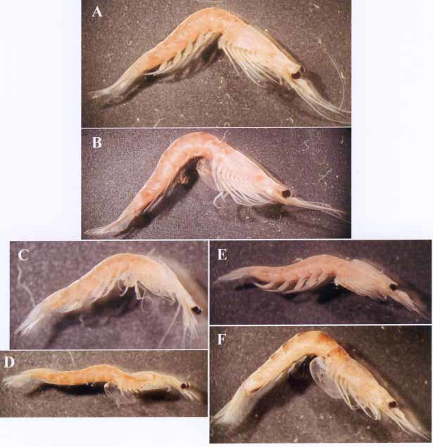
Tenagomysis longisquama sp. nov. ( Figs 1A, B View FIGURE 1. A, T , 2–5 View FIGURE 2 View FIGURE 4 View FIGURE 5 )
Material examined
All material is from Timaru, South Island, New Zealand.
Holotype: NIWA 3927 (H878), ɗ ( 23.1 mm), dissected, stn TM065, 44°38.7´S 171°26.0´E, 13 Feb. 2002, 7 m, epibenthic sled. Paratypes: NIWA 3928 (P1433), 1 Ψ ( 26.1 mm), dissected, same data as holotype; NIWA 3929 (P1434), 15 Ψ ( 20.6–24.6 mm), same data as holotype; NIWA 3930 (P1435), 2 ɗ (18.2, 22.5 mm) and 2 Ψ (19.0, 22.1 mm), stn TM060, 44°39.1´S 171°25.8´E, 13 Feb. 2002, 9 m, epibenthic sled; NSMTCr 16092, 1 ɗ ( 19.3 mm) and 1 Ψ ( 20.9 mm), stn TM063, 44°38.7´S 171°26.0´E, 13 Feb. 2002, 7 m, epibenthic sled.
Other material. NIWA 3931: 1 ɗ (broken), stn TM056, 44°38.7´S 171°26.0´E, 12 Feb. 2002, 6.5 m, epibenthic sled. NIWA 3932: 4 ɗ (13.8–22.0 mm), 2 imm. ɗ (12.4, 12.8 mm), 22 Ψ ( 18.7–23.6 mm) and 2 imm. Ψ (15.6, 17.6 mm), stn TM058, 44°39.2´S 171°26.0´E, 13 Feb. 2002, 9 m, epibenthic sled. NIWA 3943: 2 ɗ ( 18.6 mm, damaged), 4 Ψ ( 19.6–24.4 mm) and 5 imm. Ψ ( 13.2–19.5 mm), stn TM059, 44°39.2´S 171°26.0´E, 13 Feb. 2002, 9 m, epibenthic sled. NIWA 3944: 6 ɗ ( 17.5–19.6 mm), 4 imm. ɗ (damaged), 1 Ψ (damaged) and 6 imm. Ψ ( 15.8–17.4 mm), stn TM060, 44°39.1´S 171°25.8´E, 13 Feb. 2002, 9 m, epibenthic sled. NIWA 3945: 3 ɗ ( 18.3 mm, damaged), 10 Ψ ( 18.6–24.4 mm) and 2 imm. Ψ (15.2, 16.8 mm), stn TM061, 44°39.1´S 171°25.6´E, 13 Feb. 2002, 8 m, epibenthic sled. NIWA 3946: 2 ɗ (14.6, 15.3 mm) and 1 Ψ (broken), stn TM062, 44°39.1´S 171°25.8´E, 13 Feb. 2002, 9 m, epibenthic sled. NIWA 3947: 8 ɗ ( 13.5–17.6 mm), 12 Ψ ( 18.3–26.7 mm) and 4 imm. Ψ ( 16.4–17.3 mm), stn TM063, 44°38.7´S 171°26.0´E, 13 Feb. 2002, 7 m, epibenthic sled. NIWA 3948: 8 ɗ ( 16.2–18.8 mm), 1 imm. ɗ ( 9.8 mm), 23 Ψ ( 17.6–23.3 mm) and 4 imm. Ψ (13.4–17.0 mm), same data as holotype. NIWA 3949: 1 Ψ (damaged), stn TM070, 44°39.2´S 171°26.0´E, 13 Feb. 2002, 9 m, Shipek benthic grab.
Description
Body robust ( Fig. 1A, B View FIGURE 1. A, T ). Thoracic somites without sternal processes. Abdominal somites smooth, gradually increasing in length from first to fifth somites, sixth somite 1.7– 1.8 times as long as fifth.
Carapace anteriorly produced into long, triangular rostral plate with acute apex reaching distal margin of second segment of antennular peduncle ( Fig. 2 View FIGURE 2 A, B); anterolateral corner rounded; posterior margin not emarginate, leaving last 2 thoracic somites uncovered.
Eye large, not depressed dorsoventrally, 1.3–1.5 times as long as broad; cornea occupying distal 1/3 of eye; eyestalk without papilliform process ( Figs. 1A, B View FIGURE 1. A, T , 2 View FIGURE 2 A, B).
Antennular peduncle robust; in male third segment as long as first segment, 1.2 times as long as broad, with developed appendix masculina ( Fig. 2 View FIGURE 2 A); in female first segment 1.2 times as long as third segment, third segment 1.1 times as long as broad ( Fig. 2 View FIGURE 2 B).
Antennal scale 15 times as long as broad, acute distally, extending beyond apex of antennular peduncle by 2/3 of its length in male and by 7/10 of its length in female ( Fig. 2 View FIGURE 2 A–D). Antennal peduncle extending to proximal 2/9 of scale in male and to proximal 1/6 of scale in female, second and third segments subequal in length ( Fig. 2 View FIGURE 2 C, D). Antennal sympod with 2 spiniform processes, one at lateral distal angle and another on ventral surface ( Fig. 2 View FIGURE 2 C, D).
Labrum without frontal process.
Mandibular palp with second segment expanded in middle part, armed with numerous setae on mesial and lateral margins; third segment half as long as second segment ( Fig. 2 View FIGURE 2 E).
Mesial lobe of maxillule armed with 1 long, spiniform, 3 robust, plumose and 2 slen der setae on mesial margin, with 3 long, barbed, spiniform setae on distal margin, 8 slender setae on lateral margin, and with 13 slender setae on distal third of ventral surface ( Fig. 2 View FIGURE 2 F). Lateral lobe of maxillule armed with 13 or 14 robust spines on distal margin and with 5–7 long setae on ventral surface ( Fig. 2 View FIGURE 2 F).
Endopod of maxilla with second segment 1.1–1.2 times as long as broad, armed with 8–10 spiniform setae on distal margin ( Fig. 2 View FIGURE 2 G). Exopod of maxilla extending beyond distal margin of first segment of endopod ( Fig. 2 View FIGURE 2 G).
First thoracopodal endopod short, robust (Fig. 3A). Second thoracopodal endopod short (Fig. 3B). Third to eighth thoracopdal endopods long; carpopropodus divided into 10–16 subsegments by transverse articulations (Fig. 3C, D). Thoracopodal exopod with flagellum 9segmented in first and eighth pairs and 10segmented in second to seventh pairs; basal plate with tiny, acute process at distolateral corner (Fig. 3A–D).
Penis armed with 6 mesially directed setae on distal margin and with about 10 setae on distal half of lateral surface ( Fig. 4 View FIGURE 4 A).
Marsupium composed of 3 pairs of developed oostegites; oostegites on seventh thoracopod with small posterior lobe.
All pleopods of male developed, biramous ( Fig. 4 View FIGURE 4 B–E, H). First pleopod with endopod reduced to unsegmented lobe, exopod 6segmented ( Fig. 4 View FIGURE 4 B). Second and third pleopods with 5segmented endopod, and 6segmented exopod longer than endopod ( Fig. 4 View FIGURE 4 C–E). Fourth pleopodal endopod 5segmented, extending to distal end of antepenultimate segment of exopod ( Fig. 4 View FIGURE 4 E). Fourth pleopodal exopod 7segmented; proximal segment with small mesial lobe, which is curved distally and spinulated on proximal margin ( Fig. 4 View FIGURE 4 F); antepenultimate segment armed with long, strong, spiniform seta, arising from near distal end, slightly curved mesially, and extending slightly beyond apex of terminal setae; penultimate segment long, 1.5 times longer than preceding segment, 3.7 times as long as broad, armed with long, spiniform seta, which is almost straight, arising from distal end, and extending beyond apex of strong seta from preceding segment; terminal segment short, 0.4 as long as antepenultimate segment, armed with 2 subequal, terminal setae, which are 4 times as long as own segment ( Fig. 4 View FIGURE 4 E–G). Fifth pleopod with 6segmented exopod; 5 segmented endopod with rectangular lobe armed with single seta on proximal segment in addition to usual pseudobranchial lobe ( Fig. 4 View FIGURE 4 H). Pseudobranchial lobe of all pleopods developed, slightly expanded distally in second to fifth pleopods ( Fig. 4 View FIGURE 4 B–E, H).
All pleopods of female reduced to unsegmented single lobe, flattened, gradually increasing in length from first to fifth pleopods; third to fifth pleopods knifeshaped with acute apex (Fig. 3E–L).
Tenagomysis longisquama sp. nov. A–D, ( 23.1 mm), holotype, NIWA 3927 (H878); E–L, female ( 26.1 mm), paratype, NIWA 3928 (P1433). A–C, first to third left thoracopod, posterior; D, sixth left thoracopod, posterior; E–I, first to fifth left pleopods of female, posterior; J–L, apical part of third to fifth left pleopods, posterior.
Uropodal endopod slightly longer than telson, armed on mesial ventral margin from statocyst region to near apex with 44–52 spines becoming larger distally, and on ventral surface of proximal 1/3 with 13–20 spines arranged irregularly ( Fig. 5 View FIGURE 5 A, B). Uropodal exopod 1.4 times as long as endopod ( Fig. 5 View FIGURE 5 B).
Telson 1.4 times as long as last abdominal somite, 2.6–2.7 times as long as broad, tapering towards posterior end, with apical cleft ( Fig. 5 View FIGURE 5 B). Lateral margin of telson armed with 45–48 subequal small spines on entire length ( Fig. 5 View FIGURE 5 B). Apical cleft 1/4 of telson length, narrow in anterior 2/3, diverging in posterior 1/3, with pair of plumose setae on anterior end; lateral margin of cleft convex, serrulated on entire length ( Fig. 5 View FIGURE 5 B).
Remarks
Tenagomysis longisquama is similar to T. tenuipes Tattersall, 1918 View in CoL , in having the antennal scale with an acute apex, the 10 to 16subsegmented carpopropodus of the endopods of the third to eighth thoracopods, and in the shape and armature of the telson. However, it is separated from T. tenuipes View in CoL by the character states given in Table 1 View TABLE 1 .
Tenagomysis longisquama is readily distinguished from the other species of the genus, except T. tenuipes View in CoL , by a combination of the long rostrum with an acute apex, elongate antennal scale with an acute apex, 10 to 16subsegmented carpopropodus of the third to eighth thoracopodal endopods, arrangement of spines on the mesial margin of the uropodal endopod, and shape and armature of the telson.
The female pleopods of a Tenagomysis are here described for the first time.


| NIWA |
National Institute of Water and Atmospheric Research |
No known copyright restrictions apply. See Agosti, D., Egloff, W., 2009. Taxonomic information exchange and copyright: the Plazi approach. BMC Research Notes 2009, 2:53 for further explanation.
|
Kingdom |
|
|
Phylum |
|
|
Class |
|
|
Order |
|
|
Family |
|
|
SubFamily |
Mysinae |
|
Tribe |
Leptomysini |
|
Genus |
Tenagomysis longisquama
| Fukuoka, Kouki & Bruce, Niel L. 2005 |
T. tenuipes
| Tattersall 1918 |