
Stictococcus vayssierei Richard
|
publication ID |
https://doi.org/ 10.5281/zenodo.196462 |
|
DOI |
https://doi.org/10.5281/zenodo.6207527 |
|
persistent identifier |
https://treatment.plazi.org/id/0396675F-5252-9A24-58CB-FB26FD23FF15 |
|
treatment provided by |
Plazi |
|
scientific name |
Stictococcus vayssierei Richard |
| status |
|
Stictococcus vayssierei Richard View in CoL
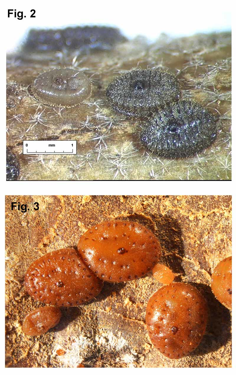
( Figs 3 View FIGURE 3 , 10 View FIGURE 10 )
Stictococcus vayssierei Richard, 1971: 592 View in CoL ; 1976: 668.
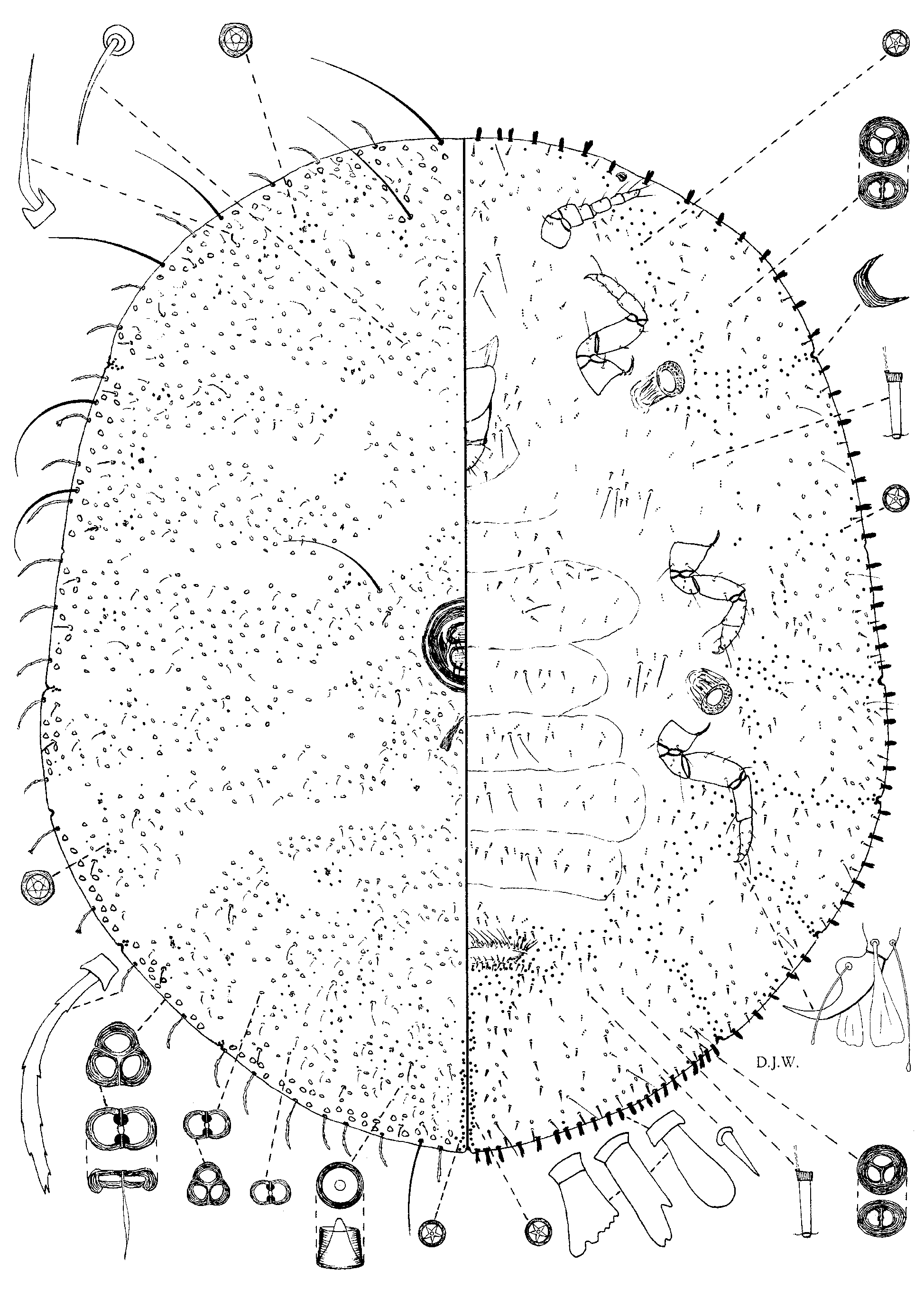
Description. Body of adult female on microscope slide broadly oval, 1.20–3.50 mm long, 1.00– 3.10 mm wide, membranous at first, becoming sclerotized at maturity. Antennae 160–200 µm long, tapering to terminal segment, 6-segmented. Legs well developed, hind coxa 65–75 µm long, hind trochanter + femur 190–230 µm long, hind tibia + tarsus 175–200 µm long, claw stout at base then curving to narrow tip, 35–40 µm long, with one slender clubbed digitule longer than claw and one widely expanded digitule. Tarsal digitules clubbed, distal inner tarsal digitules widely expanded. Ratio of lengths of hind tibia + femur to hind trochanter + femur 0.78–0.97. Ratio of lengths of hind tibia to tarsus 0.77–1.00. Labium 125–165 µm long, 100–125 µm wide, shorter than clypeolabral shield, with a ridge-like structure present on each side of basal segment extending to clypeolabral shield. Mesothoracic spiracles 100–125 µm long, 90–100 µm wide. Metathoracic spiracles 125– 175 µm long, 100–125 µm wide. Oval area of each spiraclular opening with many papilla-like structures. Vulva 550–900 µm wide. Anus oval, 200–250 µm long, 190–200 µm wide, situated near mid-dorsum. A pair of saddle-shaped apodemes opening posteriorly. Eyes oval, 30–40 µm long, 25–35 µm wide. Minute marginal clefts, each with a sclerotized plate, usually totalling 11, present opposite each spiracle, opposite each hind coxa, 2 pairs on abdomen and one at apex of abdomen. A minute cleft, without any sclerotized plate, sometimes discernible opposite each mesothoracic coxa.
Dorsal surface of body with marginal, curved barbed setae, mostly about 75 µm long, except some about 85 µm long on apparent anal lobes, and some about 110 µm long on head margin. A few long marginal flagellate setae present, each 135–175 µm long, a few on head margin about 375 µm long and some on apparent anal lobes about 350 µm long. Normal dorsal setae mostly curved and flagellate, about 30 µm long, present in more or less segmental rows. Longer flagellate setae 45–50 µm long, angled near base, present around margins and submargins and area lateral to anal ring, associated with shallow depressions, each depression also with a few discoidal pores in centre in inner polar region, each pore about 6.25 µm in diameter. Large bilocular pores, each 10.0–12.5 µm long, 7.5 µm wide, or trilocular pores about 12.5 µm wide, present in a marginal zone. Similar pores but smaller, most 10.0–12.5 µm long, but with others about 6.25 µm long, present in whorls or circles, accompanied by small circular pores with a peg-like centre, distributed over surface, some associated with shallow depressions. Minute quinquelocular pores, each about 5 µm in diameter, present in a short medial row extending forwards from apex of abdomen.
Ventral surface with a marginal series of club-shaped setae, mostly 30 µm long, about 10.0 µm wide, sometimes replaced by a few expanded setae about 25 µm long, 20 µm wide at apex and base about 15 µm wide, present near sclerotized clefts. Occasionally, these expanded setae more numerous than club-shaped setae. Minute quinquelocular pores, about 5 µm in diameter, present around vulva, then curving medially to form a row between vulva and minute sclerotized cleft at apex of abdomen. Others present lateral to vulva and extending forwards submedially to metathoracic spiracles, sparsely to mesothoracic spiracles and then anteriorly to eyes; distinct rows also present between each spiracle and margin and on abdomen to each minute marginal cleft; others sparsely distributed around margins. Thick-rimmed pores, either bilocular and 10.0 µm long, 5.0 µm wide, or trilocular pores, about 10.0 µm in diameter, present within marginal areas demarked by minute quinquelocular pores. Minute tubular ducts, each about 15 µm long, 5 µm wide at cup end, present across abdominal segments, in medial areas of head and thorax and with a few present in marginal areas. Spine-like setae, each about 20 µm long, present across abdominal segments and within marginal areas demarked by minute quinquelocular pores; others sparsely distributed in medial areas of head and thorax. Some similar longer setae, about 30 µm long, present near margins. Slender flagellate setae 75– 100 µm long, present between antennae, and others 50–75 µm long, distributed across abdominal segments and near spiracles. Thicker flagellate setae, 75–125 µm long, present on anterior edge of vulva.
Type data. Cameroun (Yaoundé, 13.xii.1969) sur Manihot esculenta (racines) déposée au Muséum national de Paris.
Material examined. Type material: labelled in ink directly on slide, left side, “ Holotype, 7611/2, MNHN – Paris.” Right side, “1er st larv. Ƥ Stictococcus vayssierei Richard 1er stade larvaire Ƥ s/ Manihot esculenta , CAMEROUN, 13 XII. 1969 (N’Jensi)” [ MNHN].
Paratypes, same data as holotype, 3 adult males on 3 separate slides, Yaoundé, 6 1st-instar Ƥ, on 6 separate slides, Yaoundé. [ MNHN].
Allotype, same data as holotype, 1 adult male [ MNHN].
Other material. Cameroon, 2 km South of Ngoila, nr Adjela, on roots of Manihot esculenta , 1.ii.2004, R. Hanna; on roots of Colocasia esculenta (cocoyam, Araceae ), 28.i.2004, R. Hanna; Aroston, on roots of Manihot esculenta (ant attended), 1.ii.2004, R. Hanna: Mboalmayo, on tubers of Manihot esculenta, 1992 , W. Hammond (all BMNH); Yaoundé, Kala, on roots of Manihot esculenta , 27.iv.1993, A. Dejean ( MNHN).
Congo (Brazzaville), Brazzaville, on unidentified plant, 1907, E. Roubaud & A. Weiss [ MNHN].
Democratic Republic of the Congo (N.W. Bass Zaire), on roots of Manihot esculenta ( BMNH).
Equatorial Guinea, Bioko, Belebou, on roots of Xanthosoma sp. ( Araceae ), 5.ii.2005, R. Hanna ( BMNH). Uganda, Ruwenzori area, Field 3, on roots of Manihot esculenta (second instars), 20.vii.2004, G. Goergen ( BMNH).
Comments. The accompanying illustration of the adult female of S. vayssierei is based on preparations of the adult female with identical data to those of the holotype. These specimens were mounted by Richard during her studies of the first instars and adult males but, although they were seen by Richard, they were not included in her description. The adult females, therefore, must be regarded as part of the original material only. The ventral marginal setae of this species are bullet-shaped as in S. formicarius but the latter species possesses flower-shaped dorsal setae and these are absent from S. vayssierei . Furthermore, the dorsal depressions are very shallow and sometimes difficult to locate in S. vayssierei whereas in all the other species the dorsal depressions are much more conspicuous.
| MNHN |
Museum National d'Histoire Naturelle |
No known copyright restrictions apply. See Agosti, D., Egloff, W., 2009. Taxonomic information exchange and copyright: the Plazi approach. BMC Research Notes 2009, 2:53 for further explanation.
|
Kingdom |
|
|
Phylum |
|
|
Class |
|
|
Order |
|
|
Family |
|
|
Genus |
Stictococcus vayssierei Richard
| Williams, Douglas J., Matile-Ferrero, Danièle & Miller, Douglass R. 2010 |
Stictococcus vayssierei
| Richard 1971: 592 |