
Simulium wygodzinskyorum Coscarón & Py-Daniel, 1989 : 318
|
publication ID |
https://doi.org/ 10.11646/zootaxa.4117.2.10 |
|
publication LSID |
lsid:zoobank.org:pub:A1FC7360-8804-4456-97BC-2B359A09D16D |
|
DOI |
https://doi.org/10.5281/zenodo.6077955 |
|
persistent identifier |
https://treatment.plazi.org/id/0396BD4F-FF8A-FFE4-2CFC-FDD640713B9E |
|
treatment provided by |
Plazi |
|
scientific name |
Simulium wygodzinskyorum Coscarón & Py-Daniel, 1989 : 318 |
| status |
|
Simulium wygodzinskyorum Coscarón & Py-Daniel
Simulium (Grenieriella) wygodzinskyorum Coscarón & Py-Daniel 1989: 319 View in CoL .
Simulium (Trichodagmia) wygodzinskyorum Crosskey & Howard 1997: 79 View in CoL ; 2004: 74; Miranda Esquivel & Coscarón 2001: 429 – 435; Coscarón-Arias 2003: 191, 206; Coscarón, Coscarón-Arias & Papavero 2008: 50; Hernández-Triana 2011: 170; Adler & Crosskey 2016.
Larva (last-instar) ( Figs 2 View FIGURE 2 A–E to 6A–I). Body length 4.6–5.0 mm (mean = 4.8 mm, SD = 0.13, n = 5); head capsule, mean lateral length 0.72–0.9 mm (mean = 0.8 mm, SD = 0.05, n = 6). General color, dorso-laterally varying from dark green to brownish green, ventrally whitish ( Figs 2 View FIGURE 2 A–E).
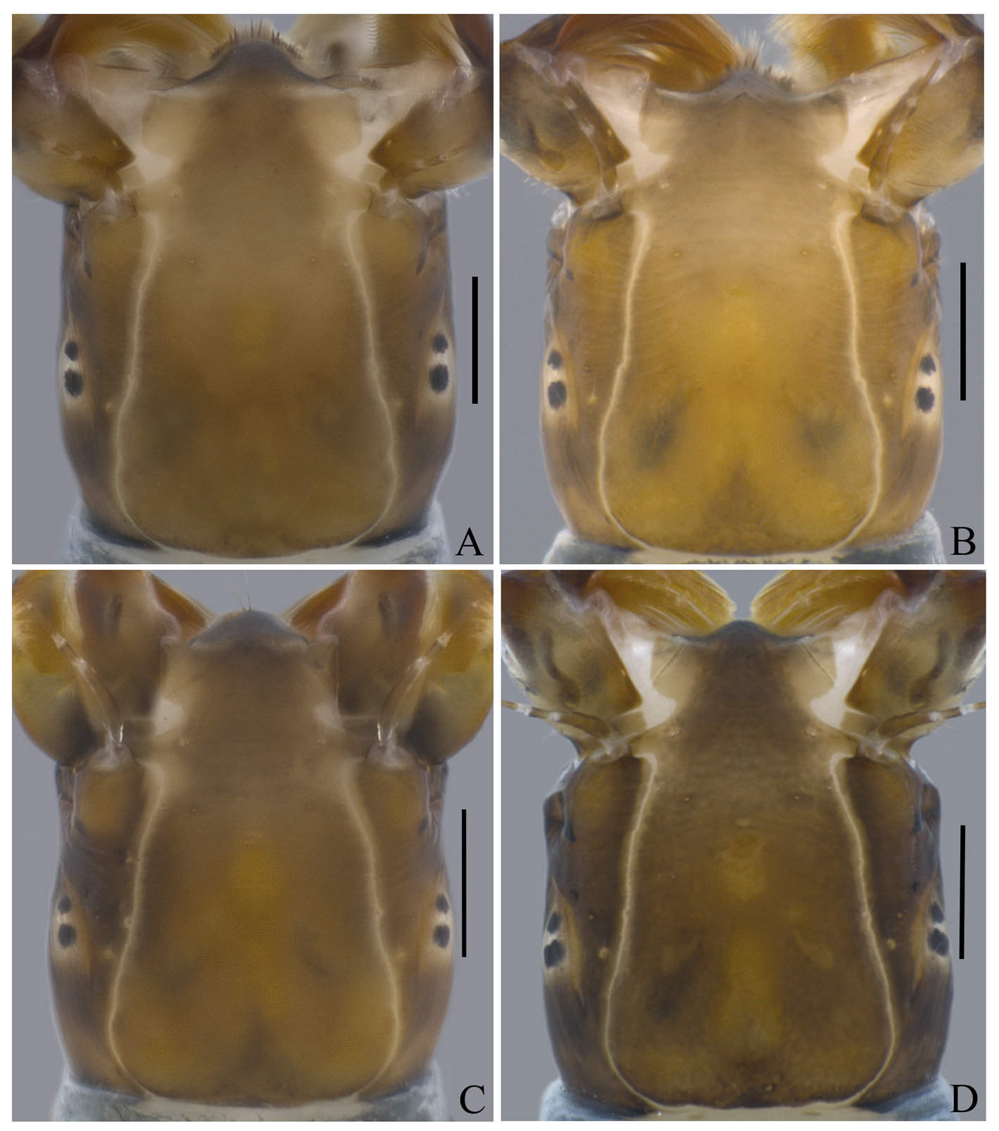
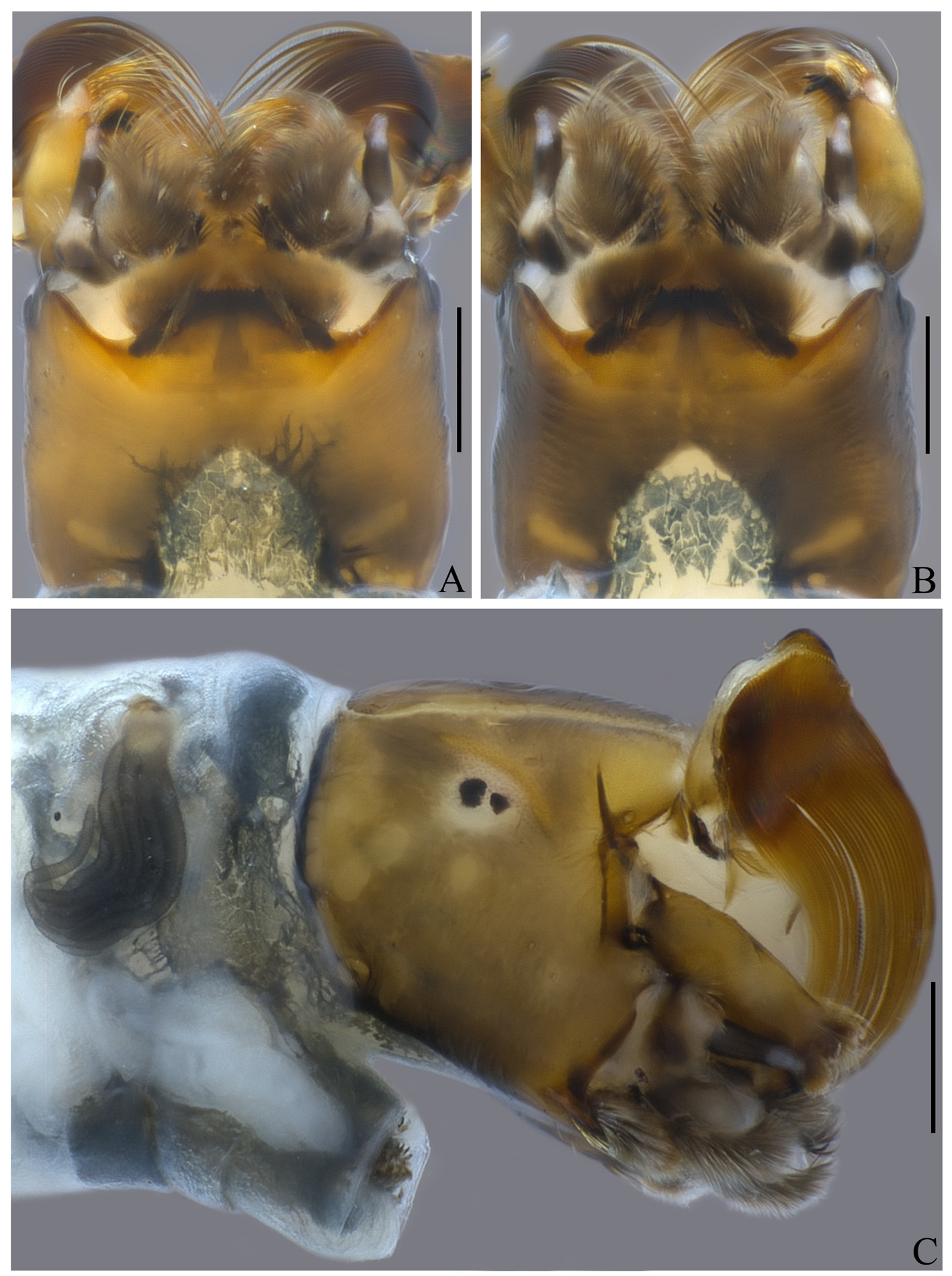
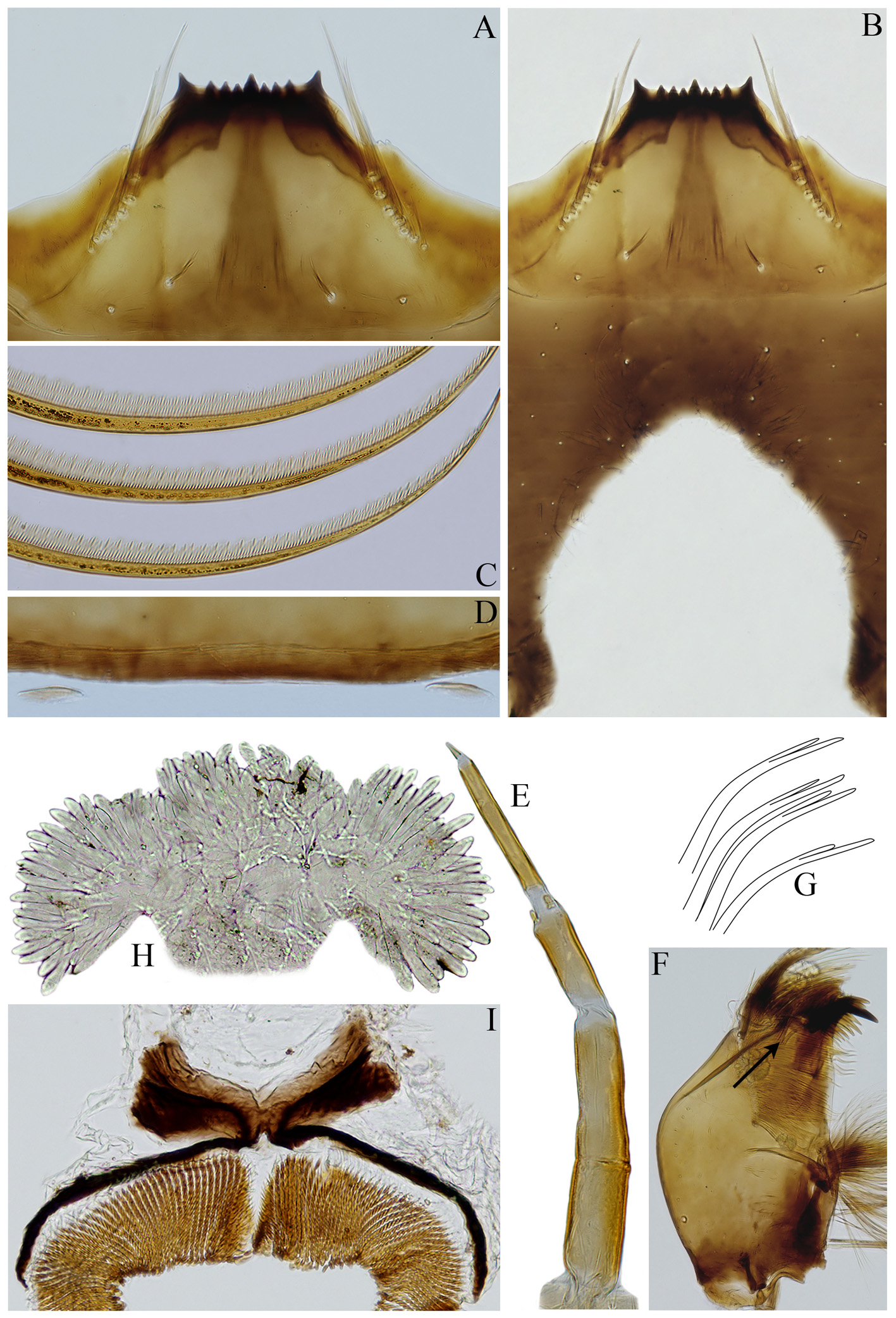
Head capsule wrinkled, with small simple setae distributed on all over it; general coloration varying from brown to yellowish brown ( Figs 4 View FIGURE 4 A–D; 5A–C); in dorsal view, frontoclypeal apotome with central area lighter and posterior margin, usually, with a brown triangular mark ( Figs 4 View FIGURE 4 B–C); with 1 + 1 lateral, short, elongated marks (more evident in some individuals) ( Figs 4 View FIGURE 4 A–D). Head, in dorsal view, with 1 + 1 small, whitish spots located above the ocelli ( Figs 4 View FIGURE 4 A- D); in lateral view with yellowish marks near posterior margin ( Fig. 5 View FIGURE 5 C); in ventral view with 1 + 1 elongated yellowish marks ( Figs 5 View FIGURE 5 A, B), located near the posterior margin. Cervical sclerites small, elliptical, free in the membrane ( Fig. 6 View FIGURE 6 D). In ventral view, postgenal cleft deep, subtriangular, with lateral margins parallel near posterior margins, covered with pigmented tissue ( Figs 5 View FIGURE 5 A, B); postgenal bridge 0.56 times the hypostoma length ( Fig. 6 View FIGURE 6 B); subesophageal ganglion not pigmented ( Figs 5 View FIGURE 5 A, B). Antenna ( Fig. 6 View FIGURE 6 E) shorter than labral fan stalk; proximal article shorter than distal and medial articles, distal article shorter than the medial one, proportions of articles (proximal: medial: distal, excluding apical sensillum) 1: 0.36–0.5: 1.33–1.75 (n= 10); general coloration yellowish brown, except base of distal article and part of half distal region of medial article whitish (see Fig. 6 View FIGURE 6 E). Labral fan with 30–42 primary rays, general coloration yellowish brown; rays with microtrichia arranged as follows: an alternating series of one long microtrichium and three slightly shorter ones ( Fig. 6 View FIGURE 6 C). Hypostoma ( Figs 6 View FIGURE 6 A, B) with anterior margin straight, strongly pigmented; medial region with a dark longitudinal stripe wide at the posterior margin; with nine simple teeth; median and lateral teeth similar in length and longer than 3 + 3 sublateral teeth; lateral teeth wider at their base than the median and sublateral teeth; with 3–5 + 3–5 lateral serrations; hypostoma with 1 + 1 lines of 7–8 sublateral seta, 1 + 1 bifid setae on basal 1/5 and 1–2 + 1–2 small setae near hypostomal groove. Mandibular teeth: one apical, two external; three subapical; 6 or 7 internal teeth; one mandibular serration and one small mandibular sensillum (sensu Craig & Craig 1986); four latero-mandibular processes thick and bifurcated on apical 1/3 ( Figs 6 View FIGURE 6 F, G). Body cuticle with sparsely distributed short, simple setae; abdomen with 1 + 1 dorso-lateral tubercles on segments II–VIII (less frequently on segments I–VIII or III–VIII), varying from well-developed to poorly developed ( Figs 2 View FIGURE 2 A–E; 3A, B); ventral tubercles absent; gill histoblast dissected with 12 filaments. Arms of anal sclerite as in Figure 6 View FIGURE 6 I, anterodorsal arms 0.45–0.60 as long as posteroventral arms, associated with some thin, short setae. Posterior circlet bearing 169– 191 rows (n= 6) with 20–29 hooks (n= 9). Rectal papilla ( Fig. 6 View FIGURE 6 H) with three branches, each with approximately 25–35 finger-shaped lobes (n= 2); total number of lobes varying from 89 to 92.
Pupa ( Figs 7 View FIGURE 7 A–B to 9A–J). Cocoon tightly woven, light brown, boot-shaped, with anterior rim slightly thickened ( Figs 7 View FIGURE 7 A, B, 8C). Thorax with 4 + 4 pairs of multiramous trichomes (3–4 branches) ( Figs 8 View FIGURE 8 D). Gills ( Figs 7 View FIGURE 7 A, B, 8A) with 12 grayish brown, stout, crenulated filaments ( Fig. 8 View FIGURE 8 B) distributed in a three-dimensional pattern: main trunk short, giving rise to four sets of primary branches, two with two secondary branches and two with four secondary branches; filaments branching at different heights; with tips pointed or slightly rounded, not sclerotized ( Figs 8 View FIGURE 8 A, B); basal fenestra evident ( Fig. 8 View FIGURE 8 A). Abdominal ( Fig. 9 View FIGURE 9 ) tergite I dark, sclerotized on proximal ¾, with one pair of anterolateral setae; tergite II with a small sclerotized area on anteromedial region, with 4 + 4 central stout setae in the medial region of the tergite ( Fig. 9 View FIGURE 9 A), two pairs of short sublateral setae; tergites III and IV each with 4 + 4 anteriorly directed pairs of stout, simple hooks on posterior margin of the central region of the tergite ( Figs 9 View FIGURE 9 B, C); tergite III with three pairs of short lateral setae on posterior margin; tergite V without setae or spines; tergite VI with comb-like groups of fine posteriorly directed microspines on antero-lateral region; tergites VII with a row of comb-like groups of fine posteriorly directed microspines near anterior margin ( Fig. 9 View FIGURE 9 D); tergite VIII with a sclerotized comb of strong spines (posteriorly directed) located along the anterior region of the tergite ( Fig. 9 View FIGURE 9 E); tergite IX with a sclerotized comb of strong spines (inward directed), located along the anterior margin of the tergite ( Fig. 9 View FIGURE 9 F), median region of the comb with scarce spines; tergite IX with one pair of short spines, posteriorly. Sternites IV–VII with comb-like groups of fine posteriorly directed microspines on central, anterior region ( Fig. 9 View FIGURE 9 H); sternite VIII with comb-like groups of fine posteriorly directed microspines on Male genitalia ( Figs 10 View FIGURE 10 A–G). Gonocoxite and gonostylus ( Figs 10 View FIGURE 10 A, B, G) yellowish brown, covered with long, thick setae; in ventral view, gonocoxite around 1.4 times wider than long, with proximal and apical region sclerotized and a central triangular region not sclerotized ( Fig. 10 View FIGURE 10 G); gonostylus longer than wide ( Figs 10 View FIGURE 10 A, G), around 1.2 times the gonocoxite length, bearing one stout spinule at the apex. Ventral plate ( Figs 10 View FIGURE 10 C, D, F) rectangular (three times wider than high), with main body and median anterior projection covered with thin setae; in dorsal view, distal margin of ventral plate body concave medially, the base of the median anterior projection is inserted posteriorly to this concave margin, represented in this view by a rounded invaginated region that occupies almost the entire length of the mesal region of the ventral plate ( Fig. 10 View FIGURE 10 C); in ventral view, median anterior projection as wide as long, slightly surpassing the distal margin ( Fig. 10 View FIGURE 10 D); in lateral view, median anterior projection elongated, anteriorly projected, with basal region wide, narrowing toward apical region ( Fig. 10 View FIGURE 10 E); in ventral view (with the apex tilted dorsally) median anterior projection 1.6 times longer than wide, apex rounded ( Fig. 10 View FIGURE 10 F); proximal margin of the ventral plate body at the median region V-shaped ( Figs 10 View FIGURE 10 C, D, F).
Material examined. Peru, Chanchamayo Province, San Ramón District , Junín Region , Uarango River (11°08'41.2"S, 75°24'11.9"W, 1017 m), 09/viii/2014, N. Hamada, J.M.C. Nascimento, R.L. Ferreira-Keppler & S.M. Clemente, cols.: 12 larvae on slides—4 ( INPA) and 8 (MHN-UNMSM), 3 pupae on slides—2 ( INPA) and 1 (MHN- UNMSM), 8 larvae in ethanol—3 ( INPA) and 5 ( MHN-UNMSM), male (pharate adult) on slide ( INPA), 5 non-last instar larvae—2 ( INPA) and 3 ( MHN-UNMSM) GoogleMaps .
Other material examined.
Simulium wygodzinskyorum Coscarón & Py-Daniel, 1989: 318 View in CoL .
Paratype male and pupal exuviae. Peru: Junin, San Ramón, Estancia El Naranjal , altitude 1000 m; 20-vii-1965 (P. & B. Wygodzinsky) (housed at the Museo de La Plata—MLP, La Plata, Argentina).
Larva on slide. Ecuador, Ningo, vii-2000, (C.L. Coscarón-Arias) (housed at the Museo de La Plata—MLP, La Plata, Argentina).


| INPA |
Instituto Nacional de Pesquisas da Amazonia |
No known copyright restrictions apply. See Agosti, D., Egloff, W., 2009. Taxonomic information exchange and copyright: the Plazi approach. BMC Research Notes 2009, 2:53 for further explanation.
|
Kingdom |
|
|
Phylum |
|
|
Class |
|
|
Order |
|
|
Family |
|
|
Genus |
Simulium wygodzinskyorum Coscarón & Py-Daniel, 1989 : 318
| Nascimento, Jeane Marcelle Cavalcante Do, Hamada, Neusa & Huamantinco-Araujo, Ana A. 2016 |
Simulium (Trichodagmia) wygodzinskyorum
| Hernandez-Triana 2011: 170 |
| Coscaron-Arias 2003: 191 |
| Esquivel 2001: 429 |
| Crosskey 1997: 79 |
Simulium (Grenieriella) wygodzinskyorum Coscarón & Py-Daniel 1989 : 319
| Coscaron 1989: 319 |