
Metapontius walteri, Johnsson, Rodrigo & Neves, Elizabeth G., 2005
|
publication ID |
https://doi.org/ 10.5281/zenodo.169725 |
|
DOI |
https://doi.org/10.5281/zenodo.5686524 |
|
persistent identifier |
https://treatment.plazi.org/id/0396CE44-BA21-FFC9-FEE0-F96FFB5CBC37 |
|
treatment provided by |
Plazi |
|
scientific name |
Metapontius walteri |
| status |
sp. nov. |
Metapontius walteri sp. nov.
Figures 1–3 View FIGURE 1 View FIGURE 2 View FIGURE 3
Material examined.— Holotype: female, associated with the octocoral, Pachyclavularia violacea , from 2 m depth, on the western side of reef, at northern end of David, Eniwetok Atoll, Marshall Islands, USA, 29 June 1969, coll. Dr. A.G. Humes. Holotype deposited in the National Museum of Natural History (Smithsonian Institution) reg. no. USNM 1074640.
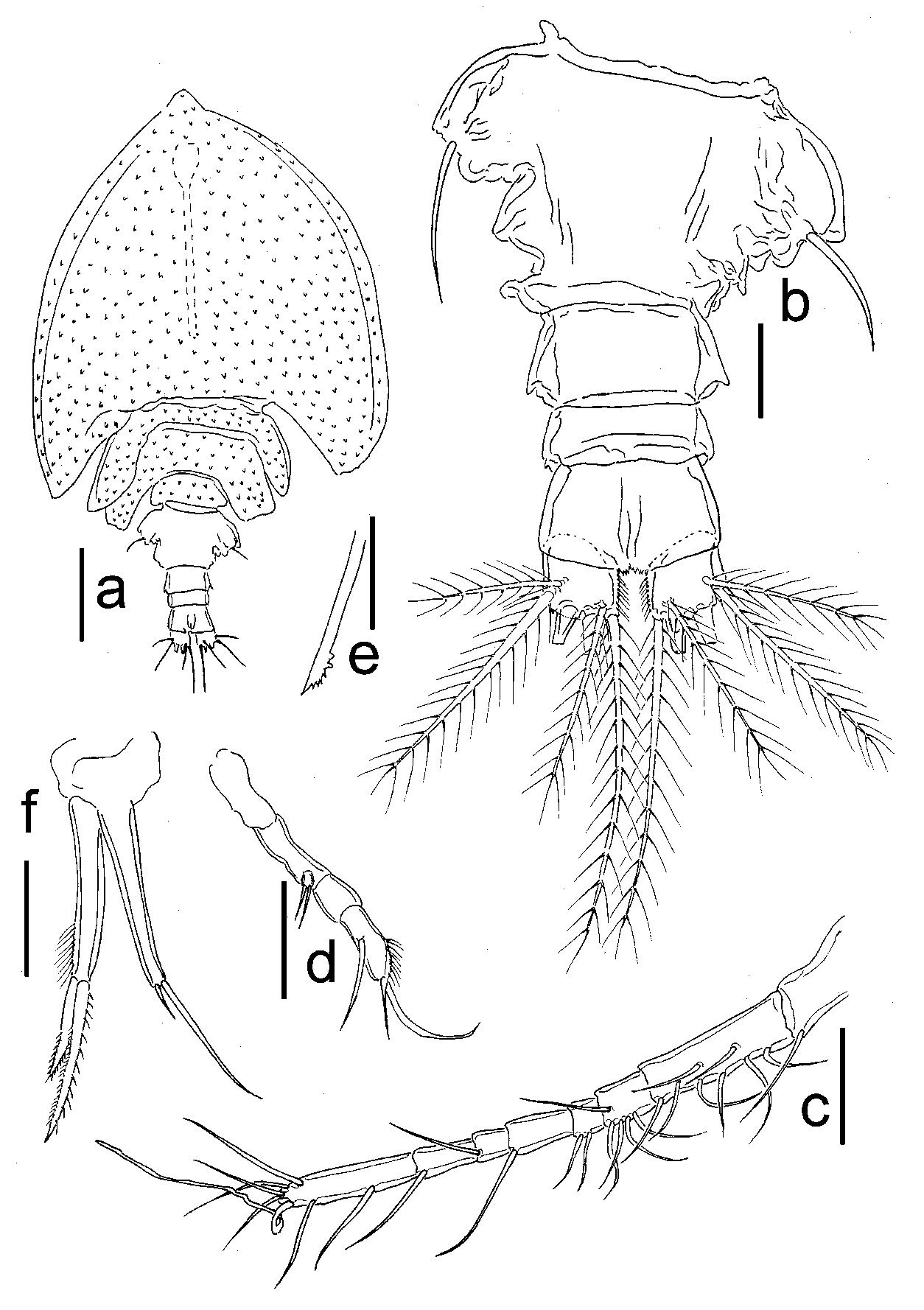
Description of female. Body length (excluding caudal setae) 1210 µm, greatest body width 775 µm, body length 1.6 times width. Body shape cyclopiform (fig. 1a), with sensillae covering prosome, cephalosome not imbricate. Cephalosome and pedigerous somites 2 and 3 with epimera pointed. Pedigerous somite 4 with epimera rounded. Prosome length:width ratio, 1.2:1. Ratio of prosome length to urosome, 3:1.
Urosome 5segmented. Genital doublesomite (fig. 1b) 134 x 237 µm, length:width ratio, 0.6:1, area of genital aperture projected laterally and armed with smooth seta. Three abdominal somites, all wider than long (56 x 113 µm; 31 x 95 µm; 61 x 100 µm), length:width ratios, 0.5:1, 0.3:1 and 0.6:1, respectively. Caudal rami 31 x 43 µm, width 1.4 times length, with row of setules on inner margin and armed with 6 setae. Seta I absent, setae IV and V broken. Length of setae II, III, VI, and VII; 68, 137, 174, and 105 µm, respectively; all plumose.
Antennule (fig. 1c) 280 µm long (not including setae) and 8segmented. Length of segments measured along their posterior margins: 30, 75, 24, 15, 29, 16, 29, and 63 µm, respectively. Segmental homologies and setation as follows. Roman numerals indicate the original segments followed by the number of setae in Arabic (Huys & Boxshall 1991): I1; IIVIII8; IXXIII5; XIV2; XVXVI1; XVIIXVIII1; XIXXX 1; XXIXXVIII6+ae. All setae smooth. Aesthetasc on segment XXI 105 µm long. Antenna (fig. 1d) 150 µm long (including distal seta), with basis 38 µm long. Endopod 2segmented; first segment 17 µm long, unarmed; second segment 35 µm long with 3 smooth setae, 1 medially placed and 2 distal, none modified as claw. Exopod 1segmented, armed with 2 distal setae.
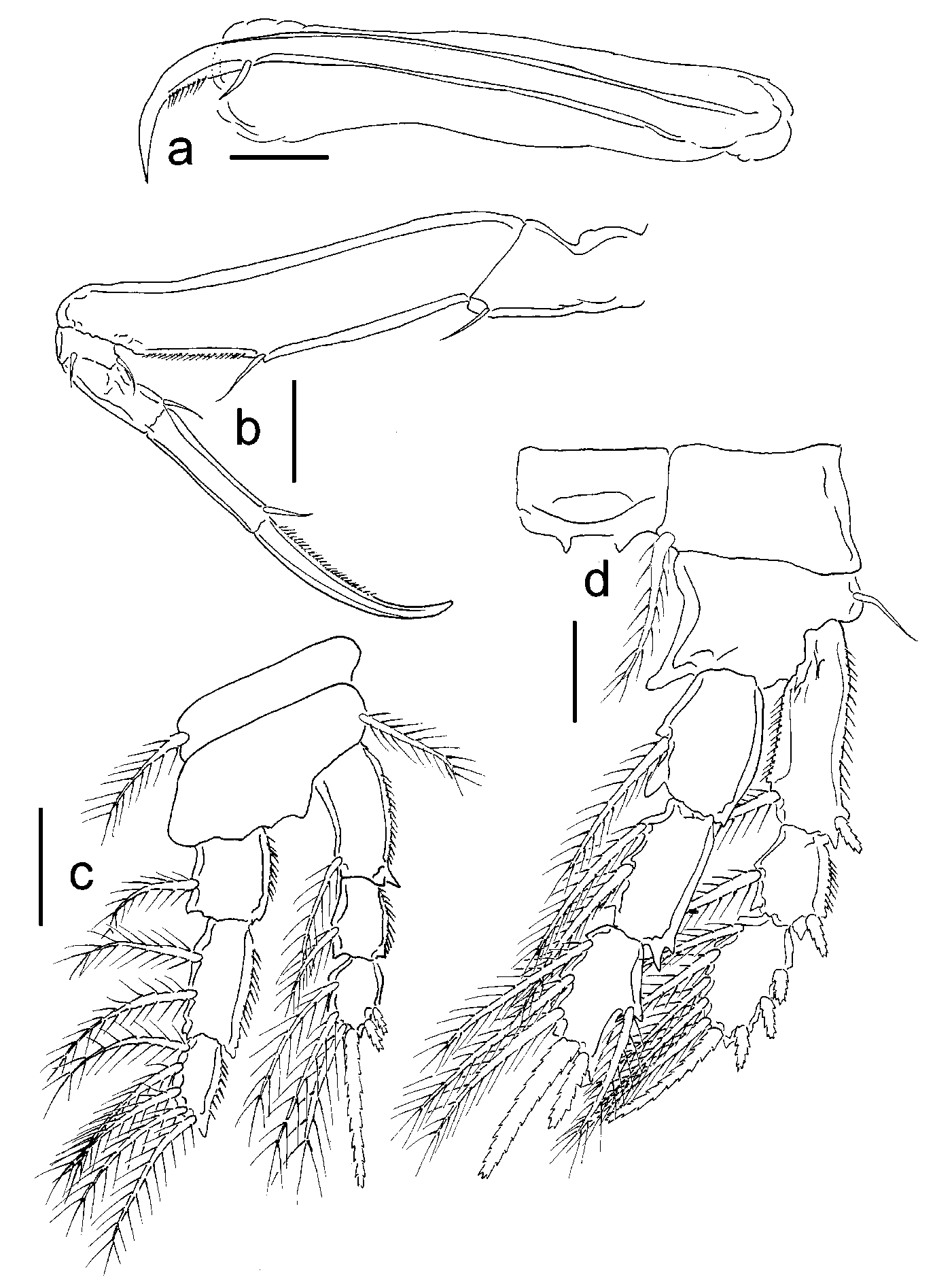
Oral cone (fig. 1a) produced into long, siphonlike distal portion, 406 µm long, 0.3 times bodylength. Mandible (fig. 1e) comprised of distally toothed stylet, palp absent. Maxillule (fig. 1f) bilobed, inner lobe 93 µm, armed with 2 stout, pinnate setae. Outer lobe 80 µm long, armed with 2 smooth setae. Maxilla (fig. 2a) with syncoxa 310 µm long, slender claw, 398 µm long, distally curved and showing small seta subdistally. Maxilliped (fig. 2b) 5segmented, comprising syncoxa 89 µm long, armed with small seta on inner margin, basis 242 µm long with small seta medially on inner margin. Endopod 3segmented, segments measuring 19, 56, and 77 µm long, respectively. First endopodal segment with 2 setae distally; second segment with 1 seta and third segment bearing curved, 110 µm long, claw, and seta.
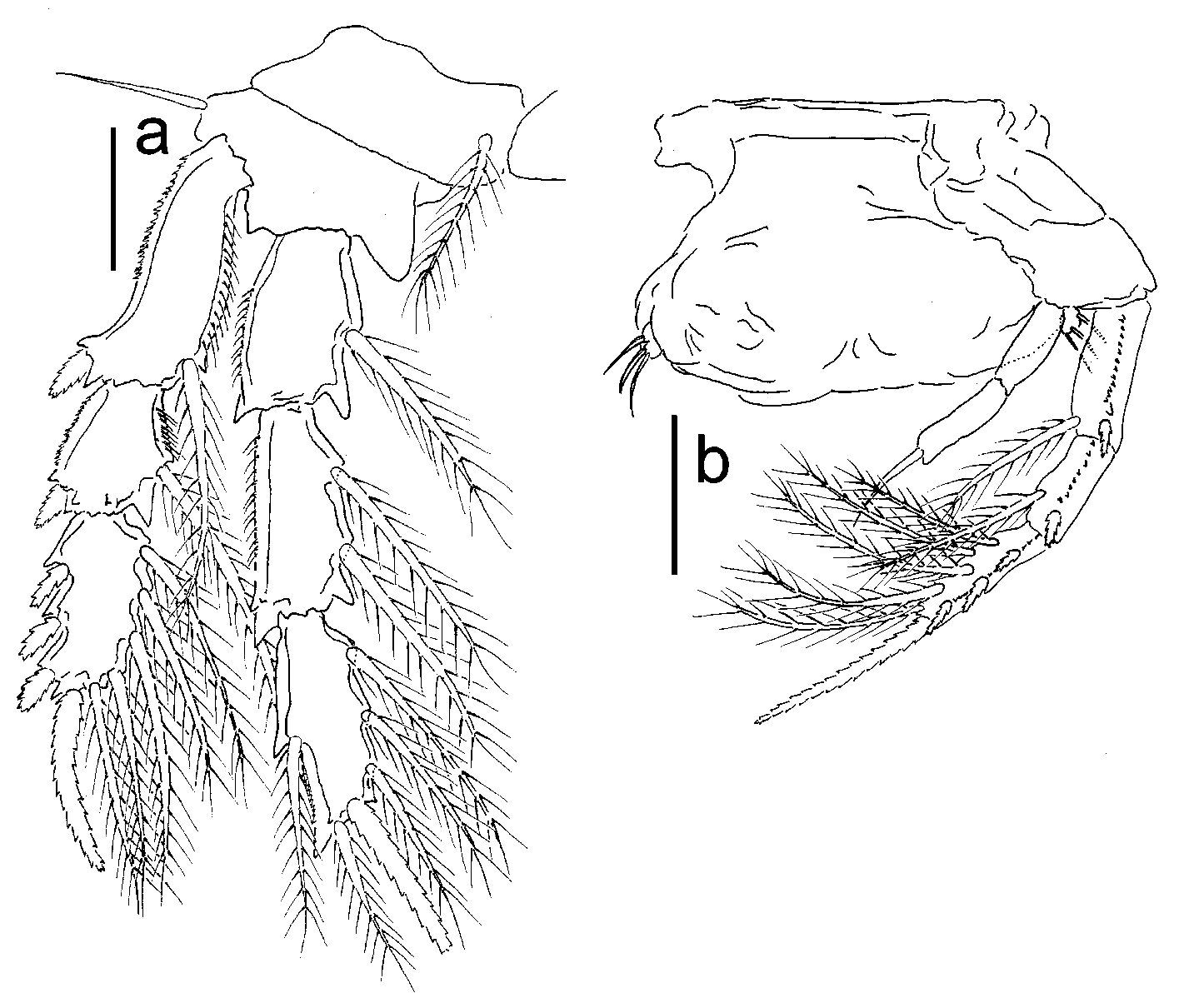
Swimming legs 1–3 (P1–P3; figs. 2c–d, 3a) biramous, all with 3segmented rami. P4 (fig. 3b) with 3segmented exopod and 2segmented endopod. Armature formula of legs 1–4 shown in table 1.
Fifth leg (fig. 3b) consisting of short, smooth seta near insertion of small, free segment, armed with 2 distal, smooth setae.
Remarks. All other siphonostomatoids collected at the same location were described by Humes (Humes 1971a & b; 1972a & b; 1973a & b; 1981).
Etymology. The specific name “ walteri ” honors Dr. Chad Walter of the National Museum of Natural History, Washington, whose efforts in rescuing and preserving Dr. Humes’ samples and notes provided the possibility that his legacy would not be wasted.
| USNM |
Smithsonian Institution, National Museum of Natural History |
No known copyright restrictions apply. See Agosti, D., Egloff, W., 2009. Taxonomic information exchange and copyright: the Plazi approach. BMC Research Notes 2009, 2:53 for further explanation.