
Sardiniella Linaldeddu, A. Alves & A.J.L. Phillips, Mycosphere
|
publication ID |
https://doi.org/10.11646/phytotaxa.627.1.1 |
|
DOI |
https://doi.org/10.5281/zenodo.10249883 |
|
persistent identifier |
https://treatment.plazi.org/id/0397879F-FC36-2902-FF64-9822FCA8FAFB |
|
treatment provided by |
Plazi |
|
scientific name |
Sardiniella Linaldeddu, A. Alves & A.J.L. Phillips, Mycosphere |
| status |
|
Sardiniella Linaldeddu, A. Alves & A.J.L. Phillips, Mycosphere View in CoL View at ENA 7: 900 (2016), MycoBank MB817511
Sardiniella View in CoL was introduced by Linaldeddu et al. (2016) with S. urbana View in CoL on a branch canker of Celtis australis View in CoL from Italy as the type species. It accommodates species that are morphologically similar to, but phylogenetically distinct from Dothiorella View in CoL and Diplodia View in CoL . Species in Sardiniella View in CoL are characterized by hyaline, aseptate conidia that become pigmented and 1-septate with age. While in Dothiorella View in CoL and some Diplodia species, namely those with aseptate brown conidia, the conidia become pigmented while still attached to the conidiogeneous cells, this has not yet been observed in Sardiniella View in CoL . Moreover, the conidial wall is thicker in Diplodia View in CoL than in Sardiniella ( Linaldeddu et al. 2016) View in CoL . So far, Sardiniella species have been reported as pathogens and saprobes of woody hosts. While S. urbana View in CoL was isolated from diseased ornamental Celtis australis View in CoL trees on the island of Sardinia, Italy ( Linaldeddu et al. 2016), the remaining species have been isolated from decaying plant material ( Hyde et al. 2017, Chen et al. 2021, Dissanayake et al. 2021). Currently Sardiniella View in CoL includes four species based on both morphological and phylogenetic analyses, viz. S. urbana ( Linaldeddu et al. 2016) View in CoL , S. celtidis ( Hyde et al. 2017) View in CoL , S. guizhouensis ( Chen et al. 2021) View in CoL and S. elliptica ( Dissanayake et al. 2021) View in CoL .
Sardiniella urbana Linaldeddu, A. Alves & A.J.L. Phillips, Mycosphere View in CoL 7: 900 (2016), MycoBank MB817512 ( Figure 16 View FIGURE 16 )
Type: ITALY, Sassari, isolated from a branch canker of Celtis australis ( Cannabaceae ), 9 Sep 2013, Benedetto T. Linaldeddu ( holotype LISE 96308, culture ex-type CBS 141580).
Sexual morph not reported. See Linaldeddu et al. (2016) for illustrations and descriptions of asexual morph.
Isolate CDP 1658. Sexual morph: Undetermined. Asexual morph: Conidiomata on palm leaflets in culture pycnidial, globose to subglobose, non-stromatic, uniloculate, black, solitary, occasionally aggregated in small groups of 2–3, superficial, or immersed in the host becoming erumpent when mature, covered with mycelial hairs, thick-walled. Conidiophores reduced to conidiogenous cells. Conidiogenous cells lining the pycnidial cavity, hyaline, smooth- and thin-walled, simple, discrete, determinate, cylindrical to broadly lageniform, straight or slightly curved, aseptate, rarely 1-septate, enteroblastic, proliferating at the same level giving rise to periclinal thickenings, or proliferating percurrently giving rise to 1–2 indistinct annelations, 5.69–13.72 × 2.71–5 μm, 95 % confidence limits = 8.12–9.39 × 3.69–4.11 μm (mean ± SD = 8.76 ± 1.77 × 3.90 ± 0.60 μm, n = 30). Conidia ellipsoid to broadly ellipsoid, few broadly obovoid or slightly reniform, rounded ends, few with subobtuse base, slightly narrower in the middle, thick-walled, hyaline and aseptate, becoming pale brown to brown and 1-septate, rarely 2-septate, with age, eguttulate, 23.71–29.42 × 9.36–11.01 μm, 95 % confidence limits = 25.67–26.63 × 9.84–10.15 μm (mean ± SD = 26.15 ± 1.34 × 9.99 ± 0.44 μm), mean ± SD conidium length/width ratio = 2.62 ± 0.21 (n = 30).
Culture characteristics: Colonies on 1/2 PDA, reaching 73 mm diam. after 7 d at 20 ℃ in darkness. Surface flat, with sparse aerial mycelium, with entire, slightly undulate margin, circular shape, whitish to pale, opaque. Reverse pale to yellowish. Turning entirely dark grey to black after about 2 w. No diffusible pigment .
Material examined: PORTUGAL, Lisbon, Parque das Nações, on foliar lesions of leaflets of Phoenix reclinata ( Arecaceae ), 8 May 2021, Diana S. Pereira (specimen HDP 093, new host and geographical record), living culture CDP 1658 ( ITS sequence OQ996239, tef1 sequence OR 233682).
Hosts: Celtis australis ( Cannabaceae ) ( Linaldeddu et al. 2016) and Phoenix reclinata ( Arecaceae ) (present study).
Distribution: Italy ( Linaldeddu et al. 2016) and Portugal (present study).
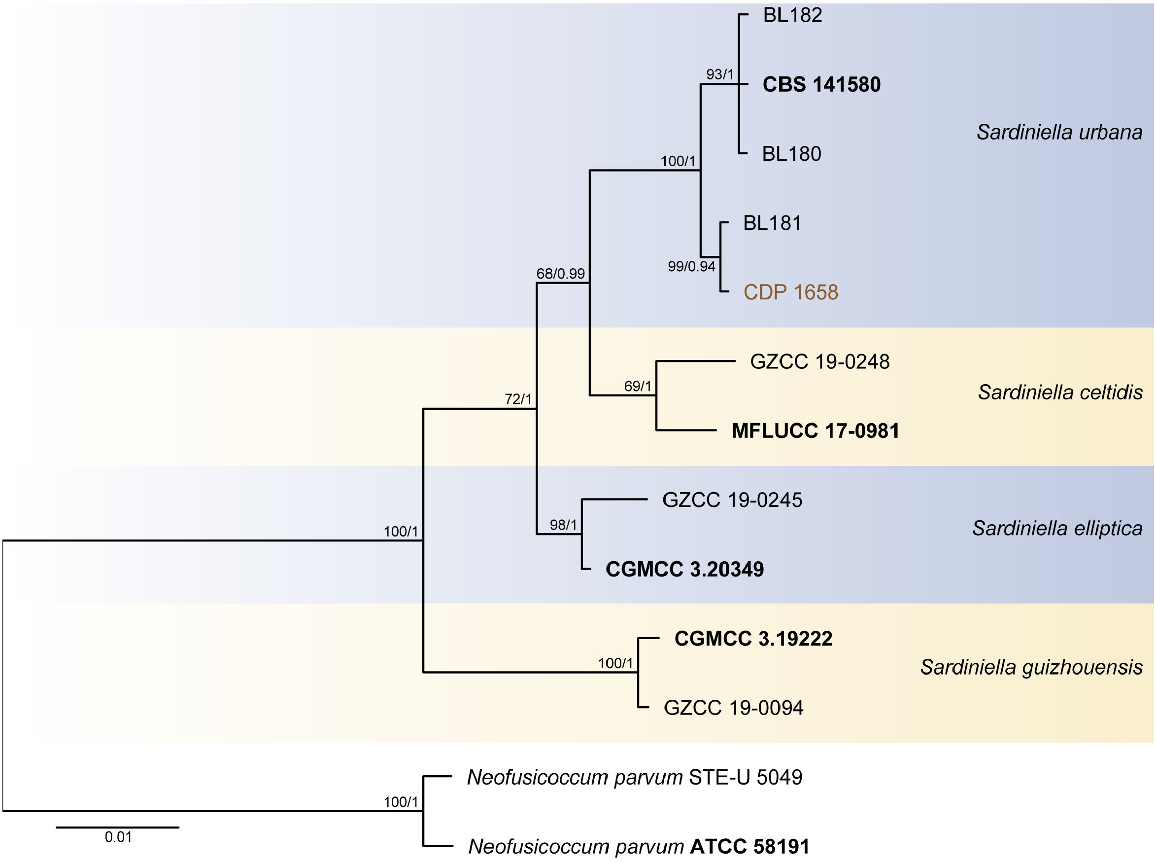
Notes: Based on the phylogenetic analyses of the combined ITS- tef1 dataset, strain CDP 1658 clustered with the ex-type strain and other strains of Sardiniella urbana with maximum ML-BS/PP values ( Figure 6 View FIGURE 6 ). Sequence comparisons with the ex-type of S. urbana ( CBS 141580) for ITS and tef1 showed 100 % and 98.81 %, respectively, sequence similarity and differences are represented by few base pair changes in tef1 partial sequence. Morphologically, CDP 1658 isolated in this study is similar to the holotype of S. urbana from a branch canker of Celtis australis in Italy ( Linaldeddu et al. 2016). Both produce globose pycnidial conidiomata with ellipsoid, thick-walled, hyaline and aseptate conidia, that become brown and 1–2-septate with age. Nonetheless, conidia of CDP 1658 are somewhat longer, but narrower than those of the ex-type of S. urbana ( CBS 141580) (mean = 26.15 × 9.99 μm versus 23.5 × 12 μm, respectively) ( Linaldeddu et al. 2016) ( Figure 16 View FIGURE 16 ). Thus, based on these morpho-molecular analyses, strain CDP 1658 was identified as S. urbana . Sardiniella urbana has only been reported from Celtis australis ( Cannabaceae ) in Italy ( Linaldeddu et al. 2016). In the present study, S. urbana is reported on Phoenix reclinata ( Arecaceae ) in Portugal, representing a new host and geographical record ( Table 5). The isolate of S. urbana studied was recorded from foliar lesions of P. reclinata , but pathogenicity has not been tested.

No known copyright restrictions apply. See Agosti, D., Egloff, W., 2009. Taxonomic information exchange and copyright: the Plazi approach. BMC Research Notes 2009, 2:53 for further explanation.
|
Kingdom |
|
|
Phylum |
|
|
Class |
|
|
Order |
|
|
Family |
Sardiniella Linaldeddu, A. Alves & A.J.L. Phillips, Mycosphere
| Pereira, Diana S. & Phillips, Alan J. L. 2023 |
Sardiniella Linaldeddu, A. Alves & A.J.L. Phillips, Mycosphere
| Sardiniella Linaldeddu, A. Alves & A. J. L. Phillips 2016: 900 |
Sardiniella urbana Linaldeddu, A. Alves & A.J.L. Phillips, Mycosphere
| Linaldeddu, A. Alves & A. J. L. Phillips 2016: 900 |