
Sarcophaga (Discachaeta) bezziana Böttcher, 1913,
|
publication ID |
https://doi.org/ 10.5281/zenodo.185607 |
|
DOI |
https://doi.org/10.5281/zenodo.6217766 |
|
persistent identifier |
https://treatment.plazi.org/id/0397C163-D94C-4354-4284-F8DE4C21A5E2 |
|
treatment provided by |
Plazi |
|
scientific name |
Sarcophaga (Discachaeta) bezziana Böttcher, 1913 |
| status |
comb. nov. |
Sarcophaga (Discachaeta) bezziana Böttcher, 1913 View in CoL , comb. nov.
Sarcophaga bezziana Böttcher, 1913b: 242 View in CoL . Type locality: Campopericoli ( Italy, Abruzzi, L’Aquila prov.).
Type material. Lectotype ɗ, herewith designated in order to promote nomenclatural stability: Campoperi- / coli 25.VII // S. Bezzii / Böttcher Typ // G. Böttcher [typewritten on a rectangular white label] // LECTOTYPE ɗ / Sarcophaga / bezziana / Böttcher, 1913 / des. D. Whitmore, R. Richet, T. Pape & R. Blackith 2008 ( SMF) [lectotype in good condition with terminalia extended but still attached to abdomen]. Paralectotypes: 1 ɗ: M. [?] [illegible locality name] / 27.VI.97 // G. Böttcher ( SMF) [specimen with middle and hind left legs missing, with terminalia extended and attached to abdomen]; 1 ɗ: Campoperi / coli [ Italy, Abruzzi, L’Aquila prov.] 25.VII // S. Bezzii / Böttcher Typ // G. Böttcher ( SMF) [mid right leg glued to a slip of card beneath the specimen, terminalia detached from abdomen at level of epandrium and stored in glycerine in a small tube pinned with the rest of the specimen].
Other material. Italy. 1 ɗ: Abruzzi (AQ) [= L’Aquila], Anversa degli Abruzzi, Pizzo Marcello, Stazzo Rotolo, 1437 m, 29.VII.1997, P. Cerretti, A. Tenga leg. (CNBFVR); 1 ɗ: Abruzzi, Corno Grande, 19.9.42, m 2200 [no collector] ( MZUR).
Comments. The listed type material represents the entire original type series: three males, all of which are conspecific. The lectotype was chosen from the three original syntypes for its overall good condition.
Diagnosis (male). Scutellum with a pair of apical setae; mid tibia with one anteroventral seta; hind femur with a strong subapical seta but no additional anteroventral setae; hind tibia with a few rows of long setulae with wavy tip on posteroventral surface; wing vein R1 dorsally setose; abdominal tergite 3 without median marginal setae; protandrial segment with a row of marginal setae; epandrium red; cercus with a slight dorsal excavation medially and with a distinct subapical dorsal hump; pregonite with a rounded tip, slightly widening apically; phallus: apical process of harpes well developed, perpendicular to main axis of distiphallus; juxta long and strongly sclerotized, with well-developed, membranous, spoon-shaped appendages arising from its base.
Redescription. Male (measurements refer to the lectotype, with the variation range of the species given in square brackets).
Length: 8.1mm [7.5–10.6].
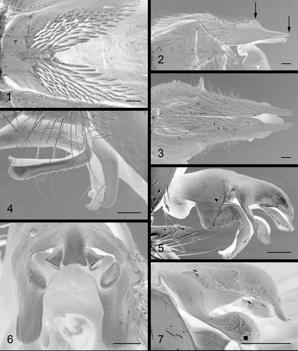
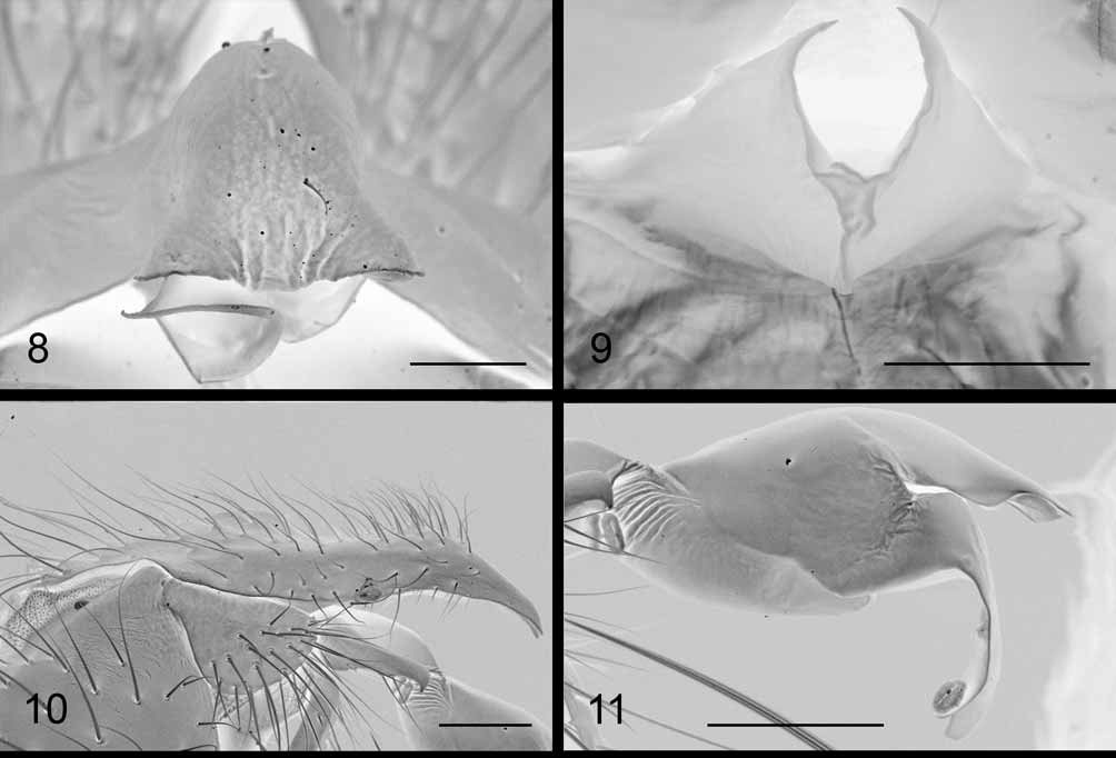
Colour. Head black, with dense light-grey microtrichosity on parafacials and fronto-orbital plate, changing with the incidence of light. Frontal vitta black. Gena, face and occiput grey-microtrichose. Antenna: pedicel black, brown at tip; postpedicel black. Prementum dark brown, palpus brown. Ground colour of thorax black, grey-microtrichose with three longitudinal dark vittae; legs black; tegula black, basicosta light yellow. Abdomen black, densely grey-microtrichose, with typical checkered pattern changing with the incidence of light, black markings becoming somewhat reduced when viewed from behind. Protandrial segment shiny black with a wide horizontal strip of grey microtrichosity across posterior 1/3–1/2; epandrium red, somewhat darkened ventrally. Cercus black; surstylus brown; phallus, pre- and postgonite brown. Head. Arista thickened on basal 1/3. Postpedicel 1.47 [1.47–1.70] times as long as pedicel. Frons at its narrowest point 0.65 [0.55–0.70] times as wide as an eye in dorsal view. Frontal vitta 0.56 [0.48–0.56] times as wide as frons at its narrowest point, visibly widening towards antennal insertion. Lateral vertical setae strong, about twice [1.2–2.0] as long as longest postocular setae. Six [5–10] frontal setae, not descending below level of middle of pedicel. Fronto-orbital plate with a more or less arranged row of fine setulae near eye margin. Parafacial with a row of fine setae close to eye margin, increasing in length and thickness ventrally. Parafacial at its narrowest point 0.36 [0.30–0.52] times as wide as eye width in strict lateral view. Lower facial margin slightly to clearly visible in lateral view below vibrissal angle. Facial ridge above vibrissa with a few decumbent setulae. Gena in profile 0.41 [0.40–0.49] times the vertical height of eye (measured in the same vertical plane as height of head); gena entirely covered with black setulae; postgenal setulae white. Two rows of black occipital setulae behind postocular setae, remaining occipital setulae white. Prementum 5.0 [4.5–5.3] times as long as wide. Thorax. Postpronotum with 3 stronger setae forming a triangle. Scutum with 2–4 [variable] + 1 (prescutellar) acrostichal, 4 + 3 dorsocentral, 2–3 intraalar, 1 posthumeral (sometimes a weak outer seta present), 1 presutural, 4 notopleural and 3 supraalar setae; postalar callus with 2 setae. Katepisternum with 3 setae. Katepimeron with fine setulae on posterior half. Scutellum with 3 pairs of marginal setae (basal, subapical, apical) and one pair of discal setae. Legs. Fore tibia with 3 anterodorsal and 1 posterior setae. Mid femur with 2–3 anterior setae near middle, several anteroventral setae in proximal two thirds, 2–3 subapical posterodorsal setae, no strong setae on posteroventral surface; mid femur without a subapical posteroventral comb. Mid tibia with 3 (sometimes 4) anterodorsal, 2 posterodorsal, 1 posterior and 1 anteroventral setae. Hind trochanter with a ventral brush of tightly spaced spine-like setae. Hind femur with a strong subapical seta but no additional anteroventral setae, and with a few stronger posteroventral setulae in basal third. Hind tibia with a row of anterodorsal setae of irregular length, 2 posterodorsal and 1 (sometimes 2) anteroventral setae; hind tibia with a few rows of long setulae with wavy tip on posterovental surface. Wing. Costal spine well developed, 1–1.5 times as long as crossvein R-M. Vein R1 with several (5–8) setae along dorsal surface. Setae on dorsal surface of vein R4+5 extending about 3/5–4/5 of the way to crossvein R-M. Second costal section 0.93 [0.89–1.09] times fourth costal section. Small spines on costa reaching about 3/4 of the way across fourth costal section. Wing cell r4+5 open at wing margin. Abdomen. Syntergite 1+2 and tergite 3 without median marginal setae. Tergite 4 with a pair of strong median marginal setae and 2–3 lateral marginal setae. Tergite 5 with a complete row of marginal setae. Terminalia. Sternite 5 ( Fig. 1 View FIGURES 1 – 7 ) strongly indented, v-shaped, with brushes of tightly spaced stout, short setae along each of its processes and a row of slightly longer and thicker setae along the inner margin of each process. Protandrial segment with a row of setulae along posterior margin. Epandrium gently curved dorsally, about 1.2–1.4 times as long as high [not measurable, damaged, in lectotype]. Cercus ( Figs 2, 3 View FIGURES 1 – 7 ) with a slight dorsal excavation medially, in lateral view with a distinct dorsal hump close to apex; length of tip, distal to hump, between 1.7 and 1.9 times height of cercus at level of dorsal hump. Surstylus ( Fig. 2 View FIGURES 1 – 7 ) sub-triangular. Pregonite ( Fig. 4 View FIGURES 1 – 7 ) with a rounded tip, slightly widening apically and with fine setulae on dorsal surface. Postgonite ( Fig. 4 View FIGURES 1 – 7 ) with a hooked tip. Distiphallus ( Fig. 5 View FIGURES 1 – 7 ): apical process of harpes long with a broad, rounded tip ( Fig. 6 View FIGURES 1 – 7 ) and projecting ventrally, perpendicular to main axis of distiphallus; juxta ( Fig. 7 View FIGURES 1 – 7 ) long in relation to rest of distiphallus, with a ventrally curved tip, and with spoon-shaped basal appendages widening apically; tip of juxta with two wide, diverging, and somewhat apically-directed processes ( Fig. 7 View FIGURES 1 – 7 ), only slightly visible in apical view ( Figs 6 View FIGURES 1 – 7 , 8 View FIGURES 8 – 11. 8 – 9 ); lateral styli funnel-shaped, widening apically ( Fig. 5 View FIGURES 1 – 7 ); vesica small, laminar, with a median longitudinal fold, appearing v-shaped in apical view ( Fig. 9 View FIGURES 8 – 11. 8 – 9 ).
Female unknown.
Biology. Unknown.
Distribution. Currently, Sarcophaga (Discachaeta) bezziana is known only from high elevations (between 1,000 and 2,000m) of the Apennines in the Abruzzi region, central Italy. The locality of one of the paralectotypes could not be identified with certainty as the label is handwritten and almost illegible; however, as all three type specimens were sent to G. Böttcher by M. Bezzi ( Böttcher 1913b: 243), it is probably from a mountain locality [“M.” on the label almost certainly stands for “Monte” (= Mount)] in Abruzzi.
Remarks. Rohdendorf (1937), who did not examine the species directly himself, placed Sarcophaga bezziana in the group “ Pierretia s. str. ” (today’s “ haemorrhoa -group” of Heteronychia ). Collart (1954) considered S. bezziana as “très voisine de Pierretia Osten-Sackeni Rohdendorf ”, without further comment. Povolný (1986) mentioned that according to Y. Verves (pers. comm.) “daß möglicherweise eine Synonymie von H. ostensackeni (Rohd.) mit Heteronychia (Heteronychia) bezziana (Böttcher, 1913) ”, and Verves (1986) synonymized “ Pierretia (Heteronychia) ostensackeni Rohdendorf, 1937 ” [= Sarcophaga (Heteronychia) infantilis ] with Sarcophaga bezziana , thus dissociating the name S. bezziana from the original identity of the species (cf. Povolný 1986: Appendix). All subsequent authors followed this synonymy (e.g., Pape 1987), but the species represented were not always Sarcophaga infantilis . Povolný (1989) confused two or three species under this name: S. benaci Böttcher, 1913 ( Povolný 1989: figs 13–14), S. infantilis ( Povolný 1989: figs 15–16) and a third species of uncertain identity ( Povolný 1989: figs 17–18); Povolný and Verves (1997) reproduced drawings from Povolný (1989) under S. bezziana , “to demonstrate genitalia variation of this species”, but they associated the phallus of S. benaci [fig. 14 in Povolný (1989)] with the cercus of another specimen and, most probably, another species [fig. 17 in Povolný (1989)]. Pape et al. (2002) tentatively removed Sarcophaga infantilis from synonymy with S. bezziana , and Pape (2004) stressed the necessity of a revision of the nominal taxon Sarcophaga bezziana Böttcher. Sarcophaga bezziana and S. infantilis are in reality remarkably different and not closely related. Sarcophaga infantilis (see Figs 10–11 View FIGURES 8 – 11. 8 – 9 ) differs principally in being much smaller (3–4 vs. 7–10mm), having much sparser microtrichosity on the thorax and abdomen, having median marginal setae on abdominal tergite 3, having a black epandrium, having a different-shaped cercus ( Fig. 10 View FIGURES 8 – 11. 8 – 9 ) and having a short juxta without juxtal appendages ( Fig. 11 View FIGURES 8 – 11. 8 – 9 ).
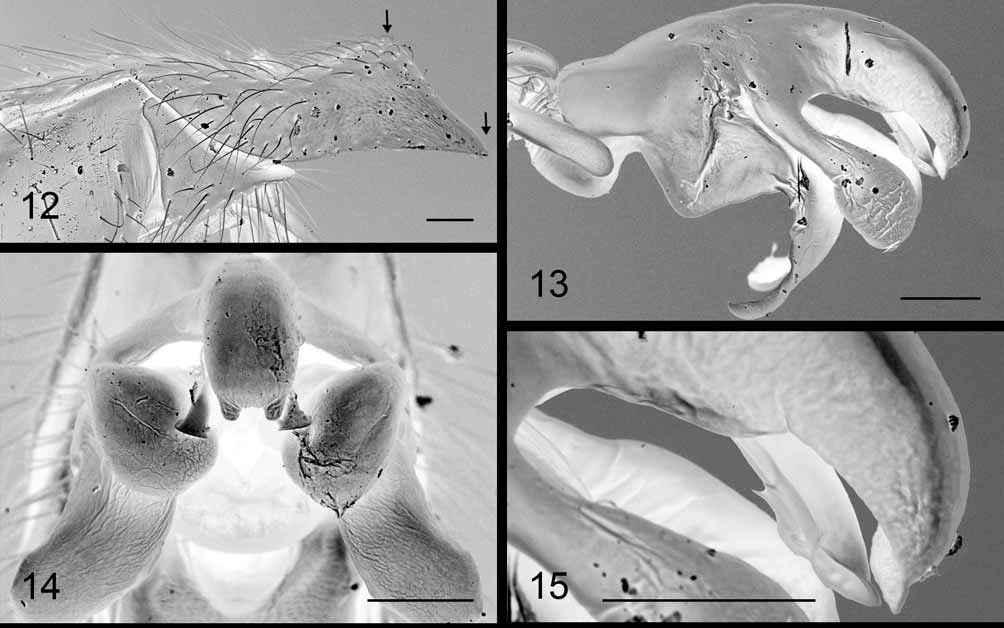
Sarcophaga bezziana is instead morphologically very closely related to S. (Discachaeta) amita , from which it differs exclusively in features of the male terminalia. The main difference lies in the length and shape of the cercus tip, i.e. the portion distal to the dorsal hump: in S. bezziana ( Fig. 2 View FIGURES 1 – 7 ), the tip is longer (between 1.7 and 1.9 times the height of the cercus at level of the dorsal hump), slightly upturned and with a distinctly concave dorsal margin; in S. amita ( Fig. 12 View FIGURES 12 – 15 ), the tip is shorter (between 0.9 and 1.3 times the height of the cercus at level of the dorsal hump), slightly downturned and with an almost straight dorsal margin. Other differences can be found in the phallus: in S. bezziana , the distiphallus, in lateral view, is curved mainly at the base and the tip and almost flat in the middle section; in S. amita , the distiphallus is more evenly curved in lateral view, almost rounded (compare Figs 5 View FIGURES 1 – 7 and 13 View FIGURES 12 – 15 ); in S. bezziana the juxtal appendages are slightly shorter and less enlarged distally than in S. amita (compare Figs 5–6 View FIGURES 1 – 7 and 13–14 View FIGURES 12 – 15 ); in S. bezziana the lateral processes at the tip of the juxta are wide, slightly diverging and somewhat directed apically, so that they are only slightly visible in apical view; in S. amita they are narrower, parallel, directed ventrally and usually well visible in apical view (compare Figs 7–8 View FIGURES 1 – 7 View FIGURES 8 – 11. 8 – 9 and 13–14 View FIGURES 12 – 15 ); finally, the lateral styli are visibly larger with respect to the juxta in S. bezziana than in S. amita (compare Figs 5–7 View FIGURES 1 – 7 and 13–15 View FIGURES 12 – 15 ).
Given their overall close similarity, a sister-species relationship is hypothesized between Sarcophaga bezziana and S. amita , and S. bezziana is tentatively removed from subgenus Heteronychia (cf. Verves 1986; Pape 1996) and placed in subgenus Discachaeta comb. nov. The original placement of S. amita in Discachaeta by Rohdendorf (1937) was questioned by Lehrer (1997), and it is not clear whether S. amita is more closely related to other species of Discachaeta or to species of Heteronychia (e.g. of the “ haemorrhoa - group”). However, this issue should be addressed through a modern cladistic analysis and is beyond the aim of the present paper.
No known copyright restrictions apply. See Agosti, D., Egloff, W., 2009. Taxonomic information exchange and copyright: the Plazi approach. BMC Research Notes 2009, 2:53 for further explanation.
|
Kingdom |
|
|
Phylum |
|
|
Class |
|
|
Order |
|
|
Family |
|
|
Genus |
Sarcophaga (Discachaeta) bezziana Böttcher, 1913
| Whitmore, Daniel, Richet, René, Pape, Thomas & Blackith, Ruth M. 2009 |
Sarcophaga bezziana Böttcher, 1913b : 242
| Bottcher 1913: 242 |