
Eilithyia singaporensis, Wang, Rong-Rong, Liang, Ai-Ping & Webb, Michael D., 2008
|
publication ID |
https://doi.org/ 10.5281/zenodo.274059 |
|
DOI |
https://doi.org/10.5281/zenodo.6228118 |
|
persistent identifier |
https://treatment.plazi.org/id/0398A373-CB27-FFD7-FF7F-F8E3F8DF9E6A |
|
treatment provided by |
Plazi |
|
scientific name |
Eilithyia singaporensis |
| status |
sp. nov. |
Eilithyia singaporensis View in CoL sp. nov.
( Figures 1 View FIGURE 1 , 3–19 View FIGURES 2 – 10. 2. E View FIGURES 11 – 19. E )
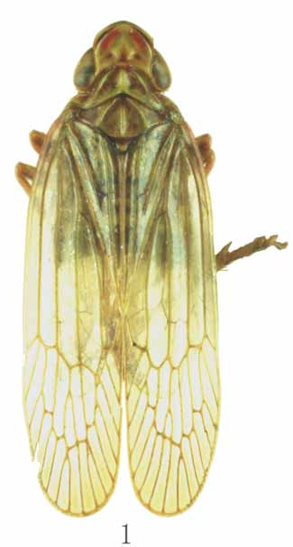
Description. Length (from apex of vertex to tip of fore wings): ɗ 8.8 mm, Ψ 9.5 mm.
General color pale brownish; vertex with lateral margins, median carina with basal half, postclypeus with basal area, clypeus with lateral margins, pronotum with anterior disc and lateral portion, meso- and metapleura, and mesonotum with anterior portion all suffused with fuscous; apex of head with one black spot, inserting between two linear blackish stripes; two blackish linear stripes along anterior margins of depression on vertex, anterior area of depression suffused with two reddish patches, posterior area of depression with a pair of fuscous spots; frons with 5 red transverse bands on each depression, carinae of frons orange, median carina of clypeus reddish distally; genae with reddish patch between eyes and lateral margin of vertex; ocelli surrounded with reddish; mesonotum with four brownish spots near basal margins; carinae of pronotum and mesonotum pale reddish; legs reddish, postfemora with distinct long reddish stripes, tarsi blackish; fore wings yellowish brown, all transverse veinlets, costal cell with basal area, middle of clavus suffused in fuscous; hind wings yellowish brown, Cu underlying apex of clavus and costal cell basally fuscous.
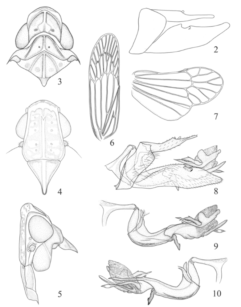
Head with eyes narrower than pronotum, broadly produced in front of eyes; apex broadly convex in dorsal view and in lateral view, forming a smooth surface interposed between the discal areas of the vertex and frons; vertex and frons not separated and their lateral carinae percurrent. Vertex ( Figs 1 View FIGURE 1 , 3 View FIGURES 2 – 10. 2. E ) subquadrate, slightly shorter than the breadth at anterior margin of eyes (0.72:1), distinctly longer than pronotum at midline; lateral margins ridged and converged anteriorly; posterior margin angulately concave; central disc between median and sublateral carinae distinctly depressed, lateral margins of depression incurving anteriorly and uniting with median carina medially; median carina ridged, abruptly becoming very broad flattened ridge at the level of anterior margin of eyes, and uniting with posterior margin. Frons ( Fig. 4 View FIGURES 2 – 10. 2. E ) longer in middle than the widest breadth (1.38:1), disc slightly depressed between median and lateral carinae, each depression with 5 indistinct gibbosities; lateral margins converging from under level of antennae to apex; posterior margin straight; median carina straight and thin, bifurcate anteriorly; lateral carinae approaching frontoclypeal suture. Postclypeus and anteclypeus ridged medially, with distinct median carina; postclypeus with lateral carinae surpassing mid-length. Rostrum long, reaching between hind coxae. Eyes oval. Ocelli small. Antennae ( Figs 4, 5 View FIGURES 2 – 10. 2. E ) short, scape cylindrical; pedicel distally expanded, about 3 times as long as scape, with microsetae extending to base.
Pronotum ( Figs 1 View FIGURE 1 , 3 View FIGURES 2 – 10. 2. E ) wider than long medially (4.56:1), distinctly shorter than mesonotum in midline; median carina broadly ridged, lateral carinae converging anteriorly; disc arched anteriorly, deeply depressed between median and lateral carina and with an impression on each side; posterior margin obtuse-angled excavated. Mesonotum ( Figs 1 View FIGURE 1 , 3 View FIGURES 2 – 10. 2. E ) tricarinate, with a distinct transverse suture separating mesoscutellum; median carina straight, reaching to transverse suture; lateral carinae curving anteriorly towards median carina; pronotum and mesonotum together medially 2.05 times as long as median length of vertex. Fore wings ( Fig. 6 View FIGURES 2 – 10. 2. E ) translucent, elongate and narrow, 3.38 times as long as maximum breadth; corium without granulation, costal cell without cross veins; Sc+R and M forking at node; Cu1 forking about level of junction of claval veins, which extending to about middle of clavus; nodal line forming a zigzag transverse line; with 5–6 subapical and 15–16 apical cells various in size. Hind wings ( Fig. 7 View FIGURES 2 – 10. 2. E ) hyaline, posterior margin strongly sinuate, with a series of disconnected transverse veins before apical area. Legs elongate; hind tibia with 3 distinct lateral spines, distally with 6 spines, basal metatarsal segment distally with 6 spines.
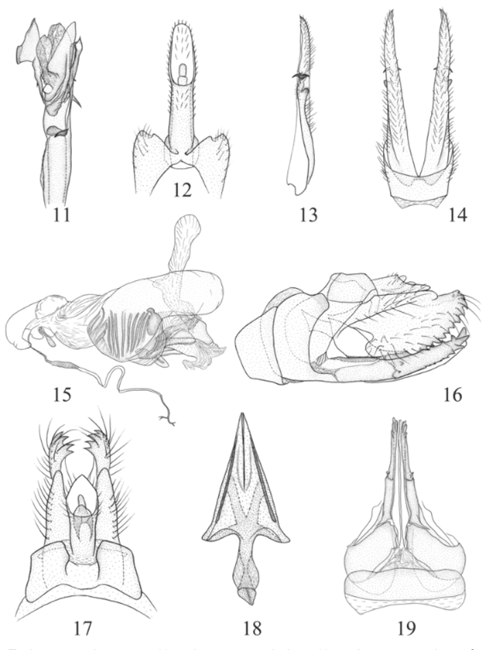
Male genitalia with pygofer ( Fig. 8 View FIGURES 2 – 10. 2. E ) narrow and high, dorsal posterior margin angulately produced posteriorly in lateral view, dorsad of which having a stout tooth, dorsa1 margin deeply excavated to accommodate anal tube. Anal tube ( Figs 8 View FIGURES 2 – 10. 2. E , 12 View FIGURES 11 – 19. E ) elongate, distinctly projected caudad, apical margin rounded, nearly parallelsided in dorsal view, anal styles relatively short and small. Gonostylus ( Figs 8 View FIGURES 2 – 10. 2. E , 13, 14 View FIGURES 11 – 19. E ) symmetrical, relatively elongate and narrow, 4.11 times as long as broad in lateral view, tapering to apex in distal half, membranously fused with pygofer at base, dorsal edge with a stubby, dorsoposteriorly directed prominence near middle and two hook-like, inward and outward respectively directed teeth beyond this prominence. Aedeagus ( Figs 9–11 View FIGURES 2 – 10. 2. E View FIGURES 11 – 19. E ) elongate, sclerotized and expended into a subcircular membranous endosoma apically, asymmetrical, basal part directed anterodorsally, then strongly curved and directed ventrally, shaft of aedeagus with three sclerotized processes: left process with a stubby knob-like prominence in dorsal margin medially, basad of which is a double-pronged processes with margin denticulated; ventral process abruptly expended into triangular plate, together with left process embracing endosoma; right process long and thin, gently curved, slightly expended apically. Structure tectiform of connective ( Figs 8–10 View FIGURES 2 – 10. 2. E ) developed; corpus connective ( Figs 8–10 View FIGURES 2 – 10. 2. E ) present; periandrium ( Figs 9–11 View FIGURES 2 – 10. 2. E View FIGURES 11 – 19. E ) well developed, asymmetrical, broadly fused with ventral base of anal segment, surrounding penis from base to middle, with two processes: one process stout and twist, directed ventrad at ventral side; the other process arising from right side, longer than three-quarters of aedeagus length, with a distinct tooth basally, apically acute.
Female genitalia with anal tube ( Fig. 17 View FIGURES 11 – 19. E ) relatively short, apical margin acute in dorsal view, anal styles relatively short and small. Gonopophyses VIII (first valvulae) ( Figs 16, 17, 19 View FIGURES 11 – 19. E ) sawlike, strongly sclerotized with about 6 blunt teeth on dorsal margin, a single relatively large tooth at apex, ventral margin denticulate with several teeth, 2–3 apical teeth blunt, laterally at base with an sinuate row of many minute teeth, and two relatively large teeth distad of teeth row. Gonopophyses IX (second valvular) ( Fig. 18 View FIGURES 11 – 19. E ) triangular, fused together on inner-lateral margin and strongly reduced but well sclerotized, apical ends not meeting together, acute at apex, gonospiculum ( Figs 16, 18 View FIGURES 11 – 19. E ) shorter than median length of triangular part 0.57:1, infundibuliform apically, flattened laterally. Gonoplac (third valvular) ( Figs 16, 17 View FIGURES 11 – 19. E ) with 11–12 teeth extending along ventral margin from apex.
Material examined. Holotype ɗ, SINGAPORE: Botanic Gardens, 25 m, 11.XII.1958 (T. C. Maa) ( BPBM). Paratype. 1Ψ, MALAYA: (W) Selangor, Ulu Gombak, 300 m, 15.VI.1961 (J. L. Gressitt) ( BPBM).
Etymology. This species is named for its occurrence in Singapore.
Distribution. Singapore, Malaya.
Remarks. This species is externally similar to E. insularis Distant, 1912 from Narkondam Islands, but can be distinguished from the latter by the characters given in the key.
| BPBM |
Bishop Museum |
No known copyright restrictions apply. See Agosti, D., Egloff, W., 2009. Taxonomic information exchange and copyright: the Plazi approach. BMC Research Notes 2009, 2:53 for further explanation.