
Lenicyamidia compta Brunnschweiler, 1962
|
publication ID |
https://doi.org/ 10.11646/zootaxa.3857.4.3 |
|
publication LSID |
lsid:zoobank.org:pub:76021E0C-7542-455B-82F4-C670A3DC8806 |
|
DOI |
https://doi.org/10.5281/zenodo.4929736 |
|
persistent identifier |
https://treatment.plazi.org/id/0398C024-7212-D50B-FF68-6B34438CF872 |
|
treatment provided by |
Felipe |
|
scientific name |
Lenicyamidia compta Brunnschweiler, 1962 |
| status |
|
Lenicyamidia compta Brunnschweiler, 1962
Figures 4 View FIGURE 4 , 7D–F View FIGURE 7 , 10A–C View FIGURE 10 , 11 View FIGURE , 13–14 View FIGURE 13 View FIGURE 14 .
1962 Lenicyamidia compta Brunnschweiler : 165–169, figs 2–3.
1966 Lenicyamidia compta Brunnschweiler —Philip: 116–117, fig. 1.
Type material. GA CPC 2827 View Materials (holotype, Figs 7D–F View FIGURE 7 , 10B View FIGURE 10 ) and GA CPC 2828 View Materials (paratype) .
Material studied. GA CPC 2827–2829, 41773–41799.
Type locality. Sample M 24 from point 221 on airphoto No. 5170 on Run 2, Moogooloo Hill (hill crest located at 23° 36′ 12″ S, 114° 44′ 14″ E; exact coordinates of sampling locality unknown); about 8 miles SSE of the Pleiades Hills, Northwest Division , Western Australia GoogleMaps .
Type stratum. Merlinleigh Sandstone, Late Eocene (see Darragh & Kendrick 2010: pp. 24–25)
ZooBank LSID. urn:lsid:zoobank.org:act:90041316-5191-4933-A119-6C0E598DD5D2
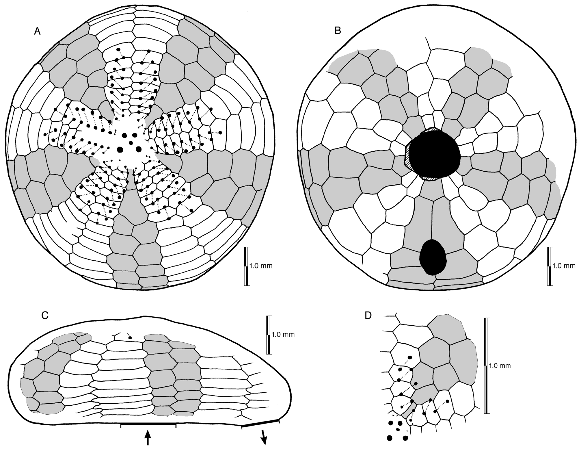
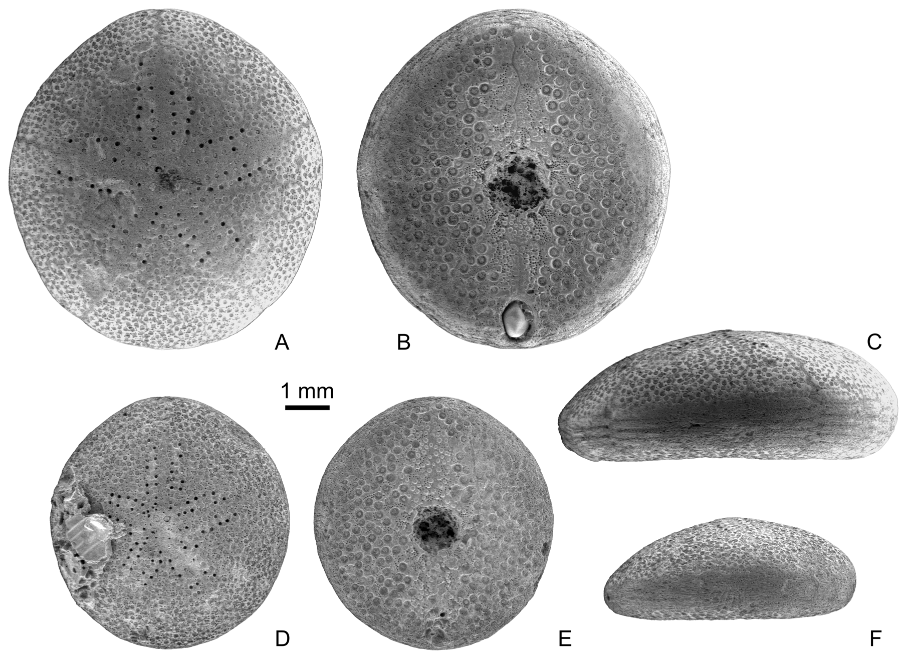
Description. Size and shape —Corona small, not exceeding 10 mm in TL among available specimens; outline in aboral view forming slightly angular circle; distinctly flattened in profile, maximum height between 30 and 40% of test length.
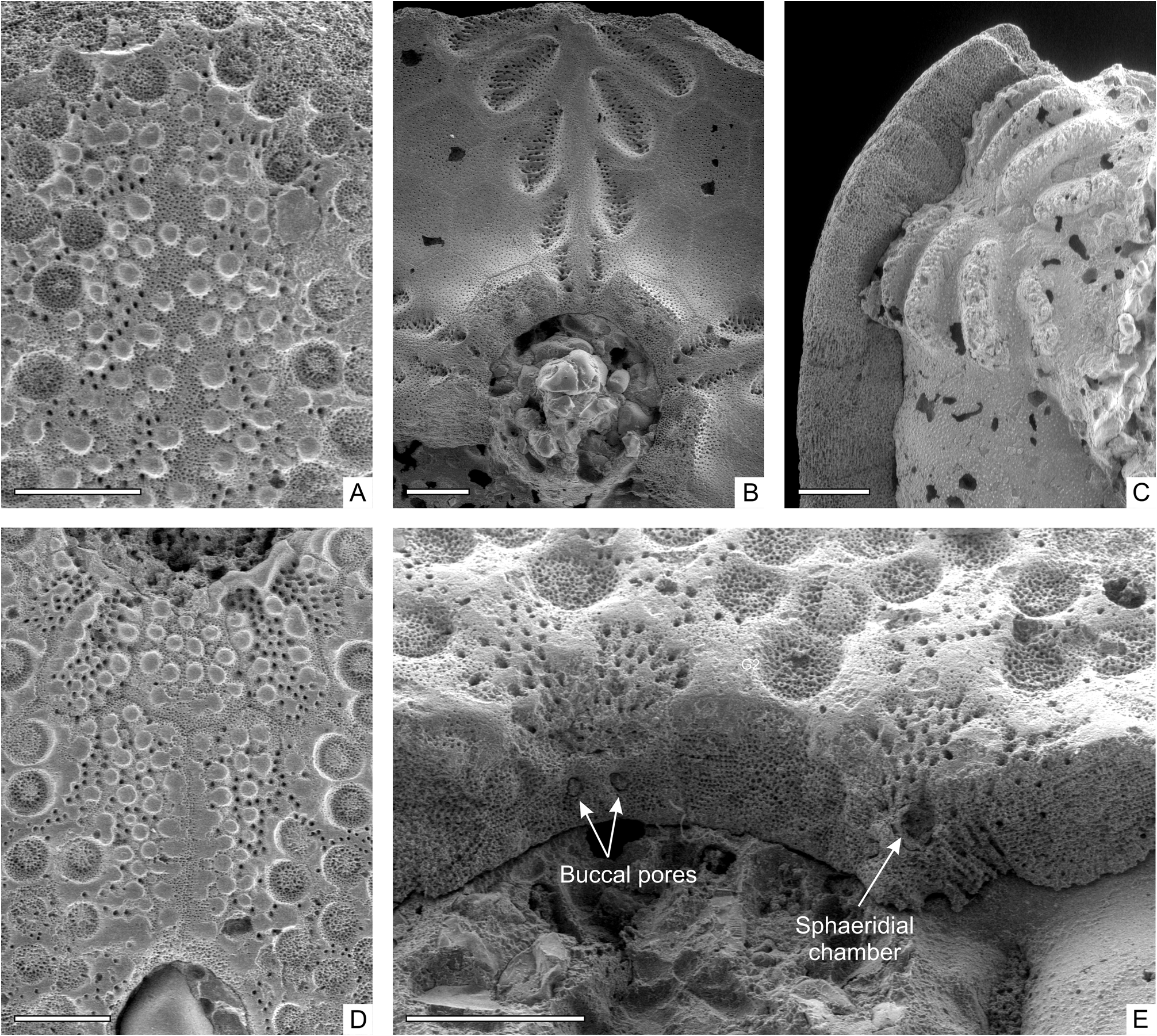
Internal buttressing —Absent, but ambulacral plates bear large pits internally and are about half as thick as interambulacral plates in their centre ( Fig. 14C View FIGURE 14 ), explaining peculiarly shaped internal casts ( Fig. 14C View FIGURE 14 ) reported by Brunnschweiler (1962: fig. 2E); pits associated with pores for accessory tube feet ( Fig. 14B View FIGURE 14 ).
Apical system —Situated centrally, at apex of corona; monobasal, with four gonopores and single central hydropore, not situated in pit or groove; ocular pores small and indistinct, lying well outside area enclosed by gonopores.
Ambulacra —Ambulacral plating simple; ambulacra expanding only slightly towards ambitus where they are barely wider than interambulacra; petals short, with up to 21 non-conjugate respiratory pore pairs in each ambulacrum; pore pairs strongly oblique, crossing ambulacral plates; interporal ridge smooth and formed by unperforated stereom; distal pore pairs becoming even more strongly oblique with distance between pores in each pair slightly decreasing; width of interporiferous zones widest halfway along petals and becoming slightly narrower again distally; petaloid region large, extending about 70% of TL; food grooves absent; buccal pores large and located close to peristomial edge, facing horizontally into peristome, externally visible only in broken specimens ( Fig. 14E View FIGURE 14 ) since they are hidden by transverse stereom bars in oral view; accessory pores evenly distributed all over test on aboral surface (including interporiferous zones and interambulacral plates), but forming patches obliquely crossing ambulacral plates and less common elsewhere on oral surface; ambitus initiating at approximately fourth to fifth pair of ambulacral postbasicoronal plates.
Interambulacra —Adapically, two unpaired plates lie in tandem adjacent to apical system; three or four postbasicoronal interambulacrals in each column visible in oral view; posterior unpaired interambulacrum expanding distinctly in region accommodating periproct; basicoronal plates usually extending only to first adjacent ambulacral plate, except in interambulacrum 2, where usually extending to second adjacent ambulacral plate on one or both sides; in largest specimen with plate sutures visible (CPC 41796), basicoronal plates of interambulacra 1, 3 and 4 also extend on one side to second adjacent ambulacral plate due to presence of large spine-bearing tubercles on margins of these plates (usually missing in smaller specimens) and fact that sutures are curved to make room for tubercles slightly modifies plate outline in such a way that they come into contact with second ambulacral plates; accessory pores evenly spread over aboral interambulacra, but less common, though present on oral interambulacral plates.
Tuberculation —Primary tubercles crenulate, perforate, homogeneously distributed on aboral surface; on oral surface, in contrast, there is a narrow area between the periproct and peristome ( Fig. 14 D View FIGURE 14 ), as well as anterior of the latter ( Fig. 14 A View FIGURE 14 ), free of primary tubercles; instead densely covered with glassy tubercles; lateral to this medial area are located primary tubercles with slightly sunken areoles, these tubercles larger than aboral tubercles.
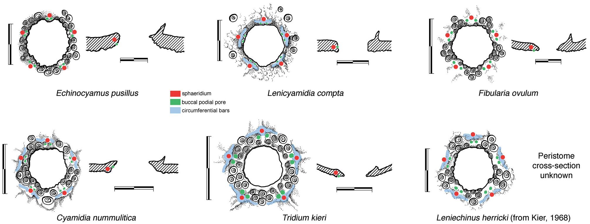
Peristome —Larger than periproct, about 18% TL; infundibulum extremely shallow but with near-vertical walls; peristomial opening facing directly downwards almost at midpoint of oral surface; framed by basicoronal circlet in which ambulacral plates only slightly longer than adjacent interambulacrals; plates of basicoronal circlet distinctly longer in posterior half of test; only scattered single tubercles present on interambulacral basicoronals adjacent to peristome, mainly on larger specimens; peristome framed by broken circle of circumferential bars overlying sphaeridial chambers ( Fig. 14E View FIGURE 14 ).
Periproct —Small, approximately 12% TL; facing down- and slightly backwards; located close to posterior margin on oral side; distinctly elongated along anterior-posterior axis; bounded by first (5.a.2, 5.b.2), second (5.a.3, 5.b.3), and one plate (5.b.4) of the third pair of post-basicoronal plates.
Perignathic girdle —Consisting of five small processes (auricles) attached to the internal surface of each interambulacral basicoronal; on their adoral side a pair of minute depressions (likely muscle scars for insertion of lantern protractors) located in each interambulacrum.
Sphaeridia —One per ambulacrum; fully enclosed ( Figs 4 View FIGURE 4 , 14E View FIGURE 14 ); situated beneath distinct transverse bar just distal to buccal pores.
Spines, pedicellariae, lantern —Unknown.
Remarks. Brunnschweiler (1962) illustrated the apical disc as being composed of four genital plus an additional central plate. However, Philip (1966) showed that the apical disc was of typical clypeasteroid monobasal structure. The illustration of an “internal mold” by Brunnschweiler (1962: fig. 3) in reality represents an internal view of the oral surface (and thus shows a mirror image of the outside plating pattern). This accounts for the apparent deviation from Lovén’s Rule in his drawing.
The peristomial region of L. compta is markedly different from that of other fibulariids studied here ( Fig. 4 View FIGURE 4 ). There are no clusters of tubercles on interambulacral basicoronals, the sides of the peristomial infundibulum are very steep and the circumferential stereom bars are very close to the peristomial edge, almost overhanging it in some cases.
The oral tuberculation of Lenicyamidia was compared with that of Lenita by Brunnschweiler (1962). While both possess a medial zone free from spine-bearing tubercles, there are two obvious differences: 1) in Lenita the tubercles have deeply incised, asymmetrical areoles (these are shallow and near symmetrical in Lenicyamidia ); and 2) large glassy tubercles are lacking in this zone in Lenita . It seems likely, therefore, that this tuberculation pattern is not homologous, but has adaptive significance causing it to have arisen independently in several neognathostome clades. In addition, Lenita , which is an early scutelline, differs in many other respects, including possession of biserial interambulacra at the apex, paired basicoronals, and other features not found in laganiforms. In contrast, Lenicyamidia , is clearly a laganiform, having two uniserial plates in the adapical interambulacra and a basicoronal circlet typical of fibulariids.


| CPC |
Culture collection of Pedro Crous |
No known copyright restrictions apply. See Agosti, D., Egloff, W., 2009. Taxonomic information exchange and copyright: the Plazi approach. BMC Research Notes 2009, 2:53 for further explanation.
|
Kingdom |
|
|
Phylum |
|
|
Class |
|
|
Order |
|
|
Family |
|
|
Genus |