
Mycomya
|
publication ID |
https://doi.org/ 10.11646/zootaxa.3815.4.4 |
|
publication LSID |
lsid:zoobank.org:pub:172C594B-7321-4F1C-B8CC-158A195A7D73 |
|
DOI |
https://doi.org/10.5281/zenodo.6134629 |
|
persistent identifier |
https://treatment.plazi.org/id/03998794-FFF4-F70F-FF40-667CFA2FFE4E |
|
treatment provided by |
Plazi |
|
scientific name |
Mycomya |
| status |
|
Key to Mycomya View in CoL View at ENA species of the South-East Asian islands
1. Coxa 2 with a spur ( Väisänen 1984a: figs. 377, 436, 445, 597, 825)............................................. 2
- Coxa 2 without a spur................................................................................. 3
2. Abdominal tergites entirely dark or light, or dark with paler posterior margins; abdominal tergite 8 with setae; tergal part of male hypopygium without a fork-like median structure; ocellar prominence not distinctly darker than posterior parts of head …............................................................ subgenus Mycomya View in CoL s. str.: M. occultans (Winnertz) View in CoL
- Abdominal tergites yellow with dark posterior margins; abdominal tergite 8 bare; tergal part of male hypopygium with a forklike median structure (e.g. Fig. 7 View FIGURE 7 D); ocellar prominence usually distinctly darker than posterior parts of head........................................................................ subgenus Calomycomya Väisänen : M. shimai sp. n.
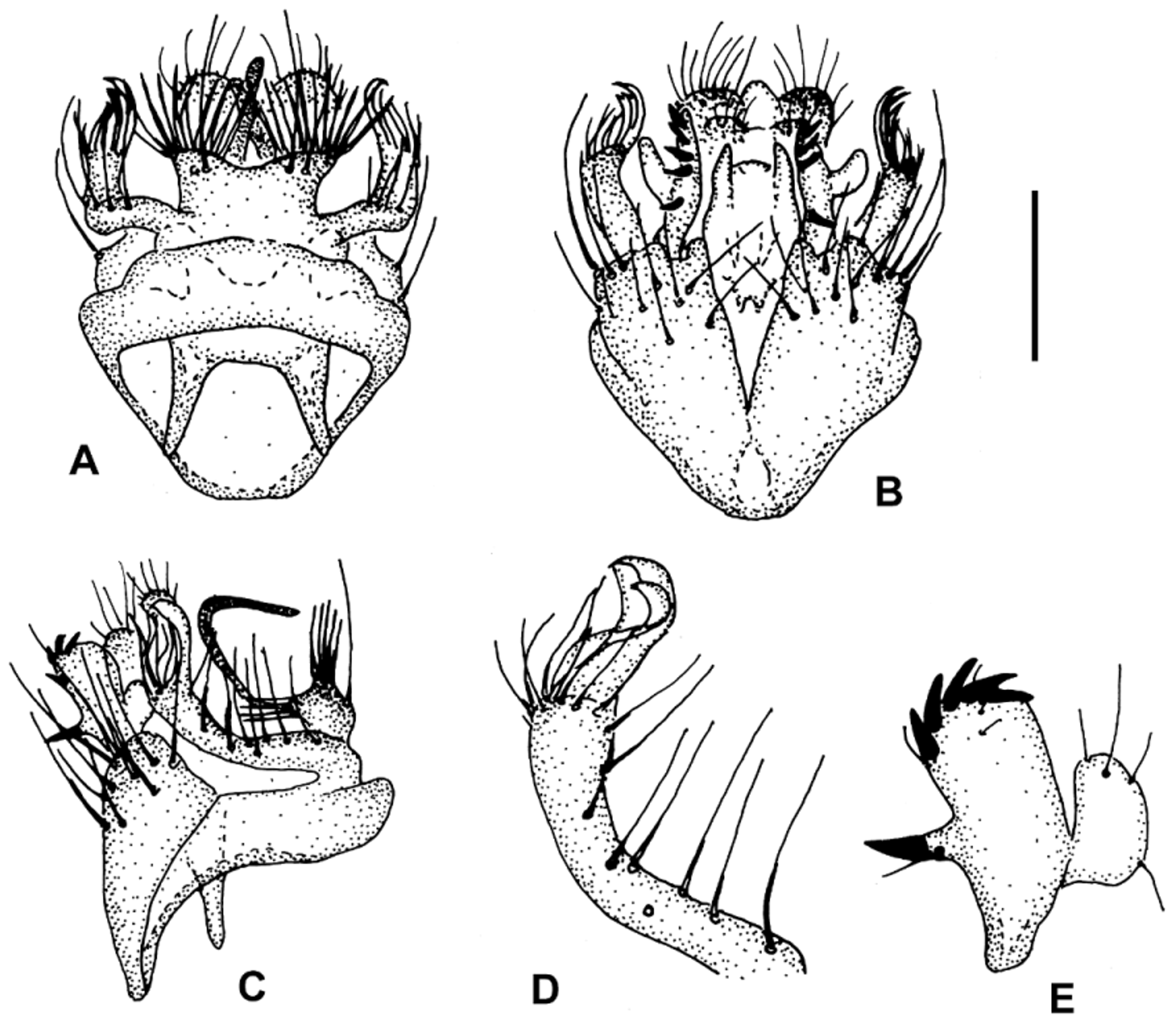
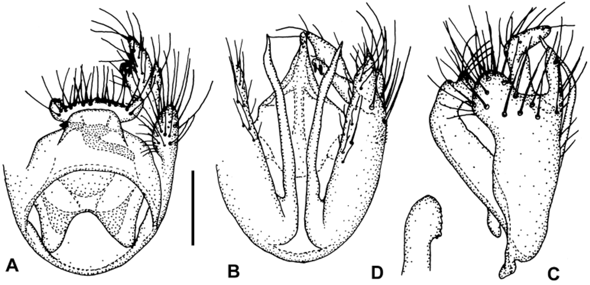
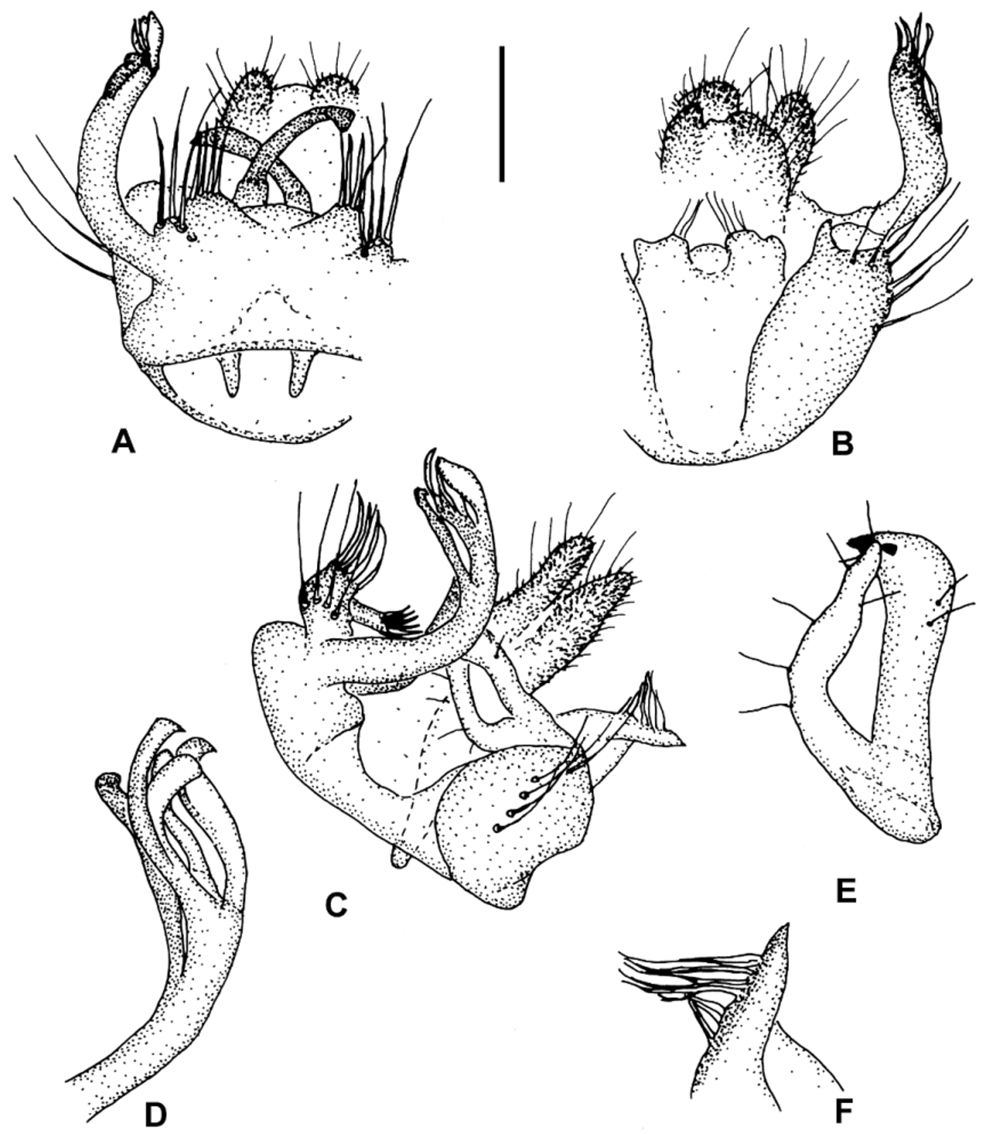
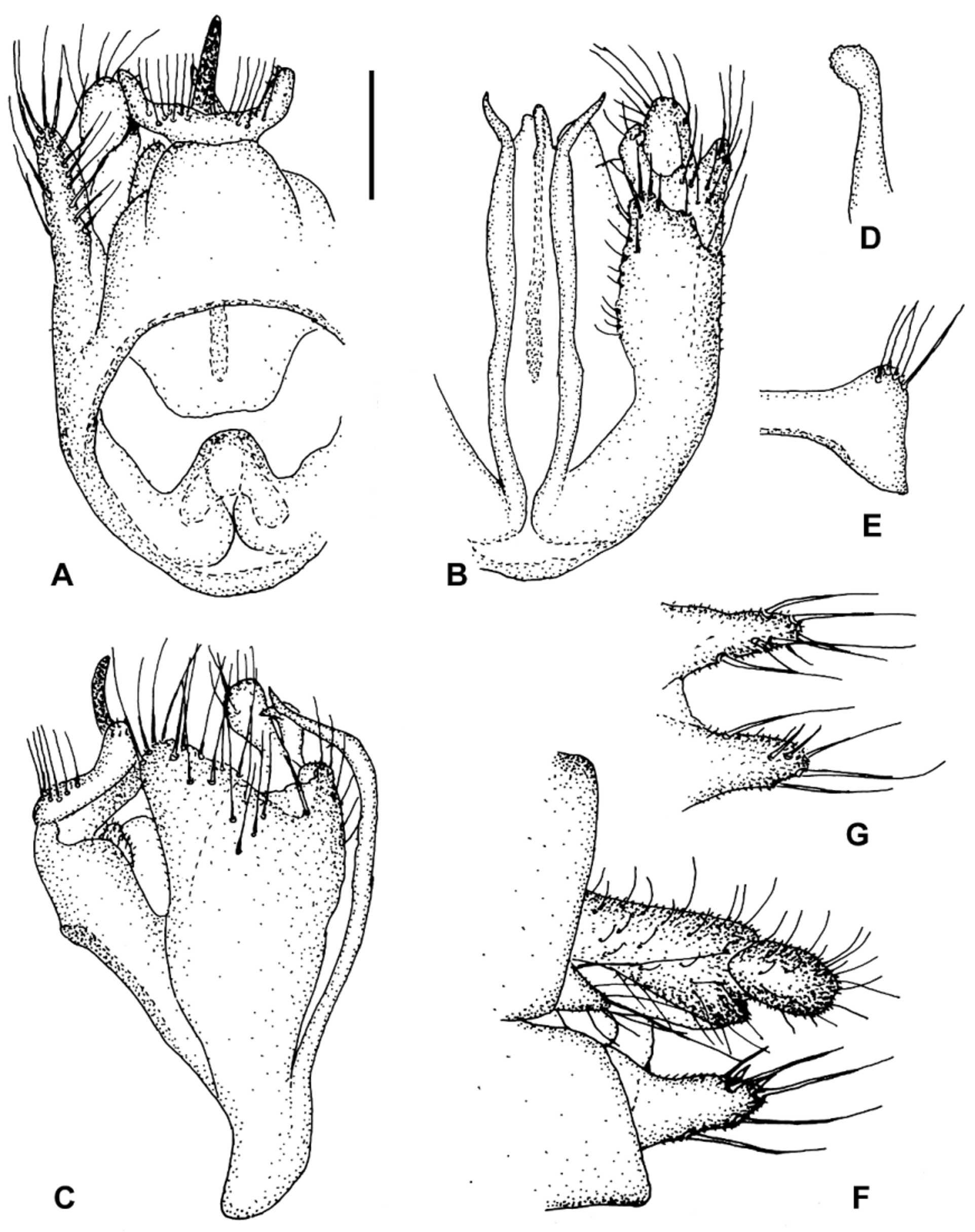
3. Sternal synsclerite of male hypopygium with long, wide, sternal lateral appendages (extending towards tergal part of hypopygium in lateral view; e.g. Figs. 2 View FIGURE 2 C, 4C, 5C, 8C); tergal part of hypopygium without lateral appendages (e.g. Figs. 2 View FIGURE 2 A, 4A, 5A)....................................................................... .. (subgenus Cymomya Väisänen ) ... 4
- Sternal synsclerite without large lateral appendages (e.g. Figs. 1 View FIGURE 1 B, 3B, 6B); tergal part with lateral appendages (e.g. Figs. 1 View FIGURE 1 A, 1D, 3A, 3D, 6A)....................................................... (subgenus Mycomyopsis Väisänen )...7
4. Sternal lateral appendage very long, wide and rounded, its subapical part much wider than its basal part ( Fig. 5 View FIGURE 5 C)............................................................................................... M. paraklossi sp. n.
- Sternal lateral appendage smaller, its subapical part is slightly narrower or about as wide as its basal part ( Figs. 2 View FIGURE 2 C, 4C, 8C)..................................................................................................... 5
5. Sternal lateral appendage relatively long, apically rounded, its subapical part is about as wide as its basal part, densely setose; long setae covering at least apical ½ of sternal synsclerite ( Fig. 2 View FIGURE 2 C); wing vein Sc ending in R1; M and Cu bare; wing length 2.5 mm ................................................................................ M. klossi Edwards View in CoL
- Sternal lateral appendage short, tapering towards apex, sparsely setose; long setae covering apical 1/3 of sternal synsclerite
( Figs. 4 View FIGURE 4 C, 8C); Sc ending in C or R1; M and Cu bare or with small setae; wing length 4.0– 4.5 mm ..................... 6 6. Sc ending in C; M and Cu bare; small cell and apex of wing weakly infuscated; sternal lateral appendage apically blunt, triangular, tapering towards apex ( Fig. 8 View FIGURE 8 C).......................................................... M. yatai sp. n.
- Sc ending in R1; M and Cu with some small setae; wing hyaline; sternal lateral appendage rounded, tapering towards apex ( Fig. 4 View FIGURE 4 C)................................................................................. M. nakanishii n. sp.
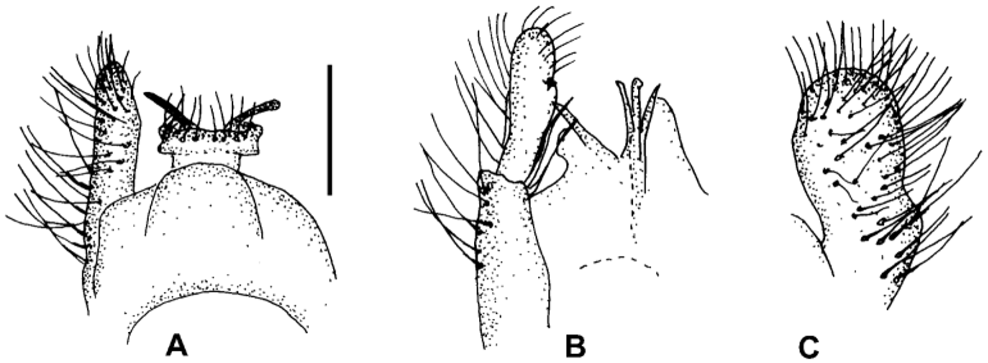
7. Gonostylus short and wide, about 2x as long as its width, with a spur in its middle part ( Fig. 3 View FIGURE 3 E)...... M. minutata Edwards View in CoL
- Gonostylus long and slender, 5– 6 x as long as its width, without a spur ( Figs. 1 View FIGURE 1 E, 6E).............................. 8
8. Outer tergal combs of tergal part of hypopygium both with about 5 spines ( Fig. 6 View FIGURE 6 A); basal and middle part of tergal lateral appendage bare ( Figs. 6 View FIGURE 6 C–D)................................................................ M. pongo sp. n.
- Outer tergal combs of tergal part of hypopygium both with about 10 spines ( Fig. 1 View FIGURE 1 A); middle part of tergal lateral appendage sparsely setose, without distinct bare area ( Figs 1 View FIGURE 1 C–D).......................................... M. apoensis sp. n.


No known copyright restrictions apply. See Agosti, D., Egloff, W., 2009. Taxonomic information exchange and copyright: the Plazi approach. BMC Research Notes 2009, 2:53 for further explanation.