
Leuceruthrus ksepkai n., 2022
|
publication ID |
https://doi.org/10.1645/22-36 |
|
DOI |
https://doi.org/10.5281/zenodo.7753988 |
|
persistent identifier |
https://treatment.plazi.org/id/039987AB-2165-FFE5-16D3-4A81AEBA979F |
|
treatment provided by |
Felipe |
|
scientific name |
Leuceruthrus ksepkai n. |
| status |
n. |
Leuceruthrus ksepkai n. View in CoL View at ENA sp.
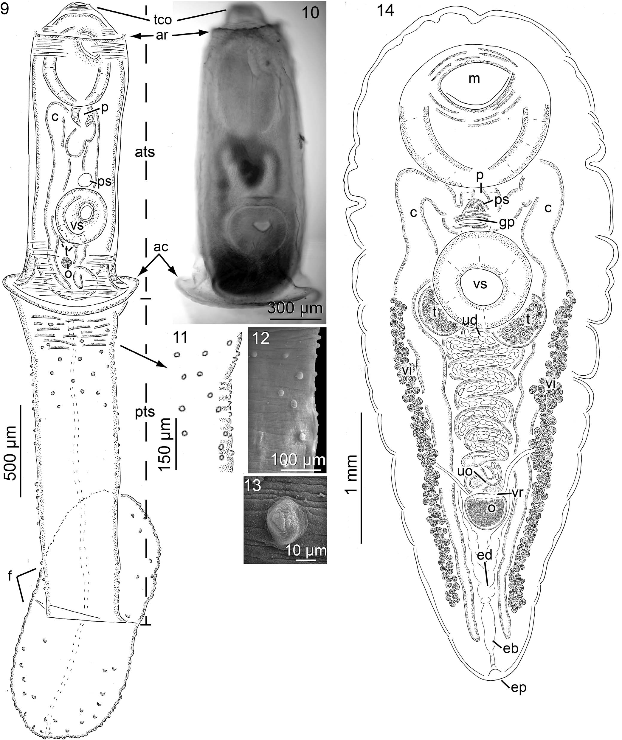
( Figs. 9 13 View Figures 9–14 ; Table II)
Diagnosis of cercaria (based on light microscopy of 12 stained, whole-mounted, naturally shed cercariae having a withdrawn distome) ( Figs. 9 13 View Figures 9–14 ): Cercaria 3,600 4,140 (3,944, 5) long. Tail stem 2,980 3,460 (3,228, 5) long or 77 84% (82%, 5) of cercariae length, maximum width 560 740 (652, 2) or 4.3 5.7 (6, 2) X longer than wide, comprised of an anterior and posterior region ( Fig. 9 View Figures 9–14 ); anterior tail stem region (ATS) 1,480 1,800 (1,668, 5) long or 39 46% (42%, 5) of cercariae length, with anterior ridge, and posterior collar, maximum width same as reported for tail stem, primarily cylindrical, tapering anteriorly, containing distome; ATS ridge near anterior end of tail stem ( Figs. 9, 10 View Figures 9–14 ), preceding tail cavity opening, 320 490 (446, 5) wide, 120 220 (168, 5) of cercaria length from anterior end of cercaria; ATS collar 620 740 (675, 4) wide or 1.3 2.2 (1.6, 4) X wider than anterior tail stem ridge, appearing as a laterally expanded ‘‘flange like’’ tegumental ring, at confluence of anterior and posterior tail stems ( Figs. 9, 10 View Figures 9–14 ); posterior tail stem region (PTS) dorsoventrally compressed, 1,400 1,820 (1,560, 5) long or 35 44% (39%, 5) of cercariae length, anterior width 520 600 (548, 4), medial width 380 580 (473, 4) posterior width 320 540 (408), nearly uniform in width anteriorly to posteriorly ( Fig. 9 View Figures 9–14 ), bearing many minute protuberances ( Figs. 9, 11, 12 View Figures 9–14 ); PTS protuberances, minute, marginally distributed throughout entire length of PTS, also encircling anterior third of PTS ( Figs. 9, 11, 13 View Figures 9–14 ). Furcae with conspicuous black markings along margins when alive, oblong, dorsoventrally compressed, bearing many minute pored protuberances; furcal protuberances, minute, distributed marginally, and occasionally submarginally ( Fig. 9 View Figures 9–14 ); dorsal furca, 690 790 (724, 5) or 17 19% (18%, 5) of cercariae length, 600 760 (678, 5) wide or 85 98% (94%, 5) of furca length, ventral furca, 680 860 (752, 5) or 17 20% (19%, 5) of cercariae length, 600 790 (684, 5) or 80 97% (91%, 5) of furca length. Tail cavity opening at anteromedial end of cercaria, directing anteriad. Tail stem spines not evident. Excretory system with 1 primary excretory canal, extending posteriad along medial axis of the posterior tail stem, bifurcating at synthesis of furcae, extending independently through each furcae, opening via excretory pore at distal end of each furcae ( Fig. 9 View Figures 9–14 ).
Body of distome (¼cercarial body) ( Fig. 9 View Figures 9–14 ) 1,540 1,630 (1,592, 5) long or 37 45% (41%, 5) of cercaria length, 490 550 (510, 5) wide or 2.8 3.3 (3.1, 5) X longer than wider, anterior margin 30 155 (88, 5) from tail cavity opening; forebody 880 980 (918, 5) long or 54 60% (57%, 5) of overall body length; hindbody 330 360 (350, 5) long or 21 22% (22%, 5) of overall body length, 37 40% (38%, 5) of forebody length, posterior end in-curled; tegument unarmed. Excretory system not evident. Nervous system not evident. Oral sucker 420 500 (467, 5) long or 26 32% (29%, 5) of body length, 310 365 (341, 5) wide or 64 75% (67%, 5) of body width, 50 100 (75, 5) or 3 6% (4%, 5) of body length from anterior body end, 840 1,040 (976, 5) or 52 66% (61%, 5) of body length from posterior body end, posterior margin 325 440 (373, 5) from anterior margin of ventral sucker ( Fig. 9 View Figures 9–14 ). Ventral sucker in posterior half of body, 260 330 (296, 5) or 16 21% (18%, 5) of body length, 270 320 (294, 5) or 51 63% (58%, 5) of body width, 55 71% (64%, 5) of oral sucker length, 79 94% (86%, 5) of oral sucker width ( Fig. 9 View Figures 9–14 ). Pharynx ovoid, 100 150 (125, 5) long or 6 9 % (8%, 5) of body length, 110 125 (112, 5) wide ( Fig. 9 View Figures 9–14 ). Esophagus extending posteriad from mouth before bifurcating posterior to pharynx, esophageal branches arching posterolaterad before joining with intestinal ceca; dextral cecum 1,005 1,290 (1,173, 5) or 62 84% (74%, 5) of body length, laterad cecum length 245 360 (288, 5), descending cecum length 740 960 (885, 5), prececal space, 450 650 (526, 5) or 28 40% (33%, 5) of body length from anterior end of body, postcecal space, 55 120 (74, 4) or 3 7% (5%, 4) of body length from posterior end of body; sinistral cecum 1,050 1,330 (1,190, 5) or 64 82% (75%, 5) of body length, laterad cecum length 230 340 (266, 5), descending cecum length 775 1,000 (924, 5), prececal space 430 710 (532, 5) or 27 44% (33%, 5) of body length from anterior end of body, postcecal space, 50 100 (74, 4) or 3 6% (5%, 4) of body length from posterior end of body ( Fig. 9 View Figures 9–14 ).
Testes abreast, round to oval ( Fig. 9 View Figures 9–14 ); dextral testis 55 65 (60, 4) or 3 4% (4%, 4) of body length, 50 70 (58, 4) or 0.85 1.2 (1, 4) X wider than long; sinistral testis 50 80 (61, 4) or 3 5% (4%, 4) of body length, 65 70 (66, 4) or 0.77 1.10 (0.92, 4) X wider than long; pretesticular space 1,000 1,160 (1,075, 4) from anterior end of body or 61 75% (67%, 4) of total body length; posttesticular space 300 420 (3,489, 4) from posterior end of body or 19 26% (22%, 4) of total body length. Prostatic sac 65 75 (71, 5) long, 60 85 (100, 5) or 0.85 1.3 (1, 5) X wider than long ( Fig. 9 View Figures 9–14 ). Genital atrium circular in ventral view, 35 45 (28, 2) in diameter. Genital pore 835 960 (877, 5) of body length from anterior end of body or at 52 59% (55%, 5) of body length. Features of terminal male genitalia (i.e., seminal vesicle, pars prostatica, ejaculatory duct, sinus organ) appearing as anlagen, difficult to discern.
Ovary near posterior end of body or 160 310 (210, 5) of body length from posterior end of body, 50 75 (59, 5) long or 0.80 1.50 (1, 5) X longer than wide, 50 60 (55, 5) wide ( Fig. 9 View Figures 9–14 ). Fine details of the female genitalia (i.e., oviduct, Laurer’s canal, ovovitelline duct, oötype, and Mehlis’ gland) difficult to discern. Uterus 430 655 (543, 2) long or 34 37% (35%, 2) of body length, 13 20 (17, 2) wide, extending in a straight line from anterior margin of ovary, passing between testes, difficult to discern distally; metraterm not evident. Vitellarium lacking in distome.
Taxonomic summary for Leuceruthrus ksepkai n. sp.
Type host: Elimia floridensis (Reeve, 1860) , rasp elimia.
Other hosts: Elimia sp. 1 and M. salmoides .
Type locality: Holmes Creek ( 30°36 ′ 25 ′′ N, 85°44 ′ 49 ′′ W), Florida GoogleMaps .
Other locality: Chocktawhatchee River ( 30°30 ′ 51 ′′ N, 85°50 ′ 55 ′′ W), Florida GoogleMaps .
Specimens and sequences deposited: Holotype (cercaria, USNM 1593586 About USNM ), paratypes (cercariae, USNM 1593587 About USNM , 1593588 About USNM ), ITS2 (cercaria from E. floridensis , GenBank ON877233 View Materials ).
Prevalence and intensity of infection: 5 of 134 (4%) E. floridensis and 12 of 210 (6%) of Elimia sp. 1 from Holmes Creek and Chocktawhatchee River, respectively, were infected.
ZooBank registration: urn:lsid:zoobank.org:act:.
Etymology: The specific epithet ‘‘ ksepkai ’’ is for Steven P. Ksepka, Aquatic Parasitology Laboratory, Auburn University, and is in recognition of his work exploring the parasites of centrarchid fishes in the southeastern United States.
Taxonomic remarks on Leuceruthrus ksepkai n. sp.
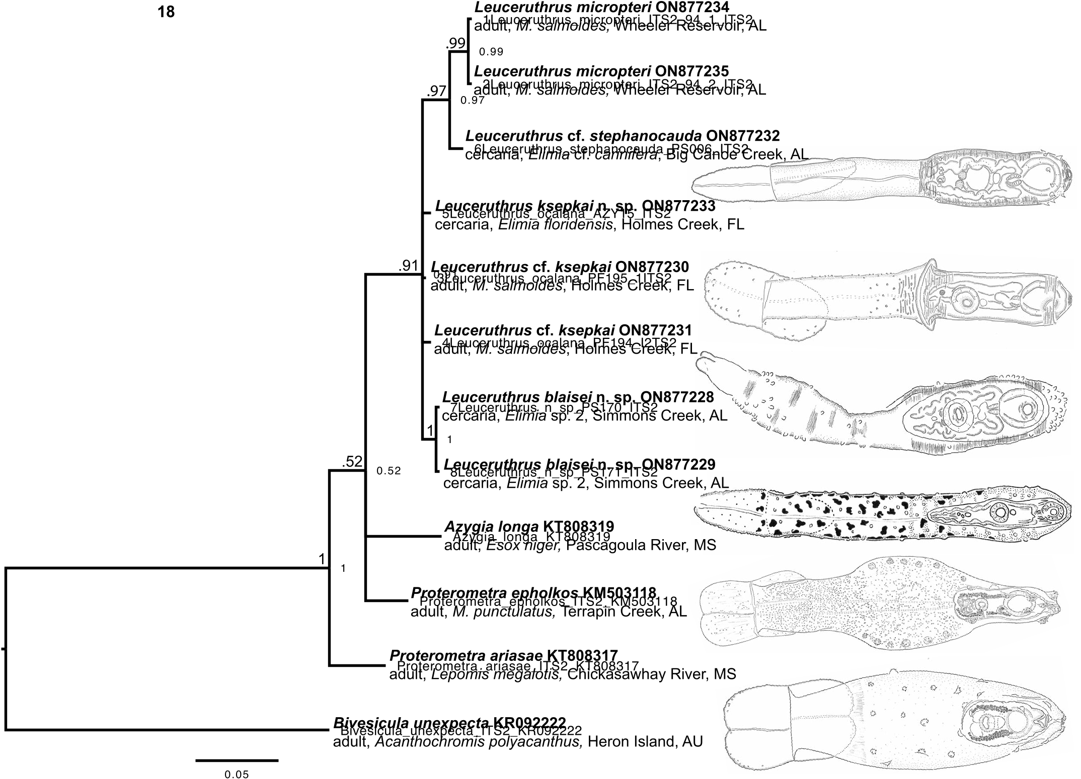
We studied only naturally shed, actively swimming cercariae of L. ksepkai (Table II). Cercariae from both snail hosts ( E. floridensis and Elimia sp. 1 ) were morphologically identical and regarded as conspecific herein. Leuceruthrus ksepkai differs from all nominal congeners by the combination of having broadly rounded furcae (with slight pigmentation along margin in living specimens), no spines on the tail stem, distinct anterior and posterior ridges that flank the tail stem portion accommodating the withdrawn distome, minute protuberances occupying the lateral margin of the tail stem for its entire length, and protuberances that encircle the anterior third of the posterior tail stem (immediately posterior to the tail stem portion containing the withdrawn distome). Leuceruthrus ocalana is the only other species of Leuceruthrus that has broadly rounded furcae (which superficially resemble those of Proterometra spp. ; Fig. 18 View Figure 18 ). As indicated above, we consider L. ocalana a species inquirendae because its original description is deficient and incomplete.
No known copyright restrictions apply. See Agosti, D., Egloff, W., 2009. Taxonomic information exchange and copyright: the Plazi approach. BMC Research Notes 2009, 2:53 for further explanation.
|
Kingdom |
|
|
Phylum |
|
|
Class |
|
|
SubClass |
Digenea |
|
Order |
|
|
Family |
|
|
Genus |