
Sacura sanguinea, Motomura & Yoshida & Vilasri, 2017
|
publication ID |
https://doi.org/10.11646/zootaxa.4306.2.10 |
|
publication LSID |
lsid:zoobank.org:pub:09E43F72-01FE-45BD-82E4-3D9A93801362 |
|
DOI |
https://doi.org/10.5281/zenodo.6021224 |
|
persistent identifier |
https://treatment.plazi.org/id/039987E3-FFDC-FFE7-C8C2-DDE5FE49FCB1 |
|
treatment provided by |
Plazi |
|
scientific name |
Sacura sanguinea |
| status |
sp. nov. |
Sacura sanguinea n. sp.
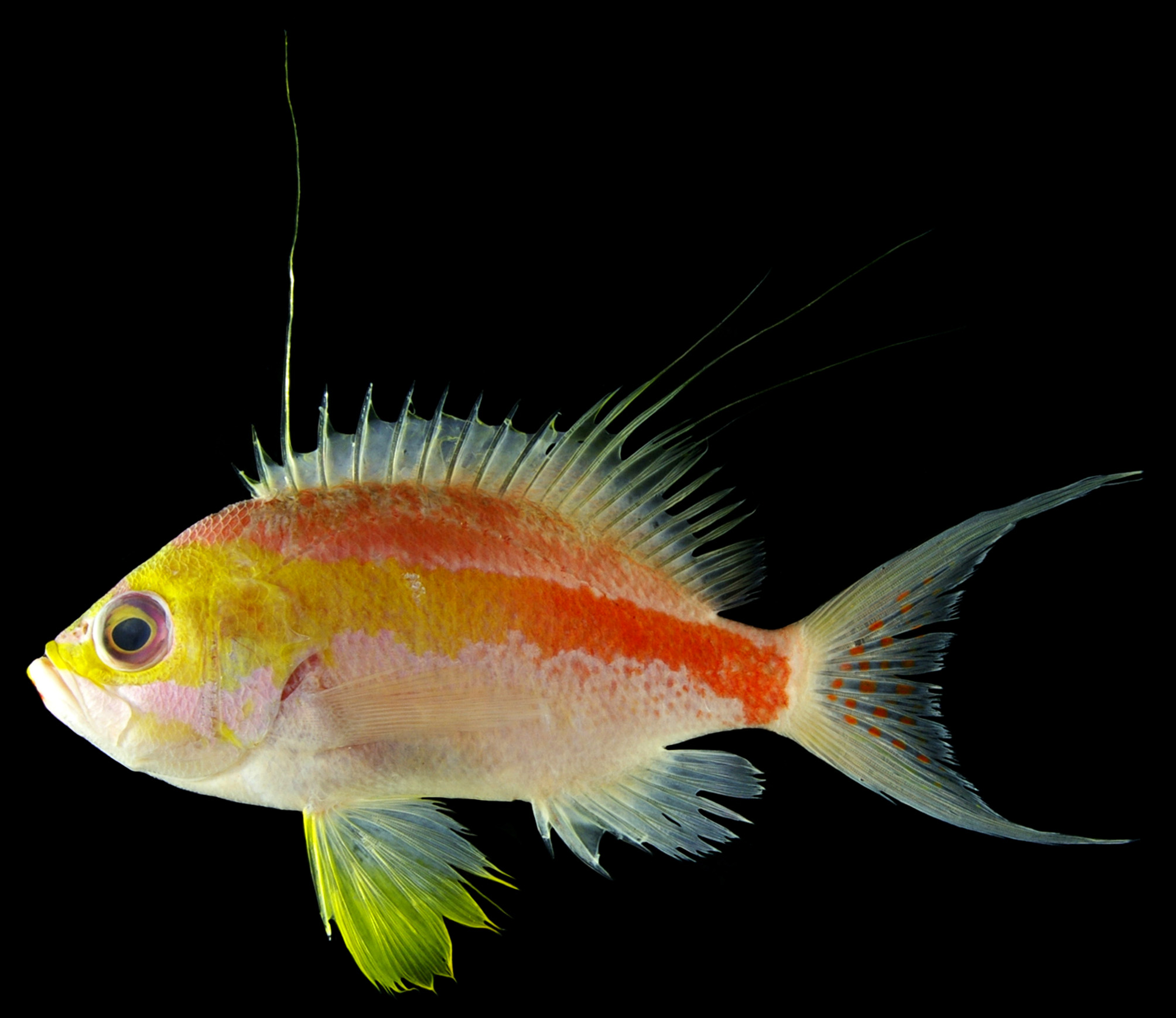
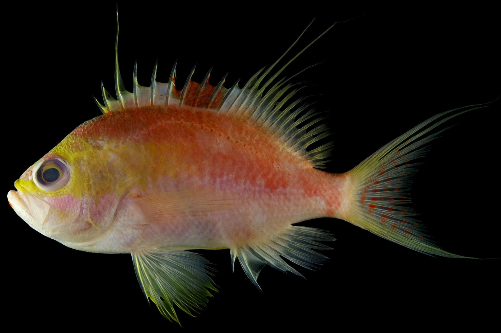
[New English name: Andaman Deepwater Anthias] Figs. 1–2 View FIGURE 1 View FIGURE 2 ; Table 1
Holotype. THNHM-F 14852 , male, 124.1 mm SL, Andaman Sea , Thailand (purchased at Pak Nam Ranong Fishing Port, Ranong Province, 09°56ʹN, 98°35ʹE), trawl, 7 December 2010.
Paratypes. KAUM –I. 33316, female, 116.3 mm SL, KAUM –I. 33317, male, 111.2 mm SL, collected with holotype. Diagnosis. A species of Sacura with the following combination of characters: dorsal-fin rays X, 15; pored lateralline scales 34; gill rakers 8 + 23 = 31; body depth 42.6–44.7% of SL; head length 39.5–41.4% SL; pectoral-fin length 32.4–33.1% SL; poorly defined broad yellow band from anterior profile of head to middle of body, the band gradually becoming red around middle of body and ending at caudal-fin base; no vertical bands or lines on body; pelvic fins yellow; caudal fin yellowish with distinct red spots scattered centrally; large dark red blotch posteriorly on spinous portion of dorsal fin in female.
Description. Meristic and morphometric data for the holotype and paratypes of S. sanguinea are shown in Table 1. Data for the holotype are presented first, followed by paratype data in parentheses (if different).
Holotype Paratype Paratype
THNHM-F 14852 KAUM–I. 33316 KAUM–I. 33317 Male Female Male
Standard length (mm; SL) 124.1 116.3 111.2
Dorsal-fin rays X, 15 X, 15 X, 15
Anal-fin rays III, 7 III, 7 III, 7
Pectoral-fin rays 17 17 17
Pelvic-fin rays I, 5 I, 5 I, 5
Procurrent caudal-fin rays (upper + lower) 6 + 6 6 + 6 6 + 6
Segmented unbranched caudal-fin rays (upper + lower) 2 + 2 2 + 2 2 + 2
Segmented branched caudal-fin rays (upper + lower) 7 + 6 7 + 6 7 + 6
Pored lateral-line scales 34 34 34
Transverse scale rows above lateral line 7 7 7
Transverse scale rows below lateral line 18 16 17
Gill rakers 8 + 23 = 31 8 + 23 = 31 8 + 23 = 31
% of SL Mean Body depth 42.6 44.2 44.7 43.8 Head length (HL) 39.5 40.2 41.4 40.4 Pectoral-fin length 32.4 33.1 32.9 32.8 Pelvic-fin length 31.0 28.6 35.0 31.5 Caudal-peduncle length 15.8 17.1 18.1 17.0 Caudal-peduncle depth 13.3 13.9 13.6 13.6 1st dorsal-fin spine length 6.2 7.1 7.4 6.9 2nd dorsal-fin spine length 9.5 10.2 10.9 10.2 3rd dorsal-fin spine length 56.1 32.2 26.5 38.3 4th dorsal-fin spine length 14.7 14.5 15.2 14.8 3rd dorsal-fin soft ray length 35.6 40.8 33.4 36.6 Anal-fin length 27.6 26.7 30.7 28.4 1st anal-fin spine length 8.6 7.9 9.3 8.6 2nd anal-fin spine length 14.5 16.2 17.1 15.9 2nd anal-fin soft ray length 20.6 20.8 21.4 20.9 Pelvic-fin spine length 18.7 18.4 20.0 19.0 % of HL
Snout length 21.4 21.8 20.6 21.3 Orbit diameter 28.2 28.2 30.2 28.8 Interorbital width 24.5 24.6 22.8 23.9 Postorbital length 53.5 53.6 54.2 53.8 Upper-jaw length 41.8 42.3 42.5 42.2 Maxillary depth 16.5 17.3 14.8 16.2 Mouth moderately large, posterior end of maxilla just short of (reaching to in KAUM–I. 33317) vertical through posterior margin of pupil; mouth strongly oblique, forming angle of 40° to horizontal axis of head and body; lower jaw slightly projecting; posterior margin of maxilla slightly concave, upper corner rounded, lower weakly pointed. Pair of widely separated, downward-projecting canine teeth at front of upper jaw, followed by outer row of slender conical teeth, more posterior teeth curved forward; inner wide band of small slender teeth; 1 (or 2) stout canine teeth at each anterior end of premaxilla, pointing towards vomer. Pair of widely separated, stout canines (2 canines at right side in KAUM–I. 33316; no canines in KAUM–I. 33317) at front of lower jaw, these canines between upper-jaw canine teeth when mouth closed; 2 (or 3) large canines at each side of lower jaw about one-third distance from lower-jaw symphysis; villiform teeth band between anterior and posterior canines; 2 (or 1) rows of slender teeth on lower jaw posterior to posterior canines. Vomer with patch of conical teeth, toothed area in shape of equilateral triangle with its base slightly bowed inwards; no teeth below vomer. Palatines with band of villiform teeth. Tongue triangular and not sharply pointed, upper surface with small papillae. Gill rakers long and slender with minute projections on inner edge; longest raker below angle shorter than longest gill filaments, and less than orbit diameter.
Anterior nostril membrane tube with skin flap posteriorly, the flap reaching posterior nostril when reflected; anterior nostril at level with center of eye. Posterior nostril vertically elongate, without skin flap. Opercle with 3 flat spines, lower 2 spines acute and tips exposed, upper spine with somewhat rounded tip embedded in scales; middle spine largest, at level of center of eye. Upper edge of preopercle with serrae, progressively larger ventrally, with 3 or 4 (3–5) strong spines at angle. Lower edge of subopercle smooth. Ctenoid scales on head and body, except for lips, isthmus, anterior lacrimal, and around nostrils; no accessory scales; no scales on pelvic fin, all other fins with scales basally. Lateral line continuous, highly arched over pectoral fin, highest below sixth dorsal-fin spine base. Formula for configuration of supraneural bones, anterior neural spines, and anterior dorsal pterygiophores /0+0/2/1+1/1/1/1/1/1/1/. Vertebrae 10 + 16. Third spine and second to fifth (or fourth) soft rays in dorsal fin prolonged. No prolonged spines and rays in pelvic and anal fins. Dorsal-fin origin just above upper end of gill opening and anterior to vertical through upper end of pectoral-fin base. Base of soft-rayed portion of dorsal fin shorter than base of spinous portion. Posterior margin of opercular membrane extending beyond vertical through fourth dorsal-fin spine base. Upper end of pectoral-fin base just above pelvic-fin origin. Posterior tip of pectoral fin extending beyond vertical through third anal-fin spine base. Tip of depressed pelvic fin extending slightly beyond anal-fin origin. Anal-fin origin posterior to last dorsal-fin spine base. Caudal fin lunate, deeply forked, with elongate slender lobes.
Color when fresh. — Based on color photographs of the male holotype ( Fig. 1 View FIGURE 1 ) and female paratype ( Fig. 2 View FIGURE 2 ): head pinkish with vivid yellow area on upper half of head, extending from anterior portion of upper lip and snout to opercular margin along ventral margin of orbit, and from above posterior half of upper margin of orbit to above opercle; interorbital region between anterodorsal margins of orbits vivid yellow; anterior surface of snout between nostrils yellow (pink in paratype); lower half of cheek pale yellow. Anteroventral surface of lower jaw red. Body reddish pink dorsally, whitish pink ventrally, with broad yellow band, its width more than orbit diameter, from head to middle of body, the band gradually becoming red around middle of body (indistinct in paratype), ending at caudal-fin base; pored scales dark red. Dorsal-fin membranes whitish, pinkish basally; membranes along posterior margins of anterior spines yellowish; soft rays pale yellow distally. Tip of prolonged third dorsal-fin spine black (not black in paratype). No blotch on spinous portion of dorsal fin (dark red blotch between sixth and tenth spines in paratype). Pectoral fin semi-transparent white. Pelvic fin yellow anteriorly and distally, otherwise white. Anal fin white, pale yellow basally. Caudal fin pale yellow, with white posterior margin (indistinct in paratype) and a number of distinct red spots centrally
Color of preserved specimens. — Head and body uniformly brownish white (with black blotch between sixth and tenth dorsal-fin spines in female paratype).
Distribution. Known only from the Andaman Sea. No data for water depth or habitat are available. Etymology. Derived from the Latin sanguinea meaning “blood red”, in reference to a red longitudinal broad band on the posterior body and red spots on the caudal fin of the species.
Remarks. The generic classification of the anthiadin fishes has been very confused ( Randall & Heemstra 2006) and the generic limits of Sacura have not yet been clearly defined ( Heemstra & Randall 1979). In their revision of Sacura, Heemstra & Randall (1979: 1–2) gave a large number of characters as diagnostic of the genus and all their characters agreed with those of the Andaman Sea specimens described here. These characters are almost the same as those diagnostic of Odontanthias Bleeker 1873 given by Randall & Heemstra (2006). Although no characters separating Sacura from Odontanthias have been published, the absence of teeth below the vomer path distinguishes the genus Sacura from Odontanthias and, on other hand, the pointed snout and large eyes of the Andaman Sea specimens also indicate that the new species should be included in Sacura .
Sacura sanguinea n. sp. can be easily distinguished from all congeners (and all members of Odontanthias View in CoL ) by its unique coloration: a poorly defined broad yellow band from the anterior profile of the head to the middle of the body, the band gradually becoming red around the middle of the body and ending at the caudal-fin base, and the caudal fin yellowish with distinct red spots scattered centrally ( Fig. 1 View FIGURE 1 ). Sacura sanguinea n. sp. and S. speciosa View in CoL share 15 dorsal-fin soft rays and 34 (in S. sanguinea ) and 33 (in S. speciosa View in CoL ) pored lateral-line scales (vs. 14 rays and 28–31 scales in S. boulengeri View in CoL , 16–18 and 26–30 in S. margaritacea View in CoL , 17 and 34 in S. parva View in CoL ; Heemstra & Randall 1979). In addition to the above-mentioned color characters, S. sanguinea n. sp. further differs from S. speciosa View in CoL in having a lower count of gill rakers (31 vs. 40 in the latter), a shallower body depth (42.6–44.7% of SL vs. 50%), and a shorter pectoral fin (32.4– 33.1% of SL vs. 42%) (values for S. speciosa View in CoL from Heemstra & Randall 1979).
Females of S. sanguinea have a large dark red blotch posteriorly on the spinous portion of the dorsal fin and the blotch is supposed to disappear as individuals transition to being male. This sexual dichromatic change is also known in S. margaritacea View in CoL and S. parva ( Heemstra & Randall 1979) View in CoL . The color characteristics of females of S. boulengeri View in CoL and S. speciosa View in CoL are unknown.
| KAUM |
Kagoshima University Museum |
No known copyright restrictions apply. See Agosti, D., Egloff, W., 2009. Taxonomic information exchange and copyright: the Plazi approach. BMC Research Notes 2009, 2:53 for further explanation.
|
Kingdom |
|
|
Phylum |
|
|
Class |
|
|
Order |
|
|
Family |
|
|
Genus |
Sacura sanguinea
| Motomura, Hiroyuki, Yoshida, Tomohiro & Vilasri, Veera 2017 |
Sacura sanguinea
| Motomura & Yoshida & Vilasri 2017 |
Sacura sanguinea
| Motomura & Yoshida & Vilasri 2017 |
S. sanguinea
| Motomura & Yoshida & Vilasri 2017 |
S. sanguinea
| Motomura & Yoshida & Vilasri 2017 |
S. sanguinea
| Motomura & Yoshida & Vilasri 2017 |
S. speciosa
| Heemstra & Randall 1979 |
S. speciosa
| Heemstra & Randall 1979 |
S. parva
| Heemstra & Randall 1979 |
S. speciosa
| Heemstra & Randall 1979 |
S. speciosa
| Heemstra & Randall 1979 |
S. parva (
| Heemstra & Randall 1979 |
S. speciosa
| Heemstra & Randall 1979 |
Odontanthias
| Bleeker 1873 |