
Paracentrophyes sanchezae, Sørensen, Martin V. & Landers, Stephen C., 2017
|
publication ID |
https://doi.org/ 10.11646/zootaxa.4242.1.3 |
|
publication LSID |
lsid:zoobank.org:pub:C8299651-B344-4287-82F9-061C100F70BF |
|
DOI |
https://doi.org/10.5281/zenodo.5625765 |
|
persistent identifier |
https://treatment.plazi.org/id/64B718A2-E11D-4E49-996C-043EB261C55D |
|
taxon LSID |
lsid:zoobank.org:act:64B718A2-E11D-4E49-996C-043EB261C55D |
|
treatment provided by |
Plazi |
|
scientific name |
Paracentrophyes sanchezae |
| status |
sp. nov. |
Paracentrophyes sanchezae n. sp.
( Figs 2–5 View FIGURE 2 View FIGURE 3 View FIGURE 4 View FIGURE 5 ; Tabs 1–2 View TABLE 1 View TABLE 2 )
zoobank.org code: urn:lsid:zoobank.org:pub:C8299651-B344-4287-82F9-061C100F70BF
Material. Holotype, adult female, collected from mud on November 2, 2014, at station 001-2014 ( Fig. 1 View FIGURE 1 ), at 57 m depth on the continental shelf, south of the Louisiana coast line, and west of the submarine Mississippi Canyon (position: 28o25’48.71’’N, 090o14’10.31’’W), mounted in Fluoromount G, deposited at the Natural History Museum of Denmark, under catalogue number ZMUC KIN- 1014 GoogleMaps . Paratypic material mounted for LM includes a single adult male, collected from mud on October 20, 2009, at station 031-2009 ( Fig. 1 View FIGURE 1 ), at 54 m depth about 170 km straight south of the Louisiana – Texas state border (position: 28o12’59.76’’N, 093o24’19.08’’W), mounted in Fluoromount G, deposited at the Natural History Museum of Denmark, under catalogue number ZMUC KIN- 1015 GoogleMaps . Additional paratypes include two males, collected from mud on November 15, 2013, at stations 078-2013 and 079- 2013 ( Fig. 1 View FIGURE 1 ). Station 078-2013 is located at 142 m depth (position: 29o53’07.72’’N, 086o47’31.20’’W), and Station 079-2013 at 125 m depth (position: 29o48’20.77’’N, 086o36’02.37’’W). Both stations are located near each other, south of the Florida panhandle in the northeastern extension of the submarine De Soto Canyon. The specimens are mounted for SEM and deposited at the Natural History Museum of Denmark, under catalogue numbers ZMUC KIN- 1081 and KIN- 1082 GoogleMaps .
Diagnosis. Segments 1 and 11 composed of one tergal and one sternal plate; sternal plate of segment 1 partially differentiated into episternal and midsternal plates. Segments 2 to 10 composed of one tergal and two sternal plates. Middorsal and midlateral spinose processes present on segments 1 to 9 in both sexes; females also with middorsal and midlateral spinose processes on segment 10, and middorsal spine on segment 11; males with flexible middorsal and midlateral spines on segment 10, and midlateral penile spines on segments 10 and 11. Outer oral styles alternating in size between larger and smaller ones; large outer oral spines with 18 basal fringe tips; small outer oral spines with 10 basal fringe tips. Perispinal setae present as unpaired ones in paradorsal position on segments 5 and 9 (occasionally 7), paired ones in paralateral positions on segments 6 to 9 (occasionally 4), and paired ones in ventrolateral positions on segments 1, 3, 5 and 9 (occasionally unpaired on 7 and 8).
Etymology. The species is named after Dr Nuria Sánchez in recognition of her contributions to pycnophyid and neocentrophyid taxonomy and systematics.
Description. Adults with head, neck and eleven trunk segments ( Figs 2 View FIGURE 2 , 4 View FIGURE 4 A–B, 5A–B). For complete overview of measures and dimensions, see Table 1 View TABLE 1 . The distribution of cuticular structures, i.e., sensory spots, processes and spines is summarized in Table 2 View TABLE 2 .
The head is formed by a retractable mouth cone and an introvert ( Figs 3 View FIGURE 3 , 5 View FIGURE 5 C–D). The pharynx carries at least two rings of inner oral styles, of which the styles in one ring form elongated helioscalids. It was not possible to determine the exact number or arrangement of inner oral styles. The external mouth cone armature consists of nine outer oral styles, arranged as one style anterior to each introvert sector, except for the middorsal sector 6 ( Fig. 3 View FIGURE 3 ). The outer oral styles alternate in size: larger styles are composed of two joined units, and carry a basal fringe consisting of ca. 18 fringe tips; smaller styles are shorter, thinner, more flexible and consist of a single unit, with a basal fringe consisting of ca. 10 fringe tips ( Fig. 5 View FIGURE 5 C). Large outer oral styles are located anterior to introvert sectors 1, 3, 5, 7 and 9, whereas smaller styles are located anterior to sectors 2, 4, 8, and 10 ( Fig. 3 View FIGURE 3 ).
The exact appearance and arrangement of scalids on the introvert could only be examined for introvert sectors 1 to 6 plus 10. However, due to the usual symmetry patterns, we would expect the arrangements in sectors 7 to 9 to be identical with those in sectors 5 to 3. The introvert sectors are defined by ten primary spinoscalids in Ring 01 ( Figs 3 View FIGURE 3 , 5 View FIGURE 5 D). Each primary spinoscalid consists of a basal sheath and a flexible, distal end piece with a blunt tip. The basal sheaths have long, marginal extensions that form transverse fringes, whereas the end pieces have tiny hairs on their proximal halves, and a smooth surface more distally ( Fig. 5 View FIGURE 5 D). Spinoscalids of Rings 02 to 06 also consist of a sheath and an end piece. These sheaths also show a marginal fringe, but the fringe tips are considerably shorter than those on the primary spinoscalids. In addition, each sheath shows an elongate row of small structures that either appear as minute denticles (mostly Ring 02), or short hairs (mostly Rings 03 to 06) ( Fig. 5 View FIGURE 5 D). Rings 02 and 04 each have 10 spinoscalids, and Rings 03 and 5 have 20, meaning that the spinoscalids of these rings form a quincunx in each sector. Even numbered sectors have no spinoscalids posterior to Ring 05, whereas odd numbered sectors show a single spinoscalid in Ring 06 ( Fig. 3 View FIGURE 3 ).
The neck has seven placids, arranged as four dorsal ones and three ventral ( Figs 2 View FIGURE 2 A–B, 3). Fourteen trichoscalids are present, located as single ones in sectors 1, 2, 4, 6, 8 and 10, and two in sectors 2, 5, 7 and 9 ( Fig. 3 View FIGURE 3 ). The positions of the trichoscalids do not follow the patterns of the spinoscalids. Trichoscalids are rather slender, consisting of a single unit, densely covered with small hairs.
The first trunk segment consists of one tergal and one sternal plate; sternal plate with intracuticular lines, indicating a partial division into one midsternal and two episternal plates ( Figs 2 View FIGURE 2 A–B, 4C-D, 5F–G). Segments 2 to 10 consist of one tergal and two sternal plates, and segment 11 of one tergal and one sternal plate ( Figs 2 View FIGURE 2 A–B, 4A– B, J–L, 5A–B, I–K). Sternal plates are flattened, while tergal plates are vaulted, giving the specimens a triangular appearance in cross-sections. Segment widths are nearly constant from segments 3 to 8, which gives the specimens a parallel sided and rectangular appearance ( Figs 2 View FIGURE 2 A–B, 4A–B, 5A).
Segments 1 to 10 in females and 1 to 9 in males with spinose middorsal and midlateral processes ( Figs 2 View FIGURE 2 A–B, 4C–F). Middorsal spinose processes with nearly smooth surface, and pointed rigid tip, projecting slightly beyond segment margin. Midlateral processes with minute hairs, and more flexible tips, projecting well beyond segment margin. A minute paralateral pore, probably a glandular cell outlet, is present next to each midlateral process ( Fig. 4 View FIGURE 4 E, G). Spinose processes are progressively longer towards the posterior part of the trunk.
Markings of muscle attachment sides (muscle scars) are present as small, oval areas in laterodorsal positions on segments 1 to 10, and in paraventral positions on segments 2 to 10 ( Figs 2 View FIGURE 2 A–B). Ventral muscle scars on segment 1 are larger, much more elongate, and located in ventromedial positions. On segment 11, muscle scars are small and rounded, and were observed in ventromedial positions only. All segments, from segment 1 to 10, are generally covered with minute, triangular, scale-like hairs, except on the processes, and around sensory spots and muscle scars. Anterior segment parts with reticulated ornamentation. Free flaps posteriorly on segments with striated substructure, and scale-like hairs that appear slightly more elongate. Pectinate fringes on segment margins not present.
Segment 1 with anterolateral margins of tergal plate projecting into horn-like extensions ( Figs 2 View FIGURE 2 A–B, 4D). Anterior segment margins are denticulated ( Fig. 4 View FIGURE 4 C–D). Sensory spots on this and all following segment belong to type 1 (but see exceptions on segment 11 though), consisting of a single pore and numerous minute papillae, forming elevated and slightly oval papillated areas. Tergal plate without setae, but with six pairs of sensory spots: two pairs are located in a subdorsal position on the anterior segment half, close to the paradorsal areas; two pairs in a laterodorsal position, but very close to the subdorsal area; and two pairs are also laterodorsal, but located close to the paralateral line ( Figs 2 View FIGURE 2 A, 5G). Sternal plate with one pair of ventrolateral setae, slightly behind the transverse midline of the segment ( Figs 2 View FIGURE 2 B, 4D, 5F), and in addition with three pairs of ventromedial sensory spots: One pair is located anteriorly on the plate, near the ventrolateral area; one pair is more medial on the incompletely differentiated episternal plates, at the same level as the setae; and one pair is more anterior, and located within the limits of the incompletely differentiated midsternal plate ( Figs 2 View FIGURE 2 B, 5F).
Segment 2: tergal plate with three pairs of sensory spots in subdorsal positions, and two pairs in laterodorsal positions ( Fig. 2 View FIGURE 2 A). Sternal plates with two sensory spots in ventromedial positions ( Fig. 2 View FIGURE 2 B).
Segment 3: tergal plate with three pairs of sensory spots in subdorsal positions, and one pair in laterodorsal positions ( Fig. 2 View FIGURE 2 A). Sternal plates with ventrolateral setae and two sensory spots in ventromedial positions ( Fig. 2 View FIGURE 2 B).
Segment 4 with tergal plate as on segment 3; one male specimen and female holotype with setae in paralateral positions ( Fig. 4 View FIGURE 4 E), but this was not observed in other specimens. Sternal plates as on segment 2.
Segment 5: tergal plate with a single, unpaired paradorsal seta, two pairs of sensory spots in subdorsal positions, and one pair in laterodorsal positions. Sternal plates with ventrolateral setae ( Fig. 4 View FIGURE 4 F) and single sensory spots in ventromedial positions.
Segment 6: tergal plate with paralateral setae, three pairs of sensory spots in subdorsal positions, and one pair in laterodorsal positions. Sternal plates as on segments 2 and 4.
Segment 7: tergal plate with paralateral setae, two pairs of sensory spots in subdorsal positions, and one pair in laterodorsal positions; one male specimen furthermore with single, unpaired paradorsal seta ( Fig. 5 View FIGURE 5 H). Sternal plates as on segments 2, 4 and 6; furthermore, one male specimen (not the same as the one with paradorsal seta) with a single ventrolateral seta on the right sternal plate only.
Segment 8 with tergal plate as on segment 6, and sternal plates as on segment 7, except for the single ventrolateral seta that now appears on the left sternal plate only.
Segment 9: tergal plate with a single, unpaired paradorsal seta, paired paralateral setae, three pairs of sensory spots in subdorsal positions, and one pair in laterodorsal positions. Sternal plates with ventrolateral setae and two sensory spots in ventromedial positions.
Segment 10 with middorsal and midlateral spinose processes present in females only ( Fig. 2 View FIGURE 2 A–B); males with flexible middorsal spine and flexible (penile) midlateral spines ( Figs 2 View FIGURE 2 C–D, 4I, 5I –K). Tergal plate with two pairs of sensory spots in subdorsal positions, and one pair in laterodorsal positions. Sternal plates with ventrolateral setae, one sensory spot in ventrolateral positions, and one sensory spot in ventromedial positions ( Figs 2 View FIGURE 2 , 5 View FIGURE 5 I –J).
Segment 11 with middorsal spine in both sexes; female middorsal spine acicular and rigid ( Figs 2 View FIGURE 2 A, 4H); male middorsal spine flexible as on preceding segment ( Figs 2 View FIGURE 2 C, 4I, 5J–K). Males also with flexible (penile) spines in midlateral positions; females without any midlateral structures, neither spines nor processes. Females furthermore with short midterminal process ( Fig. 4 View FIGURE 4 J); such a process is not present in males, but SEM images reveal the presence of a very minute, somehow papillated (sensory?) structure that appears from the midventral margin of the sternal plate ( Fig. 5 View FIGURE 5 E). Both sexes with long lateral terminal spines ( Figs 2 View FIGURE 2 , 4 View FIGURE 4 A–B). Tergal plate with one pair of subdorsal sensory spots type 3, protruding from the posterior margin of the plate ( Figs 4 View FIGURE 4 I, 5J). Sternal plate with one pair of regular ventromedial sensory spots type 1, located near the posterior segment margin, and in addition, one pair of ventromedial sensory spots type 3, protruding from the posterior margin of the plate ( Figs 4 View FIGURE 4 J, L, 5I).
Remarks on diagnostic characters. The new species can only be confused with Mixtophyes abyssalis and other species of Paracentrophyes (see Discussion for further considerations regarding the generic assignment of the new species). Females of P. sanchezae n. sp. are most easily distinguished from females of other congeners by their short, midterminal process ( Fig. 4 View FIGURE 4 J) which is shared with P. quadridentatus only ( Sørensen et al. 2010). Other differential characters that are detectable in both sexes regard the fringes of the outer oral styles, and position of setae on the trunk segments. Differences in number of fringe tips at the bases of the large and small outer oral styles (oos) were first reported by Sørensen et al. (2010). According to this study, the following numbers of fringe tips were observed in the three known species of Paracentrophyes : P. anurus : large oos: 5–7 tips, small oos: 10–12 tips; P. quadridentatus : large oos: 9 tips, small oos: 7 tips; P. praedictus : large oos: 12 tips, small oos: 8 tips. P. sanchezae n. sp. appears to have more fringe tips, with 10 tips on the small oos, equivalent to P. anurus only, and 18 tips on the large oos, which is nearly twice as many as in the congeners. The use of outer oral style fringes as diagnostic character should of course be used with some caution, until the intraspecific consistency of the character has been confirmed from more specimens. Also in regard to distribution of perispinal setae, P. sanchezae n. sp. is very easily distinguished from its congeners, which all generally are much richer in setae. P. anurus and P. praedictus both have paired perispinal setae in paradorsal, paralateral and ventrolateral positions on segments 1 to 9 ( Higgins 1983; Sørensen et al. 2010), whereas P. quadridentatus has setae in these positions as well and on segment 10 ( Sørensen et al. 2010). Oppositely, paradorsal setae in P. sanchezae n. sp. are unpaired, and restricted to segments 5 and 9 (plus segment 7 in one specimen), paralateral setae are present on segments 6 to 9 only (plus segment 4 in one specimen), and paired ventrolateral setae are restricted to segments 1, 3, 5 and 9 (plus appearing unpaired on segments 7 and 8 in one specimen).
Despite its generic assignment, P. sanchezae n. sp. also shows some resemblance to M. abyssalis. However, the two species can first of all be distinguished by the generic difference that separates Paracentrophyes and Mixtophyes, namely the presence of partially developed epi- and midsternal plates on segment 1, present in species of Paracentrophyes only ( Fig. 6 View FIGURE 6 A–E, G, I). To confuse things slightly, reexamination of a specimen of M. abyssalis mounted for SEM actually revealed ventral markings on segment 1 ( Fig. 6 View FIGURE 6 H) that could indicate a partial differentiation of the sternal plate. However, these markings are more probably due to a strong contraction of the dorso-ventral muscles of the segment. This explanation is supported by the fact that light microscope images show absolutely no traces of intracuticular lines or other indications of a partial differentiation ( Fig. 6 View FIGURE 6 F).
M. abyssalis furthermore have a longer female middorsal spine on segment 11 (MDS11/TL ratio 17% in M. abyssalis vs. 7% in P. sanchezae n. sp.). In addition, M. abyssalis has no midterminal processes ( Sánchez et al. 2014). Regarding the distribution of paradorsal, paralateral, and ventrolateral setae, P. sanchezae n. sp. is actually closer to M. abyssalis than it is to its congeners, but some differences are found also. In M. abyssalis unpaired paradorsal setae are found on segments 7 and 9, opposed to 5 and 9 in P. sanchezae n. sp.; paralateral setae in M. abyssalis are present on segments 2 and 7–9, opposed to 6 to 9 in P. sanchezae n. sp., and ventrolateral setae in M. abyssalis on segments 1 (males only) 5 and 9, opposed to 1, 3, 5 and 9 in P. sanchezae n. sp. M. abyssalis is furthermore considerably larger than P. sanchezae n. sp. (769–871 µm vs. 392–559 µm). Information on details in the mouth cone and hence outer oral styles is unfortunately not available from M. abyssalis.
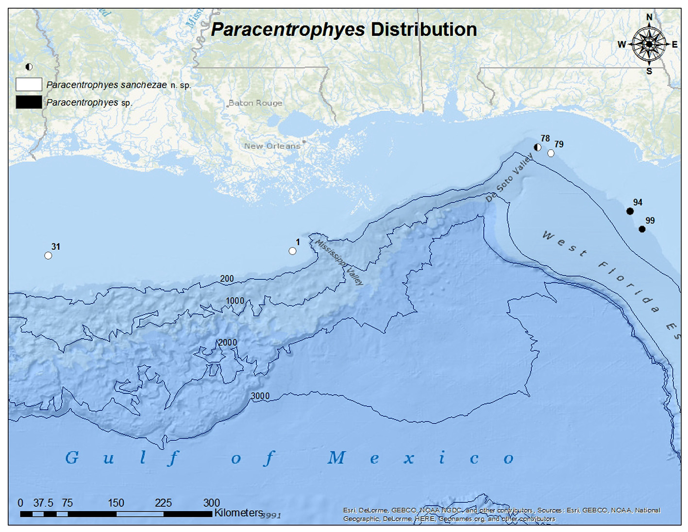


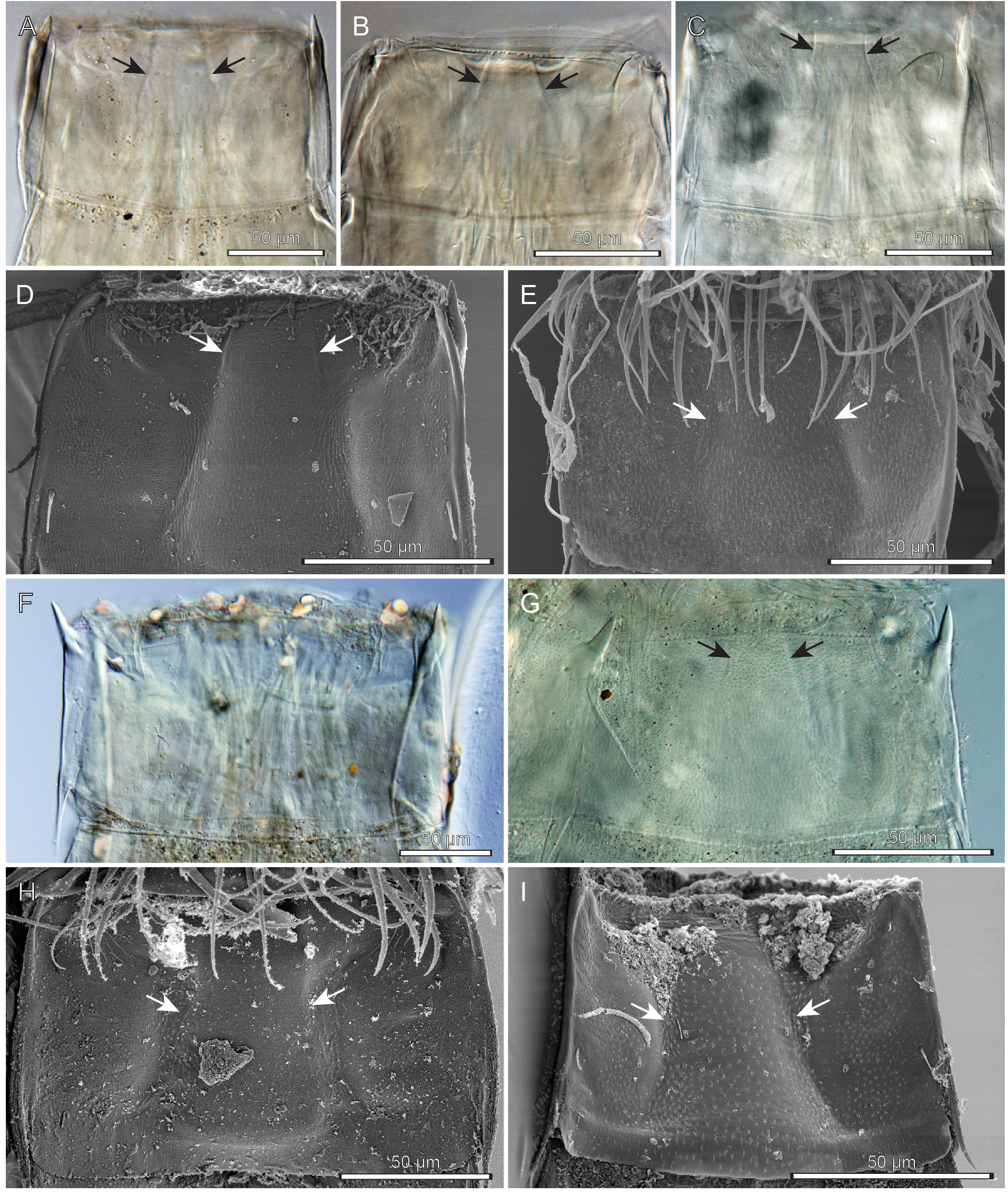
TABLE 1. Measurements from light microscopy of female holotype and male paratype of Paracentrophyes sanchezae n. sp. (in µm). Abbreviations: MD: middorsal spine; LTS: lateral terminal spine; MSW: maximum sternal width; S: segment lengths; SW, sternal widths; TL: trunk length; N / A, not applicable.
| Holotype | Paratype | |
|---|---|---|
| KIN-1014 | KIN-1015 | |
| Character | Female | Male |
| TL | 559 | 392 |
| MSW/TL | 24.7% | 32.9% |
| S1 | 74 | 73 |
| S2 | 53 | 39 |
| S3 | 48 | 43 |
| S4 | 50 | 44 |
| S5 | 52 | 47 |
| S6 | 54 | 52 |
| S7 | 62 | 53 |
| S8 | 64 | 55 |
| S9 | 65 | 56 |
| S10 | 51 | 55 |
| S11 | 24 | 25 |
| SW1 | 129 | 105 |
| SW2 | 133 | 121 |
| SW3 | 133 | 128 |
| SW4 | 134 | 128 |
| SW5 | 137 | 129 |
| SW6 | 138 | 125 |
| SW7 | 135 | 124 |
| SW8 | 131 | 121 |
| SW9 | 127 | 112 |
| SW10 | 121 | 97 |
| SW11 | 94 | 83 |
| MD10 | N/A | 53 |
| MD11 | 37 | 49 |
| LTS | 155 | 127 |
| LTS/TL | 27.7% | 32.4% |
TABLE 2. Summary of nature and location of sensory spots, setae, spines and processes arranged by series in Paracentrophyes sanchezae n. sp. Abbreviations: LD: Laterodorsal; LV: lateroventral; MD: middorsal; ML: midlateral; PD: paradorsal; PL: paralateral; SD: subdorsal; VL: ventrolateral; VM: ventromedial; ac, acicular spine; fls, flexible spine; lts, lateral terminal spine; pe, penile spines; pr, spinose process; se, seta; ss 1 / 3, sensory spot type 1 / 3; (♀), female and (♂), male conditions of sexually dimorphic characters; * marks that the structure is unpaired; ^ marks that the structure does not appear consistently in all specimens.
| Position Segment | MD | PD | SD | LD | PL | ML | LV | VL | VM |
|---|---|---|---|---|---|---|---|---|---|
| 1 | pr | ss1,ss1 | ss1,ss1,ss1,ss1 | pr | se | ss1,ss1,ss1 | |||
| 2 | pr | ss1,ss1,ss1 | ss1,ss1 | pr | ss1,ss1 | ||||
| 3 | pr | ss1,ss1,ss1 | ss1 | pr | se | ss1,ss1 | |||
| 4 | pr | ss1,ss1,ss1 | ss1 | se^ | pr | ss1,ss1 | |||
| 5 | pr | se* | ss1,ss1 | ss1 | pr | se | ss1 | ||
| 6 | pr | ss1,ss1,ss1 | ss1 | se | pr | ss1,ss1 | |||
| 7 | pr | se*^ | ss1,ss1 | ss1 | se | pr | se^ | ss1,ss1 | |
| 8 | pr | ss1,ss1,ss1 | ss1 | se | pr | se^ | ss1,ss1 | ||
| 9 | pr | se* | ss1,ss1,ss1 | ss1 | se | pr | se | ss1,ss1 | |
| 10 | pr(♀), fls(♂) | ss1,ss1 | ss1 | pr(♀),pe(♂) | se,ss1 | ss1 | |||
| 11 | ac(♀),fls(♂),mtp(♀) | ss3 | pe(♂) | lts | ss3,ss1 |
| ZMUC |
Zoological Museum, University of Copenhagen |
No known copyright restrictions apply. See Agosti, D., Egloff, W., 2009. Taxonomic information exchange and copyright: the Plazi approach. BMC Research Notes 2009, 2:53 for further explanation.
|
Kingdom |
|
|
Phylum |
|
|
Class |
|
|
Order |
|
|
Family |
|
|
Genus |