
Acrumena
|
publication ID |
https://doi.org/ 10.5281/zenodo.179102 |
|
DOI |
https://doi.org/10.5281/zenodo.6252809 |
|
persistent identifier |
https://treatment.plazi.org/id/039A0C4E-FFF2-FFBF-ECB2-C8F4FF2CA9F8 |
|
treatment provided by |
Plazi |
|
scientific name |
Acrumena |
| status |
|
Acrumena spec.
( Fig. 7 View FIGURE 7 A–C, 8A)
Locality. Loc. 2.
Material. One individual studied alive.
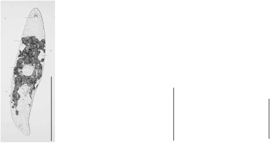
Description. This description has to be viewed as preliminary, as it is only based on observations of one live individual. No material was preserved. There were however enough details visible on the live individual and the photographs to recognise this species as belonging to the taxon Acrumena Brunet, 1965 .
The long and slender, spindle-shaped animal ( Figs 7 View FIGURE 7 A, 8A) is colourless and without eyes. The proboscis ( Figs 7 View FIGURE 7 C, 8A: p) is very small, about 1/10 of the total body length. The pharynx ( Fig. 8 View FIGURE 8 A: ph) is situated around midbody and has a diameter of approximately 1/8 of the body length.
The gonopore was not visible, but is most probably situated in the most caudal part of the body. The paired testes ( Fig. 8 View FIGURE 8 A: t) are very small and situated in front of the pharynx. The ovaries could not be discerned with certainty but are probably situated closely behind the pharynx. The vitellaria ( Fig. 8 View FIGURE 8 A: vit) are lying close together but there is no indication that they are interconnected. They extend from the level of the testes to that of the copulatory organ, which is situated at approximately 2/3 of the body. Two seminal vesicles ( Fig. 8 View FIGURE 8 A: evs) enter the copulatory bulb separately, but form a single intracapsular seminal vesicle ( Fig. 8 View FIGURE 8 A: ivs). In the most distal part of the copulatory bulb two diverticles ( Figs 7 View FIGURE 7 B, 8A: div) are present. Their exact nature is not clear and should be studied on sectioned material. These diverticles consist of two different parts: a short proximal leaf-shaped part and an elongated tubiform distal part. The latter is probably lined with a glandular epithelium. The copulatory bulb is connected with the gonopore through a very long male atrium ( Fig. 8 View FIGURE 8 A: ma). A globular uterus ( Fig. 8 View FIGURE 8 A: ut), lined with slender glands is present near the caudal end of the body.
ph Μm
25
Discussion. The elongated body shape, small koinocystidid-like proboscis, the general organisation of all organs, the presence of two seminal vesicles and the more detailed structure of the copulatory bulb indicate that this species is a representative of the monospecific taxon Acrumena (see Brunet 1965).
There are, however some clear differences between this species and A. massiliensis Brunet, 1965 (see Brunet 1965; Karling 1980). The studied individual lacks eyes, whereas these are clearly present in A. massiliensis ( Brunet, 1965; Karling: unpublished data in coll. SMNH). The vitellaria are much larger and not long and slender as in A. massiliensis . The copulatory bulb is situated more anteriorly than in A. massiliensis and the male atrium is much longer. Only two diverticles could be observed in this specimen, whereas there are clearly three diverticles present in A. massiliensis ( Brunet, 1965) .
In our view this could easily be described as a new species. We however, refrain from formally describing it as such, because there is no preserved material and some of its features should be studied on sectioned material.

No known copyright restrictions apply. See Agosti, D., Egloff, W., 2009. Taxonomic information exchange and copyright: the Plazi approach. BMC Research Notes 2009, 2:53 for further explanation.