
Tarsonemus lenticulatus Gheblealivand, Haddad and Magowski, 2018
|
publication ID |
https://doi.org/10.11646/zootaxa.4446.1.2 |
|
publication LSID |
lsid:zoobank.org:pub:2E693B67-9C59-44D0-A27E-A4DB79AA29F9 |
|
DOI |
https://doi.org/10.5281/zenodo.5971688 |
|
persistent identifier |
https://treatment.plazi.org/id/039A8799-FFC7-2064-FF7D-F8D2273CF829 |
|
treatment provided by |
Plazi |
|
scientific name |
Tarsonemus lenticulatus Gheblealivand, Haddad and Magowski |
| status |
sp. nov. |
Tarsonemus lenticulatus Gheblealivand, Haddad and Magowski sp. nov.
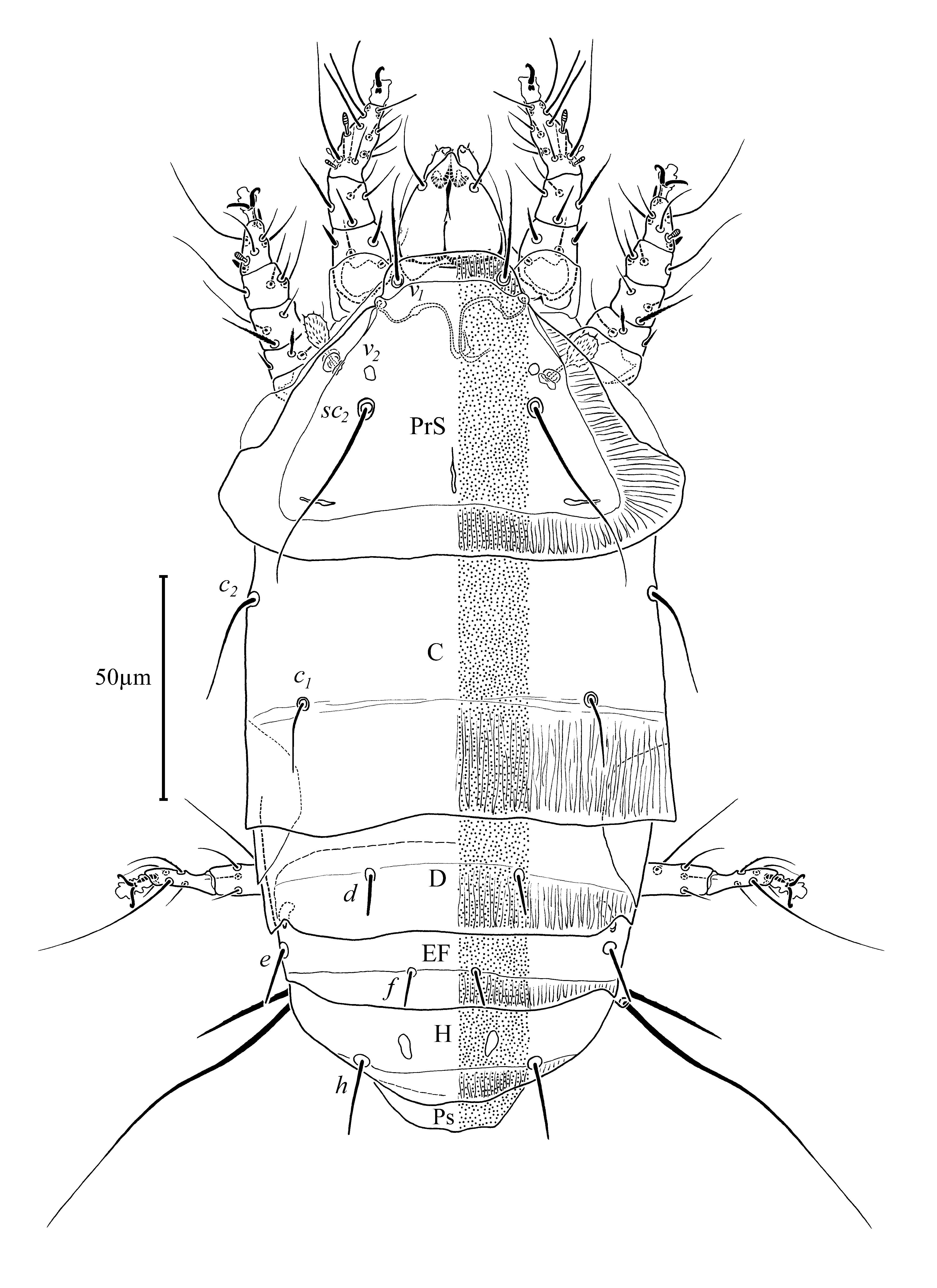
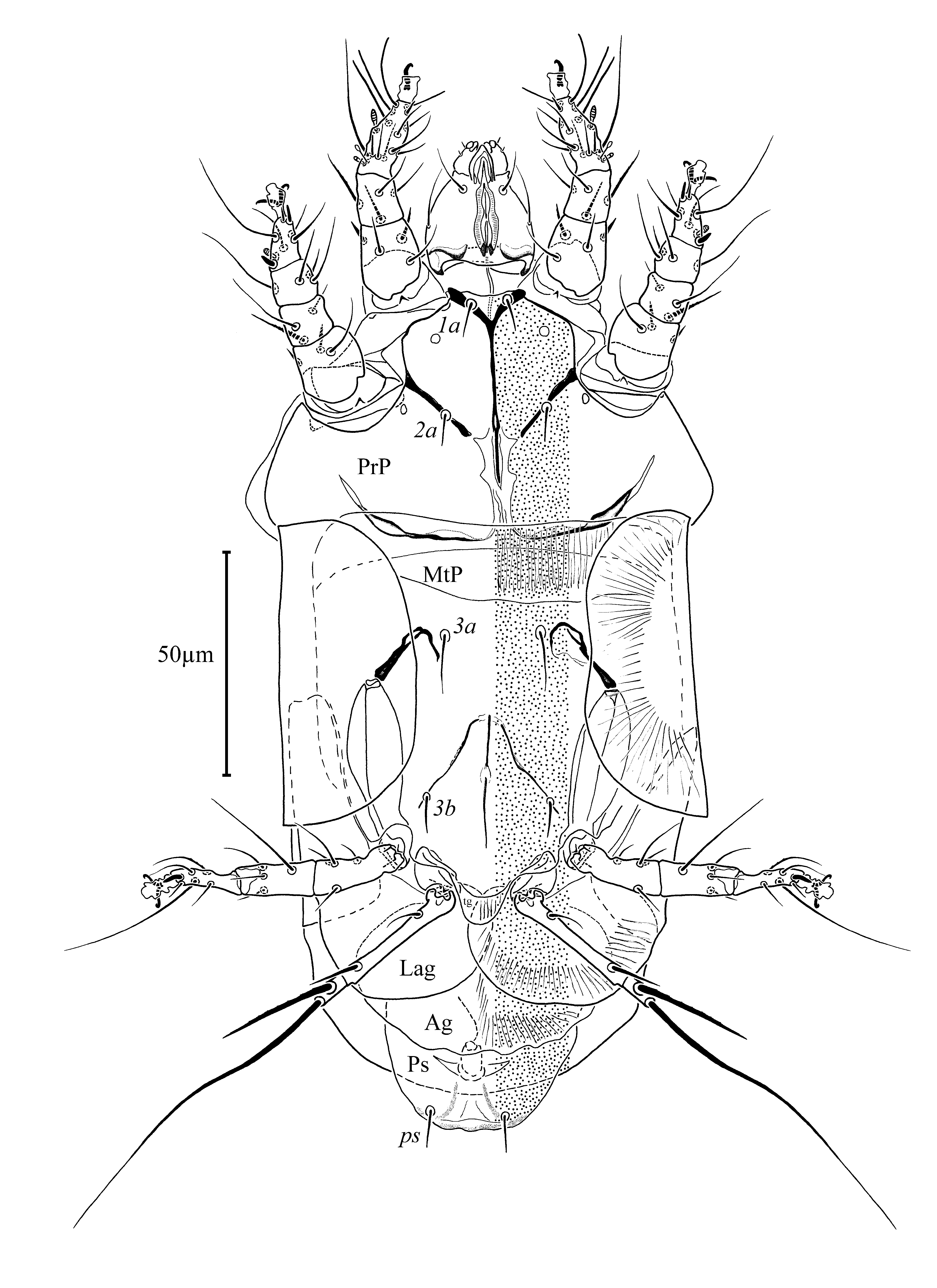
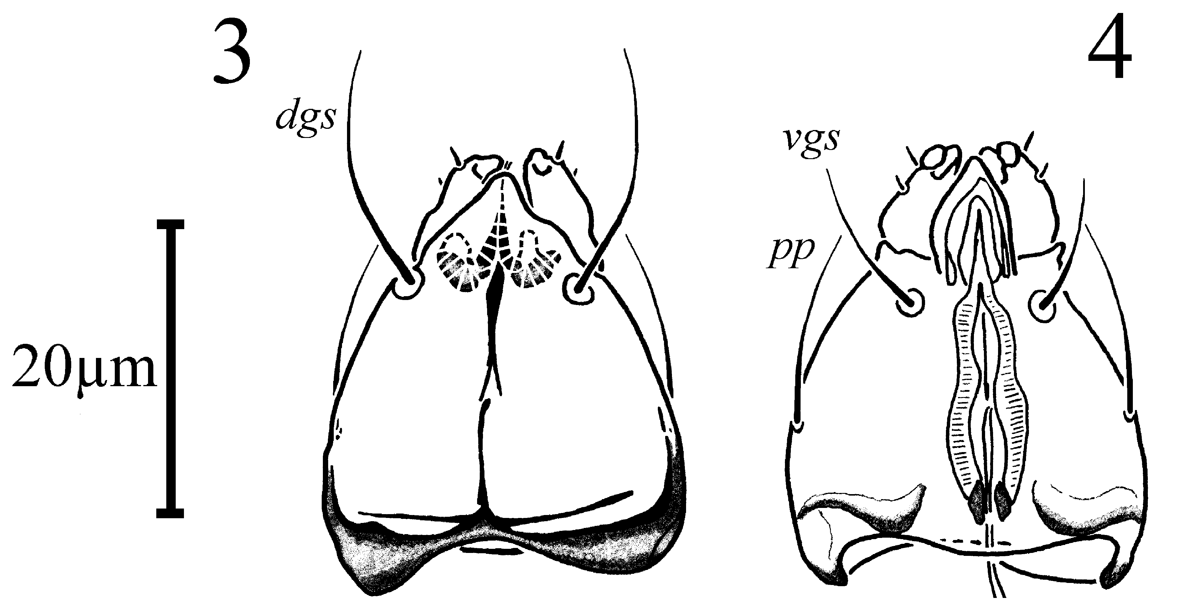
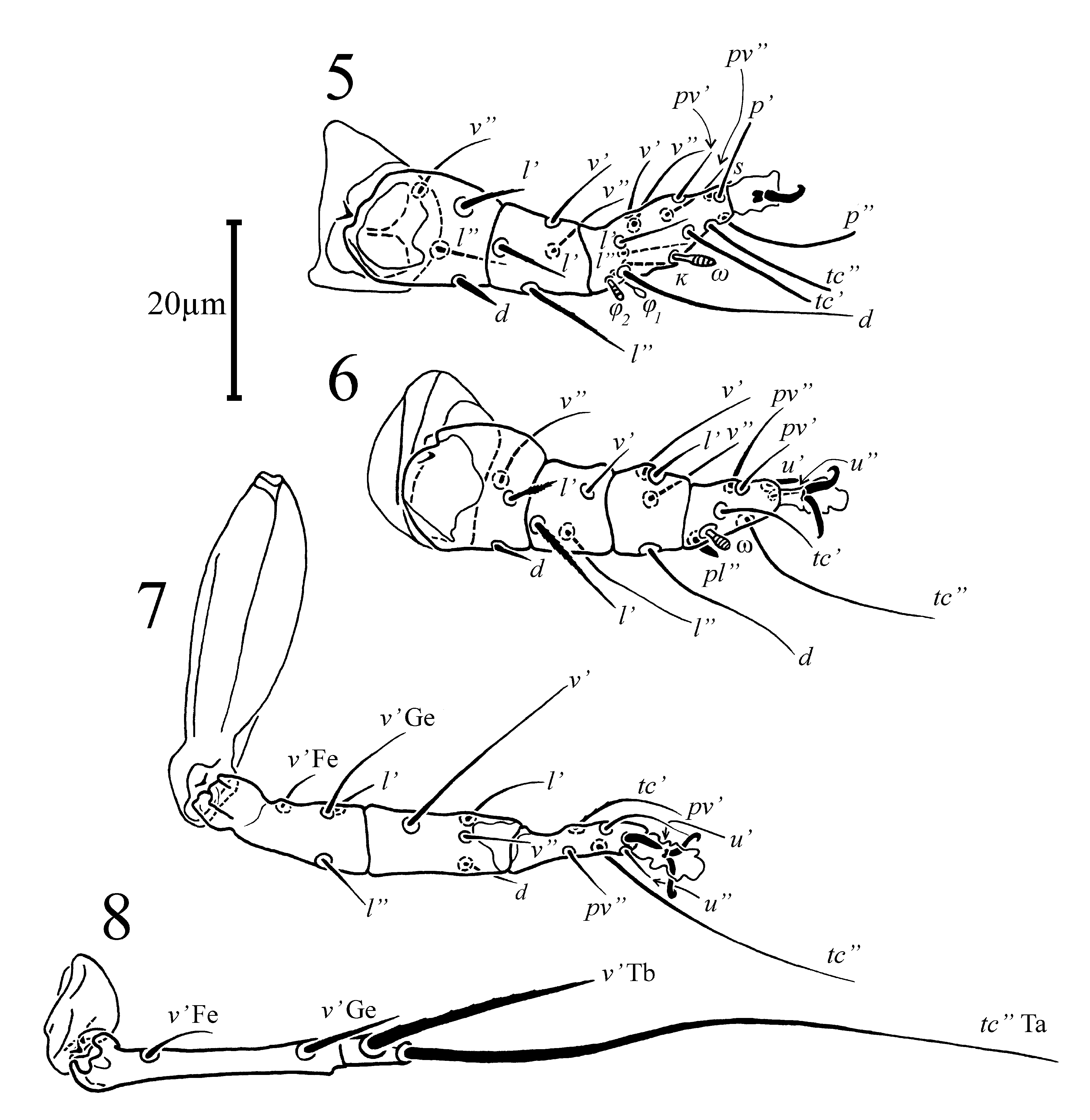
( Figs. 1–9 View FIGURE 1 View FIGURE 2 View FIGURES 3–4 View FIGURES 5–8 View FIGURE 9 , 18A, C View FIGURE 18 )
Diagnosis. Pharynx elongate- oval, with weak constriction near its mid-length. Dorsal opisthosomal setae h 2x as
long as f. Anteromedial apodeme continuous between apodemes 1 and 2. Sejugal apodeme interrupted medially, with lens-like structures and inflexions in lateral parts. Setae 1a, 2a and 3b stiff and blunt; setae 3a attenuated. Tegula semicircular in shape with rounded posterior edge. Tarsal part of Tbt I with two attenuated setae.
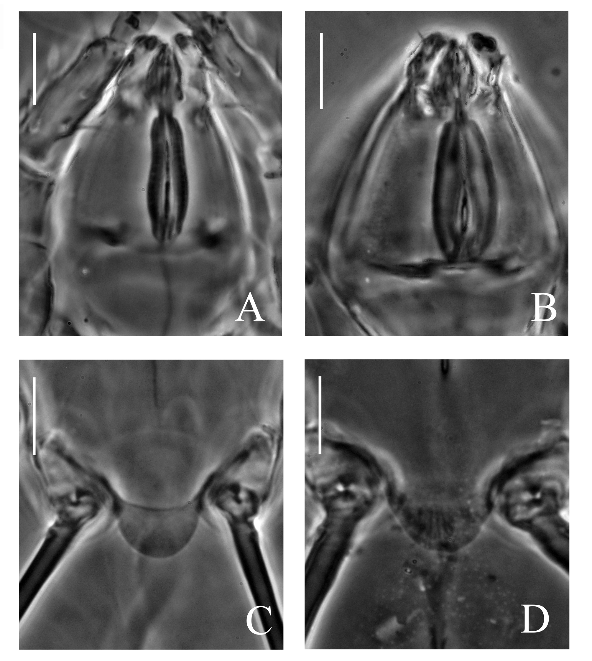
Description. FEMALE ( Figs. 1–9 View FIGURE 1 View FIGURE 2 View FIGURES 3–4 View FIGURES 5–8 View FIGURE 9 , 18A, C View FIGURE 18 ). Gnathosoma (Figs. 3,4): capsule triangular-ovoid. Pharynx ( Fig. 18A View FIGURE 18 ) elongate- oval in outline with narrowing anteriad its mid-length, finely transversely striated, with a pair of small glandular bodies, as wide as 0.24x maximum width of gnathosoma and as long as 0.7x of ventral length of gnathosomal capsule. Long postpalpal setae ( pp) evident. Setae dgs 1.7x longer than vgs. Cheliceral stylets and levers fine but pronounced. Palpi moderate in size, near parallel, convergent, each with small palptarsal processes and two minute setae.
Idiosomal dorsum (average length: 201(SD ± 19), width: 95 (SD ± 13); length = 2.1x width; Fig. 1 View FIGURE 1 ): relative lengths of setae ( v1: sc2: c2: c1: d: e: f: h): 1: 1.9: 0.6: 1.0: 0.4: 0.5: 0.4: 0.7. Rostral shieldlet over 3x wider than long, round-truncated in shape. Vertical setae ( v1) setiform, slightly barbed, separated by distance of 1x their length between their bases. Prodorsal shield (PrS) trapezoidal in outline, ca. 1.4x wider than long, with short straight prodorsomedial apodeme and two fine but defined prodorsolateral apodemes symmetrically arranged posterolaterally. Posterior edge of PrS slightly convex or straight, irregular. Stigmata located posterolaterally of setae v1, leading internally to tracheae and small atria; post-atrial segments with weakly defined expansions. Sensilla sc1 clavate, pilose, with two stronger spines apically. Pits v2 located in front of sc2, separated by same distance. Setae sc2 located at mid-length of prodorsal shield, exceeding posterior edge, separated by distance of 0.8x their length. Setae c2 slightly shorter than distance between their bases and those of c1, ca. 1.7x c1. Setae c1 slender, pointed (though somewhat less than c2), separated by a transverse distance of 4.8x their length, reaching to half of distance between their bases and posterior edge of tergite C. Posterior edge of tergite C slightly convex, irregular. Setae d, e, f and h stiff and blunt. Setae d separated by transverse distance of over 3x their length, not reaching to posterior edge of tergite D, posterolateral margin of shield D with emarginations at position of setae e on tergite E. Setae f shorter than e and d, separated by transverse interval of 1.8x their length, and reaching with tips to posterior edge of tergite EF. The distance between setae e and f on each side 1.8x that of between f. Setae h markedly longer than d, e, and f, located in distance of over 2x their length each to another. Dorsal shielding with fine, uniform, dimpled ornamentation.
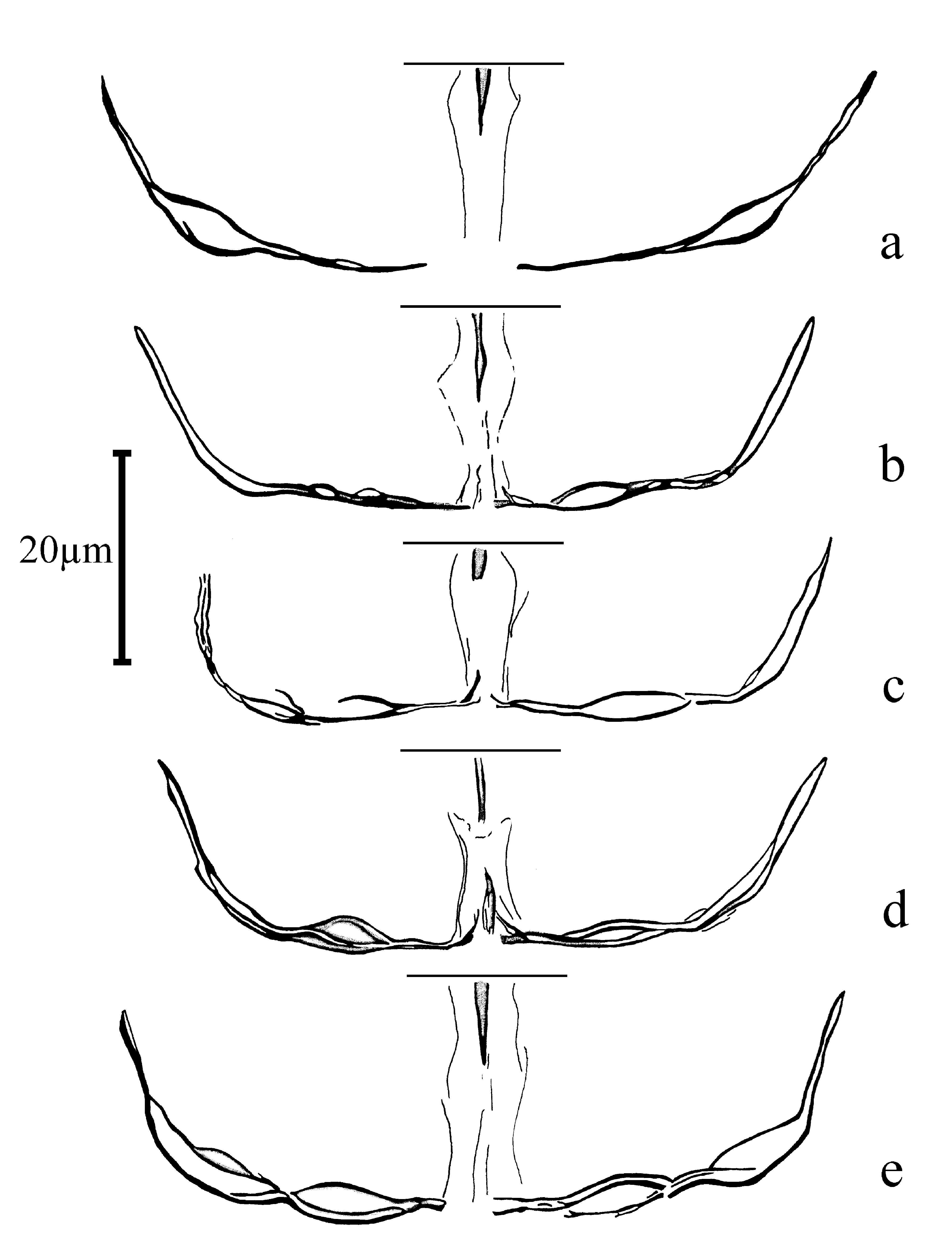
Idiosomal venter ( Fig. 2 View FIGURE 2 ): apodemes 1 well defined, united with anteromedian apodeme, the latter continuous from apodemes 1 to level of medial ends of apodemes 2, but diffusing further posteriorly in a form of indistinct breast platelet. Sejugal apodeme diffused, or discontinuous medially and sometimes with weak indentations, but well developed in lateral parts, bent anterolaterally and split in a form of two (or one) lentiform structures. Sejugal apodeme subjected to considerable variation in shape and expression among examined females ( Fig. 9 View FIGURE 9 ). Ridge of ventral propodosomal plate between trochanters I and II with angularity on each side. Setae 1a stiff, blunt, contiguous with apodemes 1 and in a distance of ca. 1.2x their length each to another. Apodemes 2 well sclerotized, not connecting with anteromedial apodeme. Stiff and blunt setae 2a located about midlength of apodemes 2, and in a distance of ca. 3.2x their lengths between their bases (one anomalous paratype specimen has doubled setae 2a, both inserted in the same alveolus on each side). Apodemes 3 extended from anterior extremities of trochanters III anteromedially and curved near bases of 3a; apodemes 4 slightly extended beyond setae 3b, not clearly uniting with each other or with posteromedial apodeme medially; posteromedial apodeme reduced anteriorly and without bifurcation. Setae 3a attenuated, separated by distance of 1.7x their length. Setae 3b stiff, shorter than 3a, separated by distance of 4.3x their lengths, somewhat wider than that of between 3a. Ventral metapodosomal plate weakly concave, almost straight anteriorly. Tegula ( Fig. 18 C View FIGURE 18 ) semicircular in shape with rounded posterior edge, 1.8x wider than long. Posterior margin of agential plate undulated. Setae ps stiff, needle like, their bases separated by distance of ca. 2x their lengths. Surface of ventral plates with fine uniform dimpled ornamentation.
Legs ( Figs. 5–8 View FIGURES 5–8 ): proportions of free segments of legs (I: II: III: IV): 1.0: 1.0: 1.1: 0.8.
Leg I: Chaetotaxy: 4–4–6(2 φ)+7(1 ω). Tarsal claw hooked, similar to claws of tarsi II and III though shorter. Seta s tapering, blunt, similar to u’ of tarsi II and III. Unguinal setae u’ and u” indiscernible. Tibiotarsus ca. 1.7x longer than wide at the base. Eupathidion p” longer than p’, both located apically; eupathidia tc’ and tc” equal and similar in length to p”, tc’ located subapically and tc” in distal 1/3rd length of the segment. Two tarsal setae pv’ and pv” slender and pointed ( pl" absent). Solenidion ω clavate, slightly larger than Ta II ω. Solenidion φ2 with striated head, shorter than φ1, famulus k as long as φ1. Seta Ge l’ stiff, weakly barbed, shortest of all on genu, seta Ge l” stiff, with minute barbs. On femur seta d short, stiff, seta l’ strong, stiff, barbed, both weakly pointed. Leg II:
Chaetotaxy: 3– 3– 4– 6(1 ω). Claws on tarsus hooked, both well developed. Seta u’ spine-like, u" weak. Spinelike seta pl” almost as long as solenidion Ta II ω, the latter located somewhat distally. Seta tc” about 2x longer than tc’ and ca. 2.5x than other tarsal setae, reaching far beyond tip of empodium. Tibial seta l’ stout, shorter than other attenuate setae on segment. Seta Ge l’ stout at the base and barbed. Femur without a lobe; seta d tapering, seta Fe l’ barbed in its half distal length. Leg III: Chaetotaxy: 1+3– 4–5. Tarsal claws as large as those of leg II. Setae u’ and u” as those on tarsus II. Seta tc” markedly (over 3x) longer than attenuate tc’, the latter weakly barbed. Seta Ge l’ simple, stiff. Leg IV: free segments of leg IV roughly equal in length to femorogenu and tibia III. Femorogenu nearly 4x longer than tibiotarsus. Genual seta longer and stouter than femoral one. Seta Tb v’ somewhat shorter than femorogenu, stout, barbed. Seta Ta tc” ca. 2x as long as whole leg IV.
Measurements: ( holotype given first followed by 7 paratypes in parentheses): body and tagmata: length of body: 226 (187–260); length of idiosoma: 197 (172–230); width of idiosoma: 97 (75–120); length of gnathosoma: 30 (25–30); width of gnathosoma: 26 (20–25); length of pharynx: 23 (16–22); width of pharynx: 5 (5–6). dgs: 13 (10–15); vgs: 8 (6–8). Dorsum - length of PrS: 68 (56–70); width of PrS: 103 (70–105); distance between stigmata: 32 (30–32). Lengths of setae: v1: 25 (20–25); sc1 15 (11–16); sc2: 45 (40-45); c2: 23 (20–25); c1: 15 (11–15); d: 11 (9–10); e: 13 (10–12); f: 10 (7–9); h: 18 (15–18). Distances between setae: v1-v1: 25 (22–25); sc1-sc1: 48 (32–45); sc2-sc2: 38 (32–37); c2-c2: 92 (83–91); c1-c1: 64 (58-65); d -d: 34 (28-34); e -e: 73 (60-73); e -f: 31 (15-30); f -f. 14 (13- 19); h -h: 37 (31-38). Venter- Lengths of setae: 1a: 6 (5); 2a: 8 (5–7); 3a: 11 (11–13); 3b: 7 (5–7); ps: 10 (7–9). Distances between setae: 1a -1a: 8 (6–7); 2a -2a: 24 (20–25); 3a -3a: 22 (18–22); 3b -3b: 28 (25–28); ps -ps: 17 (15– 18). Length of PrP: 55 (51–60); width of PrP: 97 (65–105); ap. 1–1: 17 (10–13); ap. 2– 2: 37 (30–35). Length of tegula: 7 (5–7); width of tegula: 12 (10–13). Leg segments and leg setae (lengths): Tbt I: 20 (17–20); ω I: 5 (4–5); φ1: 5 (4–5); φ2: 3 (3); қ: 5 (4–5); ω II: 4 (3–5); FeGe IV: 28 (25–31); Tbt IV: 8 (6–9); Fe v’: 7 (6–9); Ge v': 11 (10– 11); Tb v': 25 (22–25); Ta tc": 73 (50–72).
MALE and LARVA unknown.
Type material. The female holotype (slide no. SO S304) and 2 female paratypes: Sufiyan, Ivand village commune, 38°22'20"N, 46°07'38"E, 1,722 m.a.s.l., ex litter and soil, 10-October-2014 GoogleMaps ; 4 female paratypes: Zunuz , Chirchir village commune, 38°33'54"N, 45°43'35"E, 1,429 m.a.s.l., ex apple leave, 15-August-2014 GoogleMaps ; 1 female paratype: Jolfa , Iri village commune, 38°46' 35"N, 45°59'51"E, 1,085 m.a.s.l., ex Russian olive leaves, Elaeagnus angustifolia L., 30-May-2014; all coll. S. Gheblealivand. GoogleMaps
Type depositions. Holotype and 4 paratypes are deposited in the Acarology Laboratory , Department of Plant Protection , University of Tabriz, Tabriz, Iran ; 3 paratypes are kept in collection of Department of Animal Taxonomy and Ecology, A. Mickiewicz University, Poznań, Poland .
Etymology. The specific name “ lenticulatus ” (Eng. lens-like) refers to the shape of the characteristic splitting of the lateral branches of the sejugal apodeme.
Differential diagnosis. Females of T. lenticulatus n. sp. are similar to T. saccatus Livshits, Mitrofanov and Sharonov, 1979 and T. caucasicus Sharonov and Mitrofanov, 1986 in sharing the general configuration of the pharynx (without sclerotized outer part), sejugal apodeme (with medial incision) and form of the ventral metapodosomal setae ( 3a: slender and pointed, 3b stiff and blunt). It differs from both species by (1) setae 1a and 2a stiff and blunt (vs. setae 1a and 2a slender and pointed); (2) sejugal apodeme split in lateral parts in the shape of lens-like structures (vs. sejugal apodeme entire in lateral parts); (3) tegula semicircular vs. round triangular.
No known copyright restrictions apply. See Agosti, D., Egloff, W., 2009. Taxonomic information exchange and copyright: the Plazi approach. BMC Research Notes 2009, 2:53 for further explanation.
|
Kingdom |
|
|
Phylum |
|
|
Class |
|
|
Order |
|
|
Family |
|
|
Genus |
|
|
SubGenus |
Tarsonemus |