
Pseudophacopteron eastopi, Malenovský & Burckhardt & Tamesse, 2007
|
publication ID |
https://doi.org/ 10.1080/00222930701515488 |
|
persistent identifier |
https://treatment.plazi.org/id/039A87A6-FFEB-FF91-FE19-C4A9FC5DFDB7 |
|
treatment provided by |
Felipe |
|
scientific name |
Pseudophacopteron eastopi |
| status |
sp. nov. |
Pseudophacopteron eastopi View in CoL sp. n.
( Figures 4H View Figure 4 , 5E View Figure 5 , 6I View Figure 6 , 7I View Figure 7 , 8I View Figure 8 , 9J View Figure 9 , 10I View Figure 10 , 12E View Figure 12 , 13I View Figure 13 )
Description
Adult. Colour (alcohol-preserved specimens): vertex, pronotum, mesopraescutum, and mesoscutum in males entirely black, in females lighter brown with a pale ochreous midline. Genae, frons, and clypeus in males dark brown to black, in females ochreous. Lateral sclerites of thorax in both sexes dark brown to black. Antenna off-white, segments 4–8 dark brown or black apically, segments 9–10 entirely black, terminal setae white. Legs off-white or dirty yellow, metacoxa with a dark brown spot at apical margin, profemur and mesofemur with dark brown streaks near apex and base, metafemur with an oblique dark brown streak near apex, metatibia dark brown basally. Fore wing membrane clear, transparent, with pattern consisting of a dark brown band along distal half of posterior margin (from distal half of cell cu1 to the posterior tip of cell r2, leaving two small transparent crescents at wing margin in m1 and m2), smaller dark brown patch along distal half of the vein Cu1b, and irregular light brown infuscations extending across the veins M+Cu1, M, Cu1, and the basal portion of M1+2, and in cell cu2 close to fore wing base ( Figure 6I View Figure 6 ). Veins off-white, except for dark brown or black spots medially on R+M+Cu1, the base of R immediately after the branching off of M+Cu1, the M+Cu1 fork and Cu1 fork, and two spots on anal vein, which is also brown basally. Hind wing clear, transparent, veins C+Sc and A fuscous. Abdominal tergites light brown to dark brown. Sternites off-white to dirty yellow, the first visible sternite laterally with a narrow dark brown patch. Male terminalia dark brown, with parameres and apices of subgenital plate and proctiger offwhite. Female terminalia dark brown, with subgenital plate basally and proctiger in apical half ochreous.
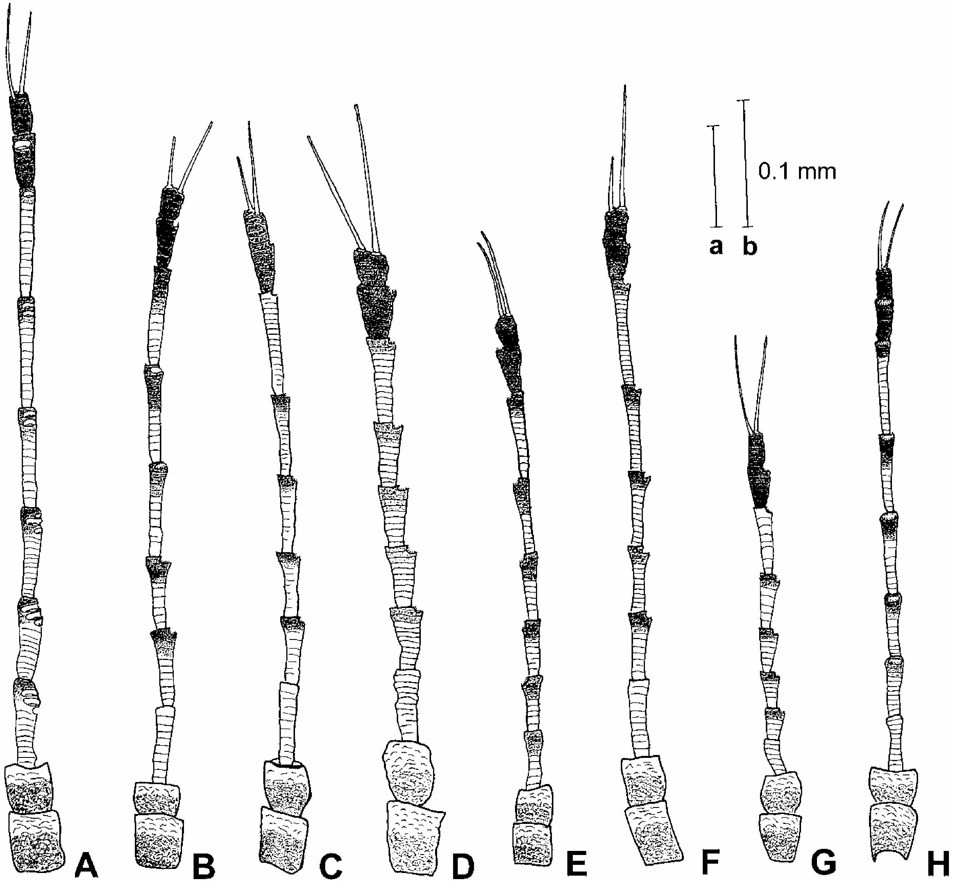
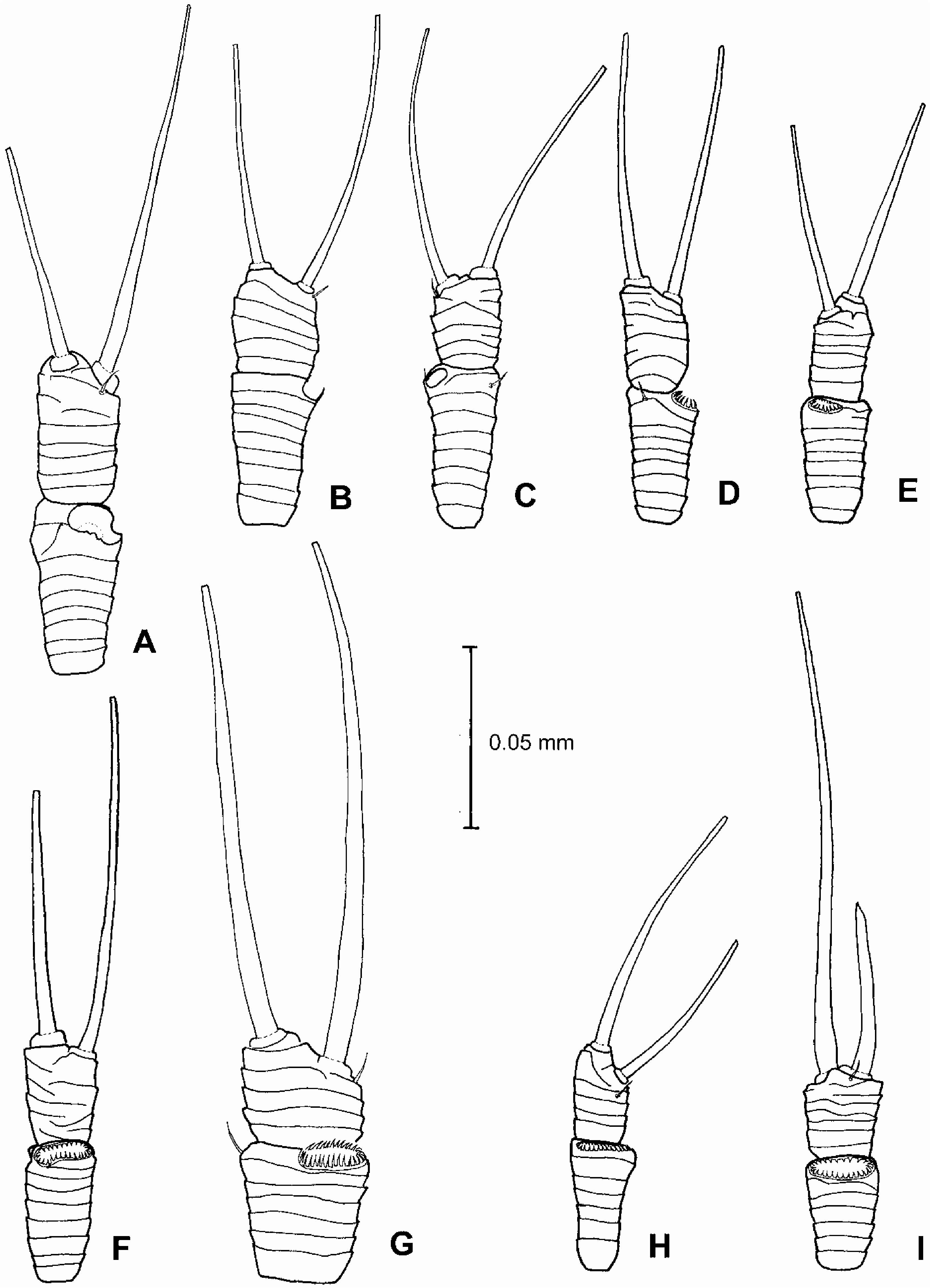
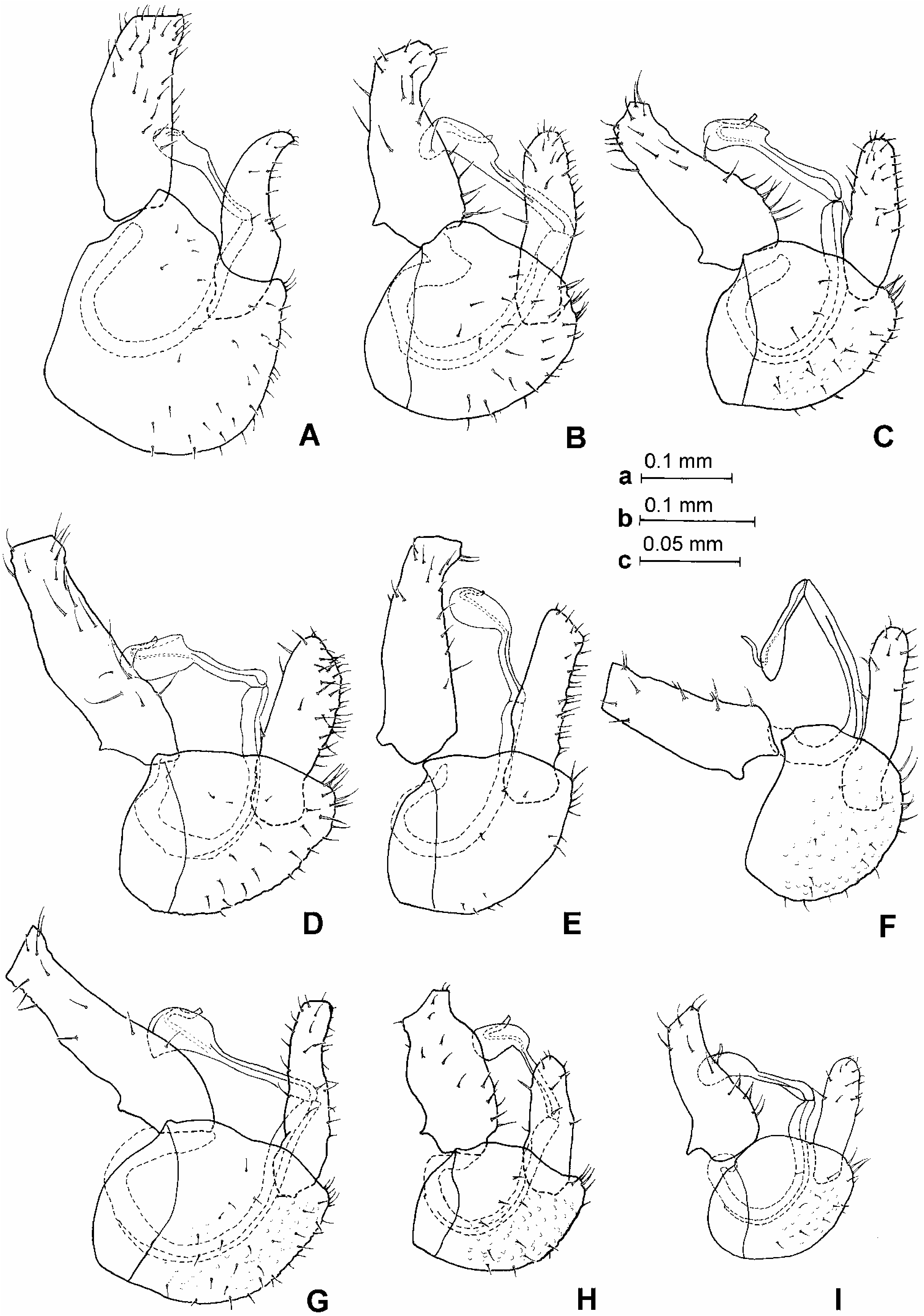
Morphology: head, in dorsal view, slightly wider than mesonotum, subglobular. Vertex in males smooth and shiny, in females with microsculpture and matt, about twice as wide as long along midline, rounded down in front. Coronal suture reduced throughout. Median ridge on vertex raised, distinct. Vertex on either side of median ridge convex, slightly bulging. Lateral ocelli lying on small tubercles slightly above plane of vertex. Occiput and preoccipital sclerite narrow, eyes not stalked, in frontal view subglobular. Genae slightly swollen. Tubercle below torulus pointed, forming an acute angle. Frons narrow, parallelsided. Clypeus subglobular. Antenna elongate, slender, segments cylindrical, weakly widening to apex ( Figure 4H View Figure 4 ); a single rhinarium on apices of segments 4–9; rhinaria elliptic with a wreath of cuticular spines; terminal setae more or less equal, approximately as long as the segments 9 and 10 together ( Figure 5E View Figure 5 ). Fore wing moderately elongate, outer anterior margin more or less evenly curved; apex unevenly rounded. Vein Rs relatively short. Surface spinules present in cells cu2, cu1, m2, m1, and distal part of r2, arranged in fields along posterior wing margin ( Figure 7I View Figure 7 ). Costal break in apical sixth or seventh of C+Sc. Hind legs relatively long and robust. Meracanthus short, acute, pointed. Metafemur constricted medially. Metatibia bearing an open crown of eight unsclerotized apical spurs and a few additional similar spurs laterally. Metabasitarsus bearing two black sclerotized spurs. Dorsal margin of abdomen, in profile, serrate; posterior margin of tergites 4 and 5 medially swollen into a prominent tubercular process. Male terminalia as in Figure 8I View Figure 8 . Proctiger relatively long, slightly narrowing to apex. Subgenital plate, in profile, as long as high, dorsal margin straight. Paramere, in profile, relatively narrow, parallel-sided, rounded apically; in posterior view, inner and outer margins parallel-sided; inner surface covered in fine setae and three stouter setae subapically, apex forming a sclerotized sinuate edge with a small tooth ( Figure 9J View Figure 9 ). Basal segment of aedeagus stout; apical segment with a slightly hooked head, broad and rounded at apex; sclerotized end tube of ductus ejaculatorius relatively long, sinuate ( Figure 10I View Figure 10 ). Female terminalia as in Figure 12E View Figure 12 . Proctiger relatively long, dorsal margin sinuate, weakly concave, apical process long; circumanal ring with two rows of pores, pores of outer row more or less contiguous. Subgenital plate, in profile, moderately long, dorsal margin slightly concave, ventral margin slightly sinuate, apex blunt; in ventral view, relatively broad basally, from the middle abruptly narrowing into apical process, apex with a small indentation ( Figure 13I View Figure 13 ). Dorsal and ventral valvulae lacking lateral teeth. Measurements and ratios in Tables I–III.
Fifth instar larva and biology
Unknown.
Host plant
Adults of the type series, including teneral specimens, were collected on Dacryodes edulis (Burseraceae) which is a possible host plant. We examined another series including young larvae collected on Sorindeia grandifolia (Anacardiaceae) (see the comments).
Distribution
Cameroon.
Material examined
Holotype: „, Cameroon: West Province, Titié (Foto), Dschang , 10 ° 049N, 5 ° 269E, 26 May 2006, Dacryodes edulis (J. L. Tamesse and V. J. Dzokou) . Dry-mounted ( NHMB) . Paratypes: Cameroon: 5♀, same data as holotype; 2 „, 2♀, South Province, Nkolandom, Ebolowa , 11 ° 099N, 2 ° 559E, 583 m, 26 May 2006, Dacryodes edulis (J. L. Tamesse and Y. P. Mveyo) ; 1 „ , 1♀, South-West Province, Mamfe , 9–10 February 1957 ( V. F. Eastop) ; 1♀, same data, but January 1957. Dry- and slide-mounted, and preserved in alcohol ( BMNH, LZUY, MHNG, MMB, NHMB) .
Material not included in the type series: Cameroon: 3 „, 3♀, seven larvae, two larval skins, West Province , Santchou (Menoua), 11 February 2006, Sorindeia grandifolia (J. L. Tamesse and V. J. Dzokou) . Dry-mounted and preserved in alcohol ( NHMB) .
Etymology
Named in honour of the British hemipterist and leading aphidologist Victor F. Eastop who collected the species for the first time.
Comments
Pseudophacopteron eastopi View in CoL sp. n. is similar to P. verrucifrons Burckhardt and van Harten, 2006 View in CoL , P. lecaniodisci View in CoL sp. n., and P. pusillum View in CoL sp. n. in having a dark band along distal half of posterior fore wing margin, the broad and more or less hooked apical dilation of the distal segment of the aedeagus, and the relatively long female subgenital plate. P. eastopi View in CoL differs from the three species in the darker head and thorax in males, as well as in the fore wing shape (outer anterior margin more evenly curved) and pattern (membrane in anterior half irregularly infuscate). P. eastopi View in CoL is similar to P. pusillum View in CoL in the small size, the head shape, the lack of dark brown patches in anterior half of fore wing and the shape of the female terminalia, but differs in the long and slender antenna with segments 4–9 cylindrical and weakly widening apically, the shorter terminal setae, the more robust hind legs, the smaller size of the dark brown patch on sides of the first visible abdominal sternite, the narrower and parallel-sided parameres, the narrower head of apical segment of aedeagus, the shape of female subgenital plate in ventral view and the lack of lateral teeth on dorsal and ventral valvulae.
The series collected on Sorindeia View in CoL differs from the type series in the slightly more expanded fore wing pattern. This is interpreted here as intraspecific variation but more material and confirmed host plant data are needed for a conclusive answer.


No known copyright restrictions apply. See Agosti, D., Egloff, W., 2009. Taxonomic information exchange and copyright: the Plazi approach. BMC Research Notes 2009, 2:53 for further explanation.
|
Kingdom |
|
|
Phylum |
|
|
Class |
|
|
Order |
|
|
Family |
|
|
Genus |
Pseudophacopteron eastopi
| Malenovský, Igor, Burckhardt, Daniel & Tamesse, Joseph L. 2007 |
Pseudophacopteron eastopi
| Malenovský & Burckhardt & Tamesse 2007 |
P. lecaniodisci
| Malenovský & Burckhardt & Tamesse 2007 |
P. pusillum
| Malenovský & Burckhardt & Tamesse 2007 |
P. eastopi
| Malenovský & Burckhardt & Tamesse 2007 |
P. eastopi
| Malenovský & Burckhardt & Tamesse 2007 |
P. pusillum
| Malenovský & Burckhardt & Tamesse 2007 |
P. verrucifrons
| Burckhardt and van Harten 2006 |