
Pseudophacopteron cuniculus, Malenovský & Burckhardt & Tamesse, 2007
|
publication ID |
https://doi.org/ 10.1080/00222930701515488 |
|
persistent identifier |
https://treatment.plazi.org/id/039A87A6-FFF2-FFAB-FE0B-C7D5FEC7F97B |
|
treatment provided by |
Felipe |
|
scientific name |
Pseudophacopteron cuniculus |
| status |
sp. nov. |
Pseudophacopteron cuniculus View in CoL sp. n.
(Figures 1A–C, 3A, B, 4A, 5A, 6A, 7A, 8A, 9A, B, 10A, 11A, 13A, 14, 21A)
Description
Adult. Colour: body grey yellow, light grey in alcohol-preserved specimens. Darker markings on head and thorax almost absent, vertex and pronotum sometimes orange or light brown laterally and in depressions, mesoscutum yellow with four pale grey bands. Genae, frons, and clypeus uniformly yellow or off-white. Antenna off-white, segments 3–8 dark brown or black apically, segments 9–10 entirely black, terminal setae white. Legs dirty pale yellow with light to dark brown markings on all femora and metacoxae; apex of metafemora dorsally narrowly black; metatibiae weakly infuscated at base. Fore wing membrane clear, transparent, with brown pattern consisting of large patches in cell cu1, around apical parts of the veins Rs and M1+2 and their touching point, a distinct narrow brown infuscation along basal half and apical third of M3+4 and an indistinct light brown infuscation across Cu1 and M close to their branching ( Figure 6A View Figure 6 ). Veins light, off-white, C+Sc dark brown in median part between base and junction of R1, anal vein brown basally, and small, well-delimited dark brown or black spots medially or in apical half of R+M+Cu1, at the base of R immediately after the branching off of M+Cu1, on the M fork and the Cu1 fork, the touching point of Rs and M1+2, at the apices of R1, Cu1a, Cu1b, and two or three spots on M1+2, M3+4, Cu1a and anal vein. Hind wing clear, transparent, veins C+Sc and A fuscous. Abdominal tergites yellow, with orange
Figure 1. Pseudophacopteron spp. (A–C) P. cuniculus ; (D) P. kala ; (E, F) P. nothospondiadis ; (G, H) P. fuscivenosum . (A) Head, dorsal view; (B, D, F, H) head, frontal view; (C) head, oblique ventral view; (E, G) head, oblique dorsal view. Scale bars: 0.05 mm (A–F); 0.1 mm (G, H).
brown and brown markings dorsally, forming a dorsal median band. Sternites pale yellow with dark brown markings of variable extent, especially on sides of first visible sternite. Male terminalia with subgenital plate pale yellow, brown apically, proctiger yellow or ochreous, parameres brown. Female terminalia with yellow proctiger, brown dorsally and apically.
Figure 2. Pseudophacopteron spp. (A, B) P. morion ; (C, D) P. lecaniodisci ; (E, F) P. pusillum . (A, C, E) Head, oblique frontal view; (B, D, F) head, oblique ventral view. Scale bars: 0.05 mm (A–F).
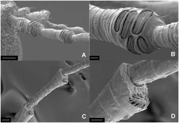
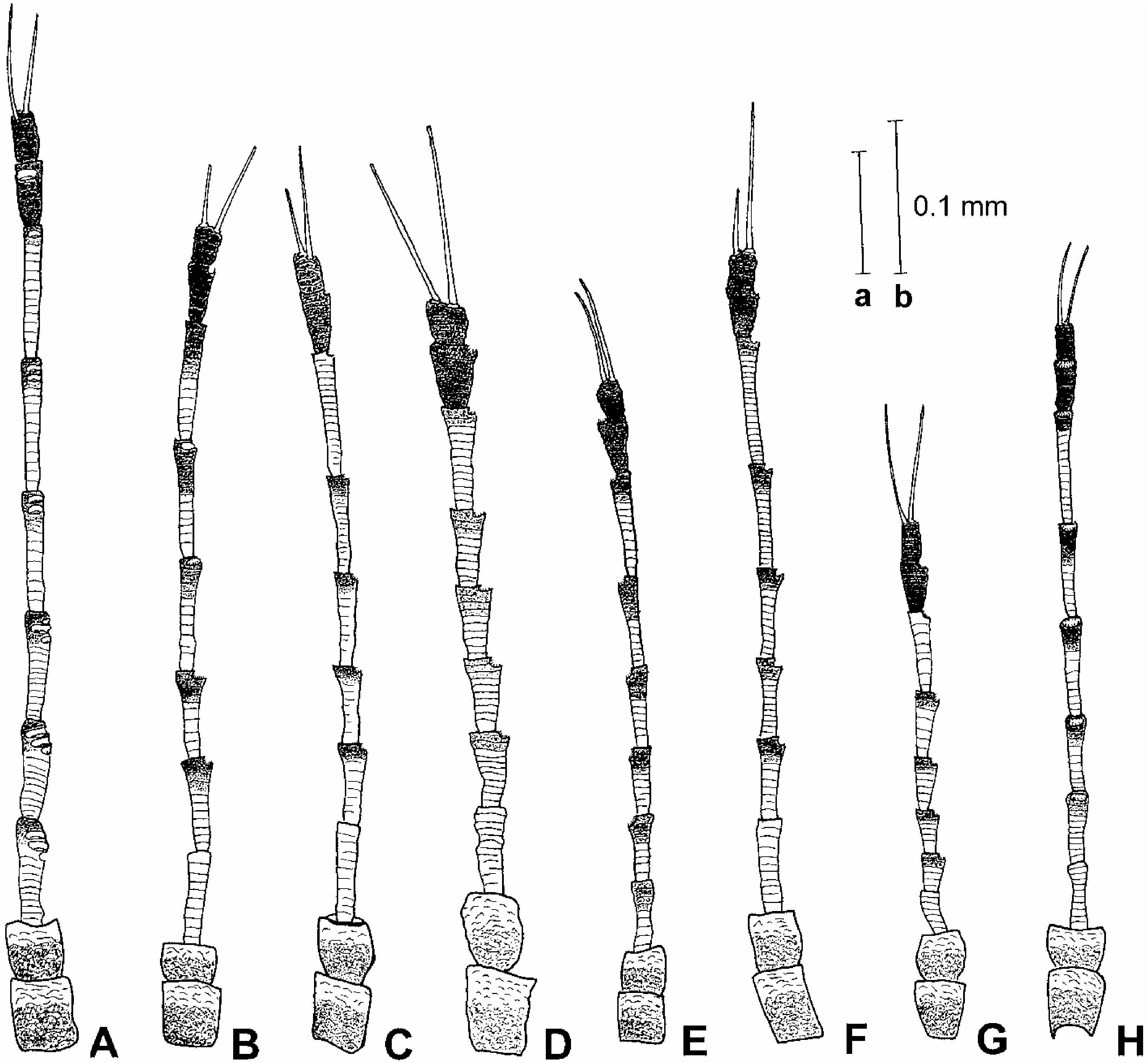
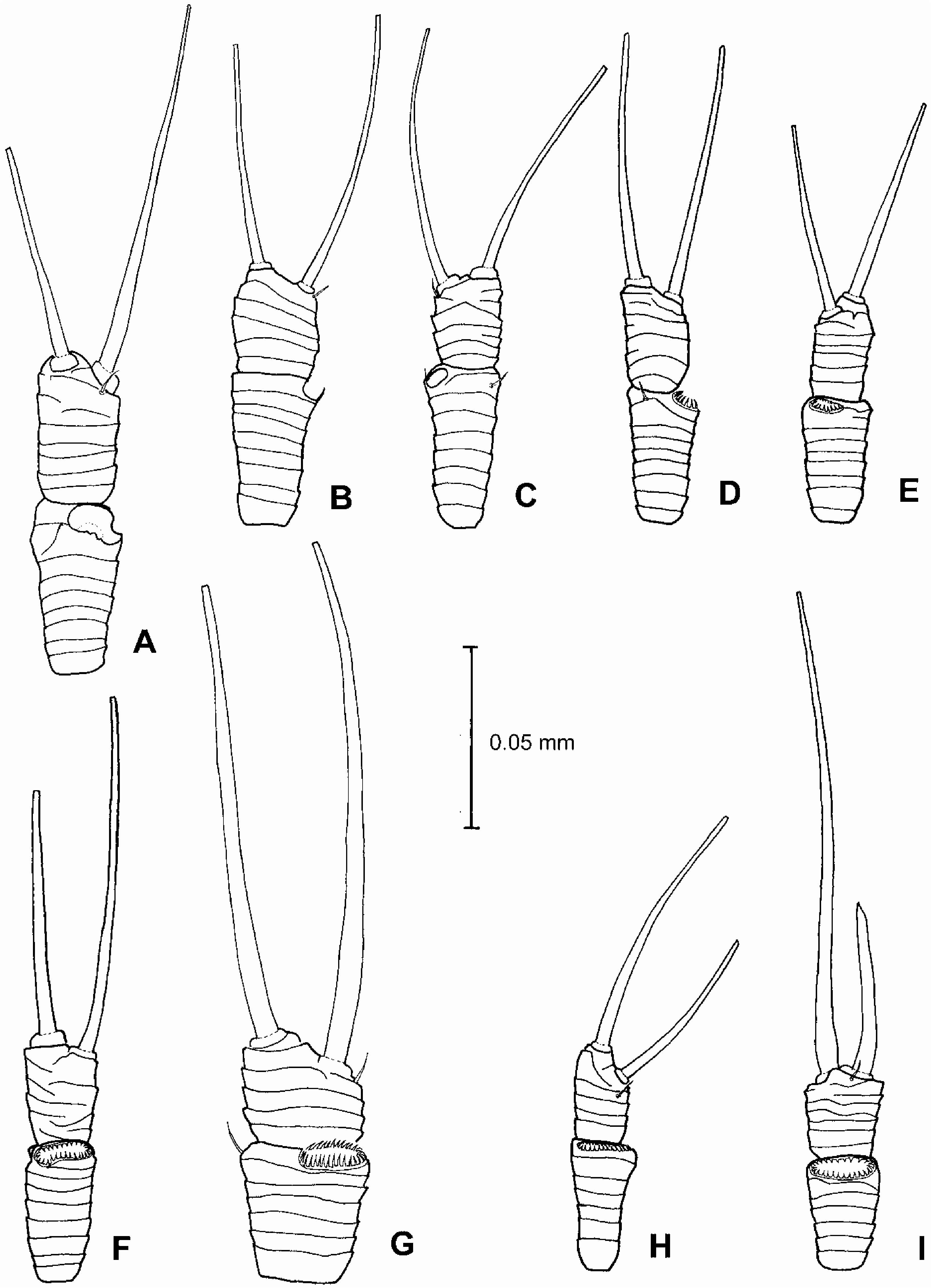
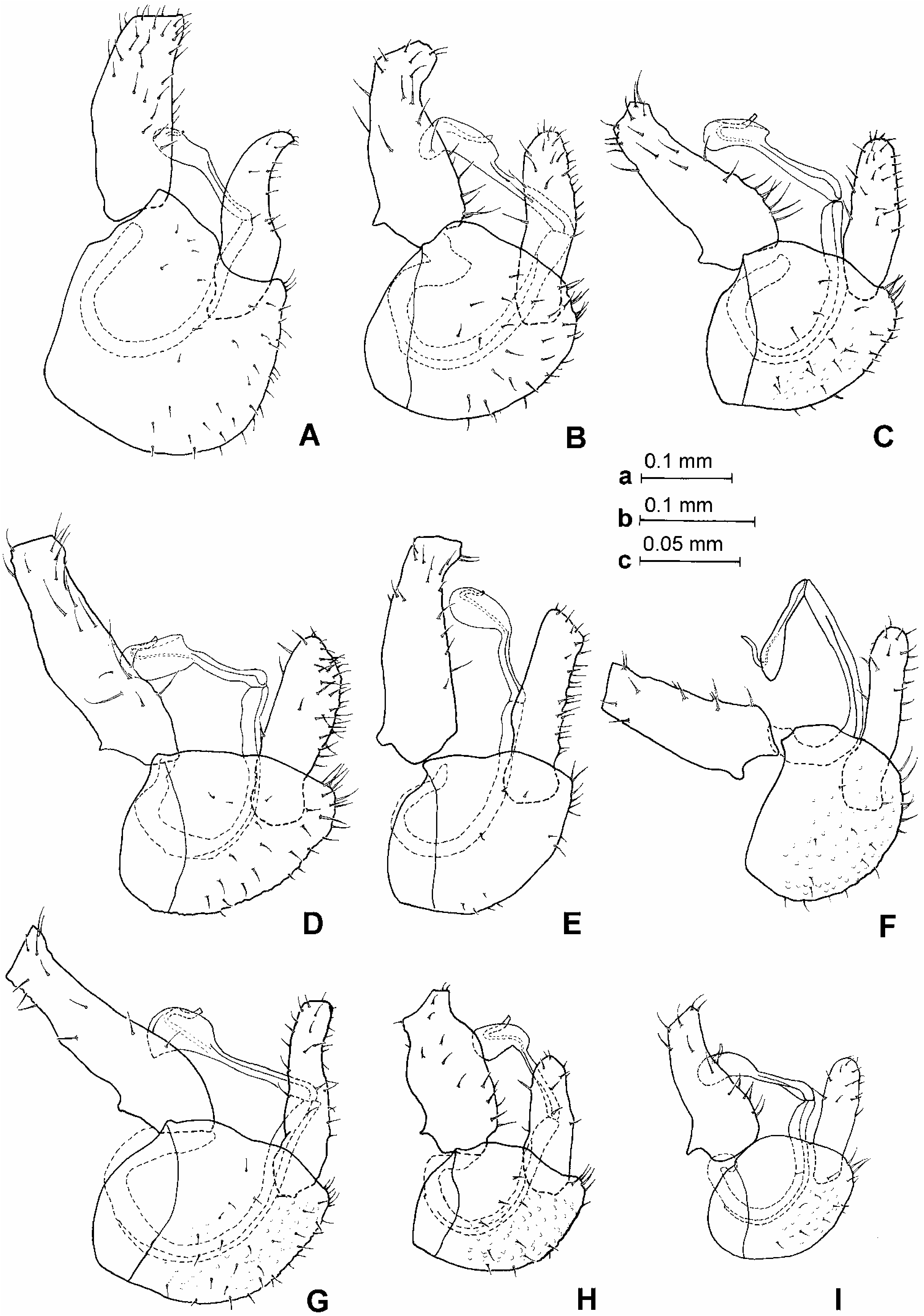
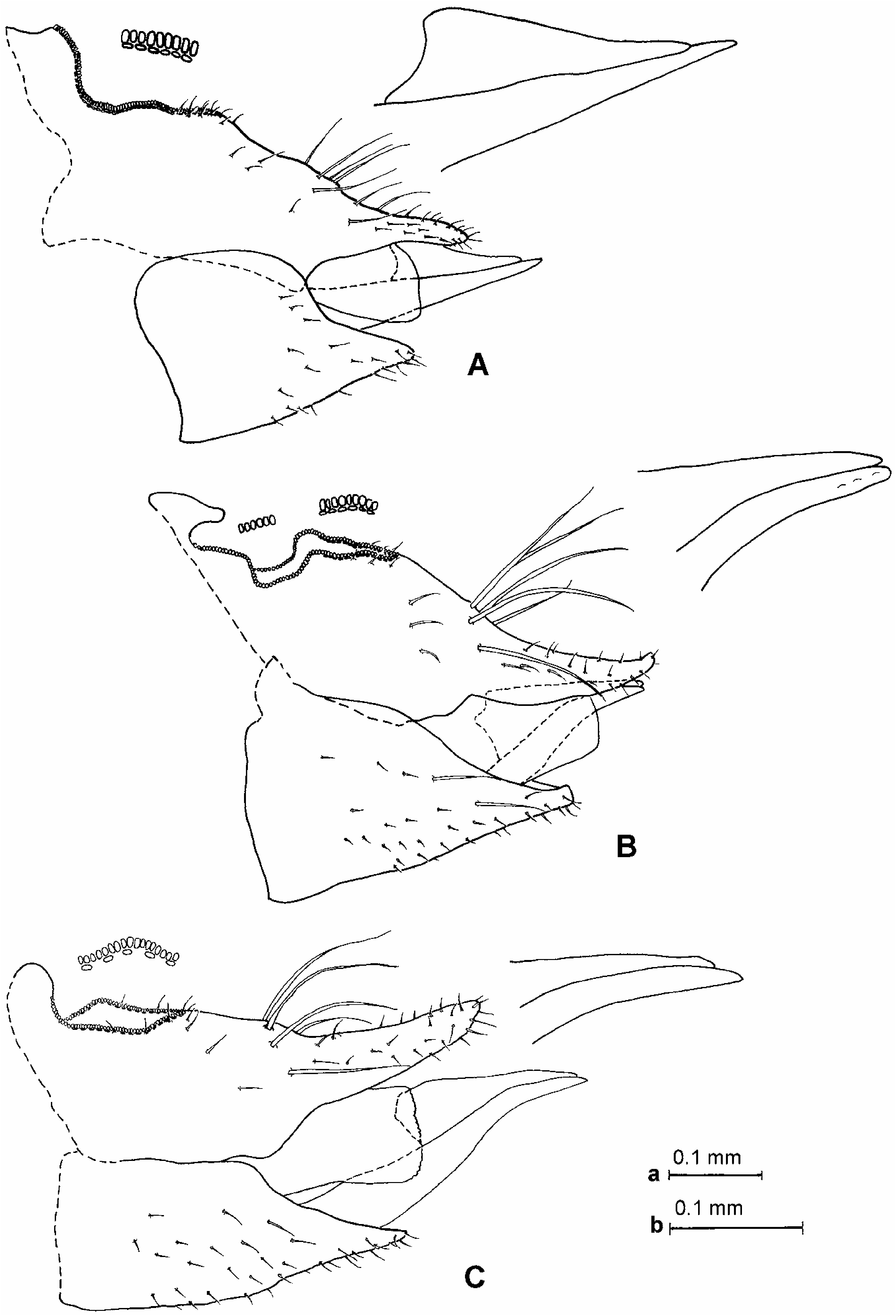
Morphology: head, in dorsal view, slightly wider than mesoscutum, subglobular (Figure 1A). Vertex with microsculpture, matt, 1.7 times as wide as long along midline, rounded down in front; in frontal view, relatively flat (Figure 1B). Coronal suture distinct in basal and apical thirds, reduced in the middle. Median ridge on vertex weakly raised, indistinct. Vertex with large anterior tubercle on either side of midline. Lateral ocelli lying in the same plane as vertex. Occiput in dorsal view narrowly triangular. Preoccipital sclerite narrow, eyes not stalked, in frontal view, subglobular. Genae distinctly swollen. Tubercle below torulus pointed, forming almost a right angle (Figure 1C). Frons narrowly pentagonal. Clypeus subglobular. Antenna long, slender, segments cylindrical, weakly widening to apex ( Figure 4A View Figure 4 ); with following numbers of rhinaria: segment 3: five to eight, segment 4: four or five, segment 5: three or four; segments 6 and 7: two, segments 8 and 9: one; rhinaria elongate, lacking cuticular spines ( Figure 3A, B View Figure 3 ); terminal setae subequal, the longer seta slightly longer than segments 9 and 10 together ( Figure 5A View Figure 5 ). Fore wing very broad, strongly widening towards apex which is truncate. Surface spinules present in all cells, densely spaced especially in the apical part, only a few spinules present in cell c+sc ( Figure 7A View Figure 7 ). Costal break in apical third of C+Sc. Hind legs long and robust. Meracanthus relatively long, acute, pointed. Metafemur not constricted medially. Metatibia bearing an open crown of 17–20 densely spaced slender, unsclerotized apical spurs, lacking similar spurs laterally. Metabasitarsus bearing two black sclerotized spurs. Dorsal margin of abdomen in profile forming large hump, with tergite 4 medially swollen and forming highest point; posterior margins of tergites 3 and 5 dorsally straight, not swollen into prominent tubercular process. Male terminalia as in Figure 8A View Figure 8 . Proctiger relatively long, cylindrical. Subgenital plate, in profile, higher than long, dorsal margin strongly angular. Paramere, in profile, curved, narrowing to apex which is turned backwards ( Figure 9B View Figure 9 ); apex forming small, strongly sclerotized tooth; in posterior view, inner margin straight, strongly indented subapically ( Figure 9A View Figure 9 ); inner surface covered in fine setae. Basal segment of aedeagus narrow; apical segment moderately long, with elongate, narrow, apically rounded, slightly pyriform head; sclerotized end tube of ductus ejaculatorius relatively short, sinuate ( Figure 10A View Figure 10 ). Female terminalia as in Figure 11A View Figure 11 . Proctiger relatively long, with dorsal margin weakly sinuate, almost straight, apical process long, parallel-sided; circumanal ring with two rows of pores, pores of outer row contiguous. Subgenital plate relatively short, in profile with dorsal margin strongly concave, ventral margin straight; apex narrowly triangular, pointed; in ventral view narrowly triangular, apical process with concave margins and narrow, truncate apex ( Figure 13A View Figure 13 ). Dorsal and ventral valvulae lacking lateral teeth. Measurements and ratios in Tables I–III.
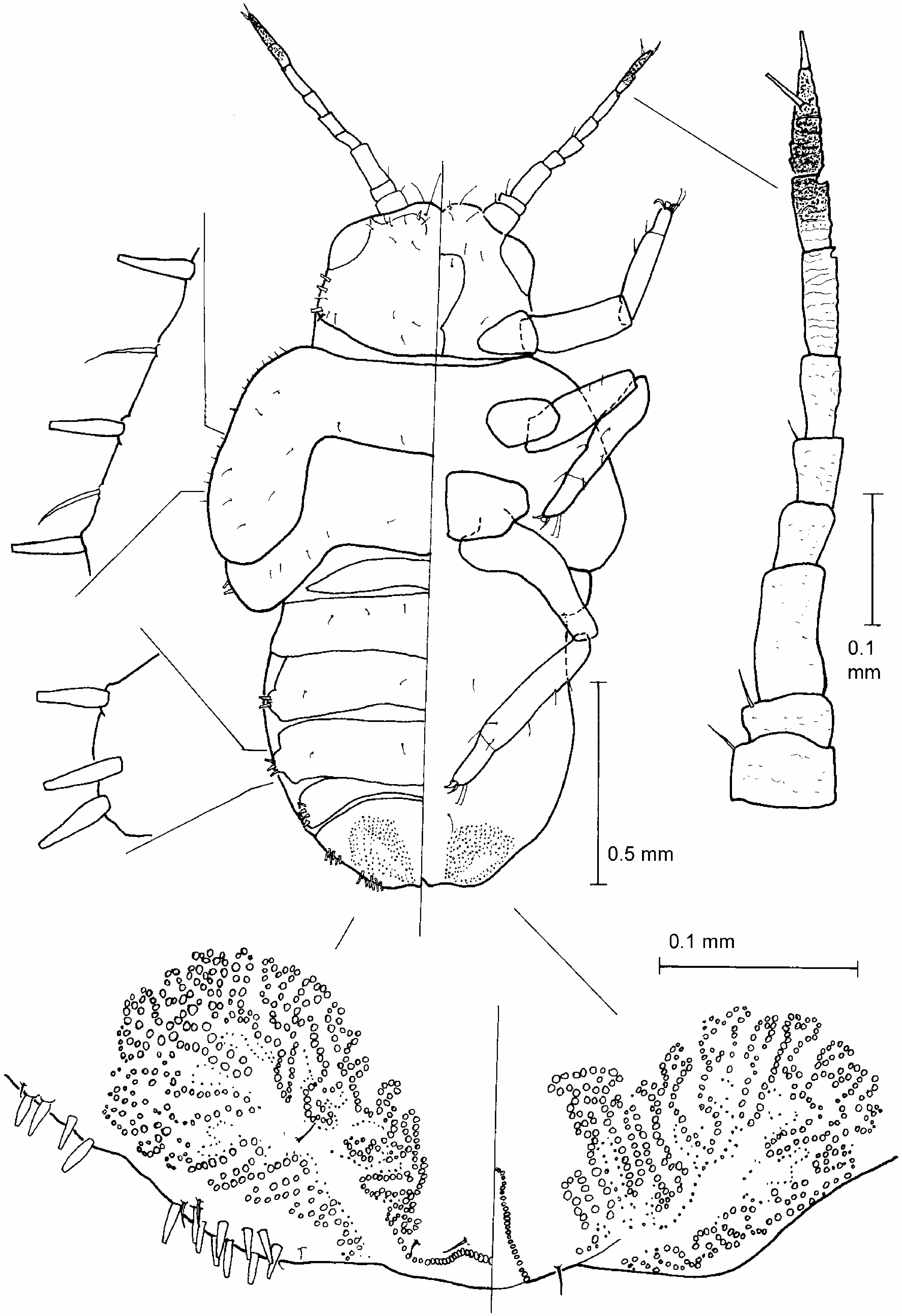
Fifth instar larva ( Figure 14 View Figure 14 ). Uniformly pale yellow, two apical segments of antenna dark brown. Body relatively slender, with long limbs. Body margin with following numbers of slender lanceolate setae, narrowly truncate at apex (one side only): head in front of insertion of antenna: two to three; cephalothorax behind eyes: four; fore wing pad: eight; hind wing pad: three; abdomen: (1–2) + (3–4) + 3 + (3–4) + (6–7) + (8–14); lanceolate setae on abdominal margin arranged in groups situated on small tubercles. Lanceolate setae on body margin alternating or accompanied by short simple setae. A small simple seta present in anterior half of ocular region. Eye with ca 50 distinct ommatidia. Anterior margin of head forming two shallow lobes. Antenna straight, relatively long, with nine segments; a single rhinarium apically on each of segments 7 and 8. Tarsal arolium very small relative to claws, with a triangular pad and indistinct petiole ( Figure 21A View Figure 21 ). Abdomen dorsally weakly sclerotized, with six hardly noticeable sclerites, caudal plate incompletely fused; apex of abdomen truncate. Anus in terminal position. Circumanal ring large and strongly sinuate, widely extending on to dorsal and ventral surface of caudal plate. Measurements and ratios in Table IV.
Host plant
Blighia unijugata , Blighia sp. (Sapindaceae) .
Biology
P. cuniculus does not induce galls on the host. The eggs are laid on the upper leaf surface along the median vein. The sucking of the larvae distorts the leaves which become necrotic in parts. The leaves dry up a few days after the emergence of the adults.
Distribution
Angola, Cameroon, Kenya, Nigeria, Tanzania, Uganda.
Material examined
Holotype: „, Cameroon: Centre Province, Soa, 3 ° 579N, 11 ° 369E, 725 m, 25 March 2004, secondary forest, Blighia unijugata (J. L. Tamesse) . Dry-mounted (NHMB). Paratypes: Angola: 1 „, 3♀, 7 miles W Gabela, 16–18 March 1972, at light (D. Hollis); 1 „, 1♀, same data but beaten from Albizzia spp. ; 1 „, 3 miles SW Salazar, 15 March 1972, at light (D. Hollis). Cameroon: 1 „, 6♀, 34 larvae, same data as holotype. Kenya: 1♀, Kakamega Forest, 18 December 1970 (A. E. Stubbs); 3 „, 8♀, Kakamega Forest, 0 ° 229N 34 ° 509E, 1600 m, 7–11 February 1999, rain forest, canopy fogging, Teclea nobilis (Rutaceae) (T. Wagner). Nigeria: 2 „, 1♀, Ibadan, June 1956 (V. F. Eastop); 4 „, 3♀, Ibadan, 18 March 1957, trapped (V. F. Eastop); 1 „, Ibadan, 25 March 1959 (F. A. Squire); 1 „, same data but April 1959; 1 „, 1♀, same data but April 1960; 1♀, same data but June 1960; 1 „, Ibadan, Moor Plantation, 9–10 April 1961 (F. A. Squire); 1 „, Ibadan, DFR Arboretum, 14 January 1964, yellow tray (M. J. White). Tanzania: 5 „, 7♀, S. Pare Mountains, hillside above Gonja, ca 3000 feet, 12–16 June 1974, beaten from Blighia sp. (D. Hollis) . Uganda: 3 „, 2♀, District Masindi, Budongo Forest near Sonso, 1 ° 459N, 31 ° 359E, 21–30 July 1995, secondary forest, canopy fogging, Rinorea beniensis (Violaceae) (T. Wagner); 2♀, same data but Trichilia rubescens (Meliaceae) ; 1 „, same data but 15–21 January 1997, Cynometra alexandri (Caesalpiniacae) ; 3 „, 1♀, and ca 200 adult specimens in alcohol, same data, but Rinorea beniensis , at night. Dry- and slide-mounted, and preserved in alcohol (BMNH, LZUY, MAKB, MHNG, MMB, NHMB, USNM).
Etymology
The Latin noun cuniculus 5 rabbit is used in apposition. The habitus of P. cuniculus is reminiscent of a rabbit because of the fore wing shape and maculation.
Comments
P. cuniculus View in CoL sp. n. resembles Pseudophacopteron caffrariense Capener, 1973 View in CoL , a species known so far only from South Africa, in the body colour, the fore wing maculation with dark brown spots on the veins, the relatively flat vertex with weakly raised and indistinct median ridge and a pair of large anterior tubercles, the relatively broad pentagonal frons, the multiple rhinaria on antennal segments 4–8, the relatively long and acute meracanthus, the unconstricted metafemur, the metatibia lacking lateral spurs, the angular dorsal margin of the male subgenital plate, the shape of apical dilation of distal segment of aedeagus, the relatively long female proctiger, and the absence of teeth on the dorsal and ventral valvulae. P. cuniculus View in CoL sp. n. differs from P. caffrariense View in CoL in the larger size, the very broad fore wing, the clear fore wing membrane near apex of vein R1, the separated patches on apex of vein Rs and touching point of Rs and M1+2, the medially reduced coronal suture on vertex, the pointed tubercle on genae below torulus, the presence of multiple rhinaria on antennal segment 3, the terminal setae on antenna subequal and as long as or slightly longer than segments 9 and 10 together, the sickle-shaped paramere, and the host-plant association.
In P. caffrariense the fore wing dimensions are as follows: WL51.42–1.77, WL/ WW52.11–2.32, the fore wing membrane bears a dark brown macula across vein R 1, and the maculae on apex of vein Rs and touching point of Rs and M1+2 are fused, the coronal suture on vertex is distinct throughout, the tubercle on genae below torulus is blunt, the antennal segment 3 lacks rhinaria, the terminal antennal setae differ markedly in length and are shorter than segments 9 and 10 together, the paramere in profile is stout, evenly convex anteriorly, straight posteriorly and not sickle-shaped, and the host-plant is Pappea capensis (Sapindaceae) . Material of P. caffrariense examined: paratypes, 8 „, 6♀, South Africa, Cape Province, Steytlerville, 16 and 18 February 1966 (A. L. Capener) ( NCIP, BMNH, dry- and slide-mounted).


| R |
Departamento de Geologia, Universidad de Chile |
| NCIP |
South African National Collection of Insects |
No known copyright restrictions apply. See Agosti, D., Egloff, W., 2009. Taxonomic information exchange and copyright: the Plazi approach. BMC Research Notes 2009, 2:53 for further explanation.
|
Kingdom |
|
|
Phylum |
|
|
Class |
|
|
Order |
|
|
Family |
|
|
Genus |
Pseudophacopteron cuniculus
| Malenovský, Igor, Burckhardt, Daniel & Tamesse, Joseph L. 2007 |
P. cuniculus
| Malenovský & Burckhardt & Tamesse 2007 |
P. cuniculus
| Malenovský & Burckhardt & Tamesse 2007 |
Pseudophacopteron caffrariense
| Capener 1973 |
P. caffrariense
| Capener 1973 |