
Microperla geei Chu, 1928
|
publication ID |
https://doi.org/ 10.11646/zootaxa.4780.3.8 |
|
publication LSID |
lsid:zoobank.org:pub:C6872B26-7639-4A12-8B8A-A5A4E02AC953 |
|
DOI |
https://doi.org/10.5281/zenodo.3857478 |
|
persistent identifier |
https://treatment.plazi.org/id/039A87C5-7A7E-AB16-D8A9-FDFB2E8EF952 |
|
treatment provided by |
Plazi |
|
scientific name |
Microperla geei Chu, 1928 |
| status |
|
Microperla geei Chu, 1928 View in CoL
Figs. 1–16 View FIGURE 1 View FIGURE 2 View FIGURE 3 View FIGURE 4 View FIGURE 5 View FIGURE 6 View FIGURE 7 View FIGURE 8 View FIGURE 9 View FIGURE 10 View FIGURE 11 View FIGURE 12 View FIGURE 13 View FIGURE 14 View FIGURE 15 View FIGURE 16 .
Microperla geei Chu, 1928 View in CoL . China J. 9(4): 197; Wu, 1938. Plecopterum sinensium, a monograph of the stoneflies of China (Order Plecoptera View in CoL ): 67; Illies, 1966. Das Tierreich 82: 19; Wu, 1973. Acta Ent. Sin. 16(2): 97; Stark & Sivec, 2000. Acta Entomol. Sloven. 8: 102.
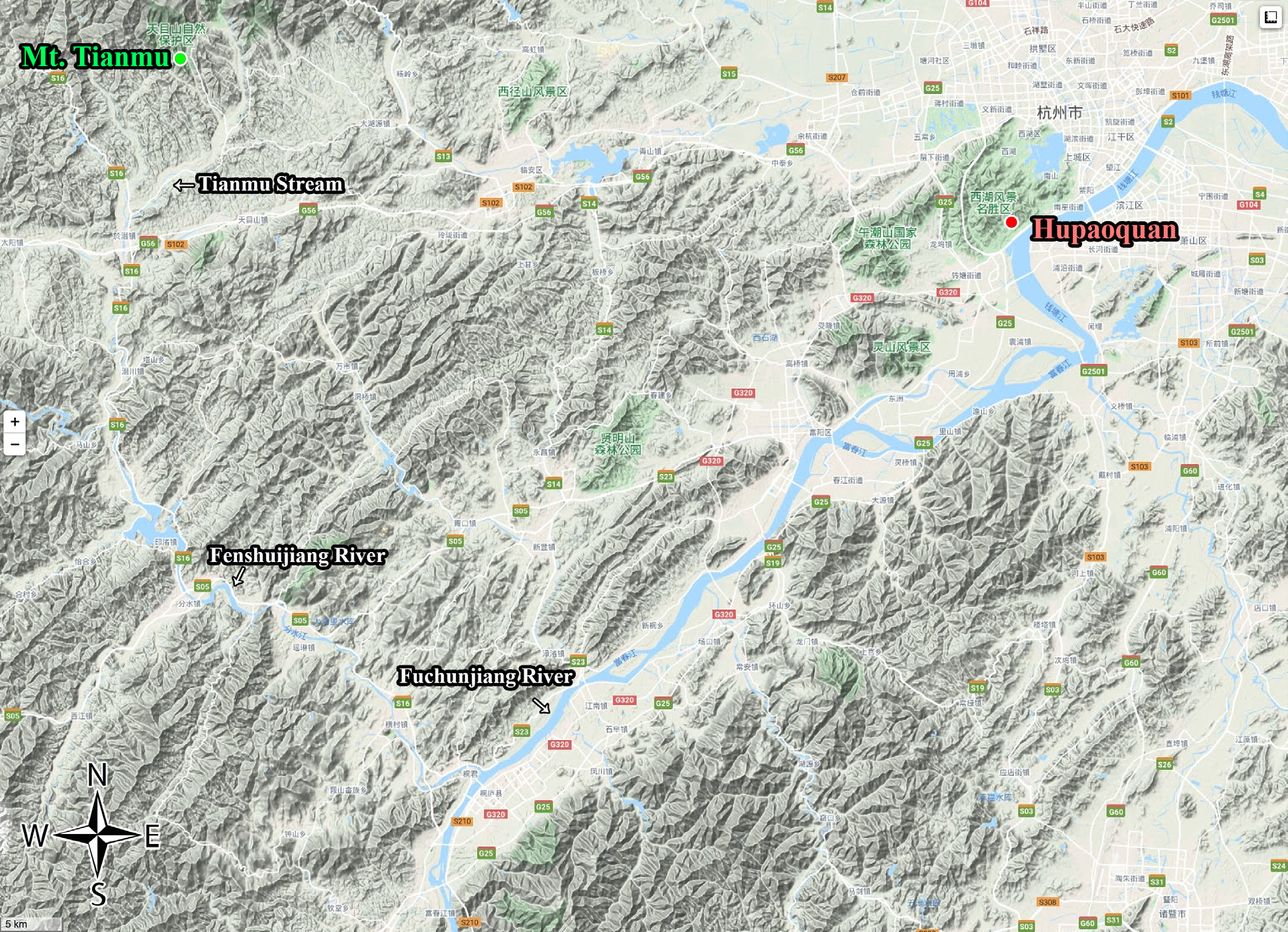
Type information. Holotype: male, China: Zhejiang Province, Fu Bao (Hu Pao Quan), Hangchow (Hangzhou City), China (lost, Wu 1962) . Paratype: one female, same locality and data as holotype (lost, Wu 1962).
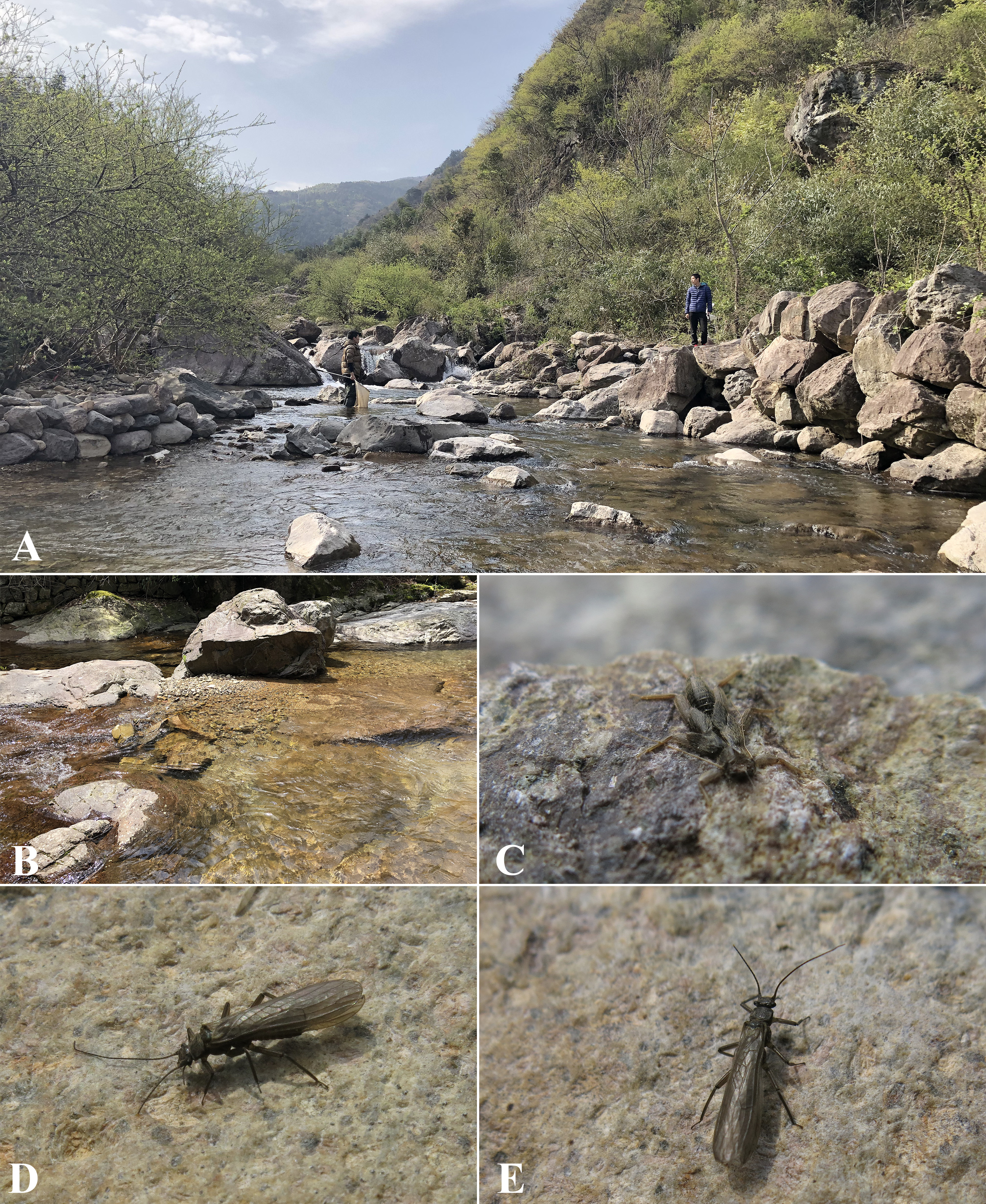
Material examined. Neotype herein designated: male, China: Zhejiang Province, Hangzhou City, Tianmu Mountain, Tianmu Stream ( Figs. 1 View FIGURE 1 , 2 View FIGURE 2 ), 30.3604 N, 119.4434 E, 580 m, 5 April 2020, leg. Zhi-Teng Chen, Xu-Hong- Yi Zheng & Zhen-Xing Ma (ICJUST) GoogleMaps . Paratypes: 7 males, 17 females, one larva, one exuvia, same locality and data as holotype (ICJUST) GoogleMaps .
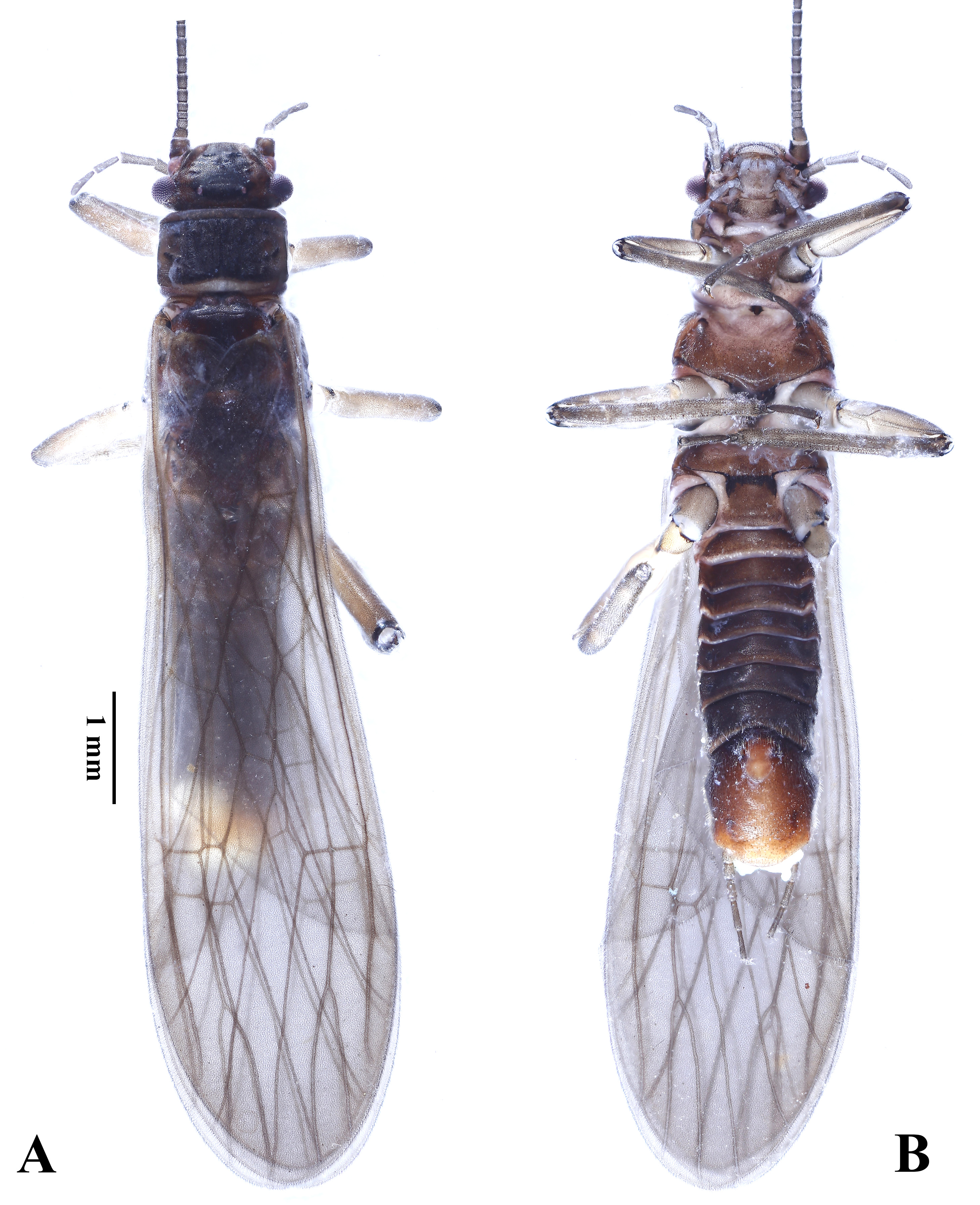
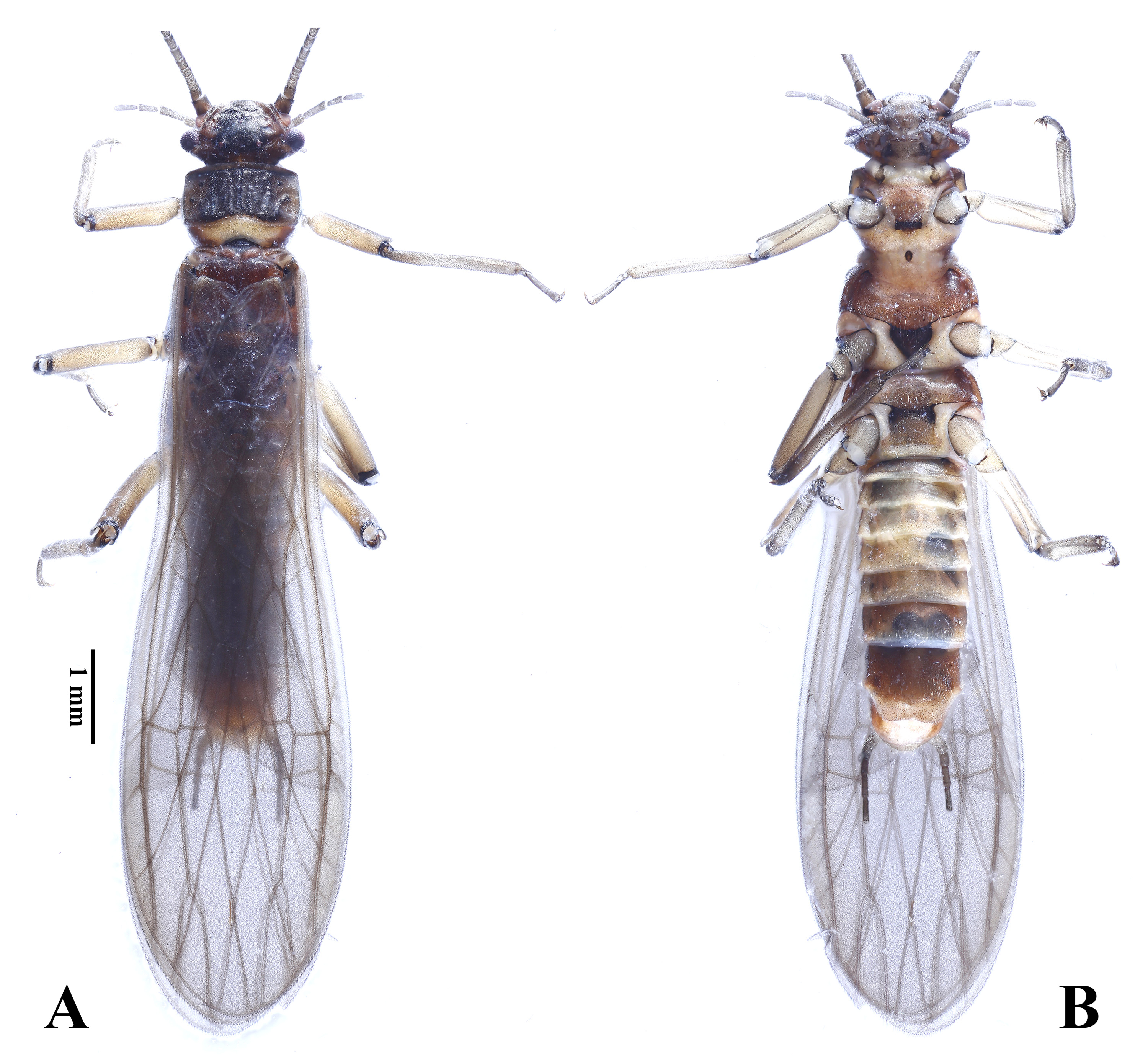
Male. Body ( Fig. 3 View FIGURE 3 ) length ca. 6.5 mm (n = 8). Body generally dark brown. Head ( Fig. 4 View FIGURE 4 ) with a dark median area. Triocellate, but anterior ocellus largely reduced; compound eyes small rounded and protruded. Antennae slen- der and dark brown. Pronotum rectangular with obtuse corners, median area dark pigmented and laterally with several dark spots on lateral pale areas; posteromedial margin of pronotum pale. Meso- and metanota mostly dark brown. Macropterous ( Fig. 5 View FIGURE 5 ); wings hairy and subhyaline, veins pale brown. Wing venation generally identical to that in Chu (1928), except for the one or two extra crossveins beyond Sc. Legs generally pale brown, joints dark.
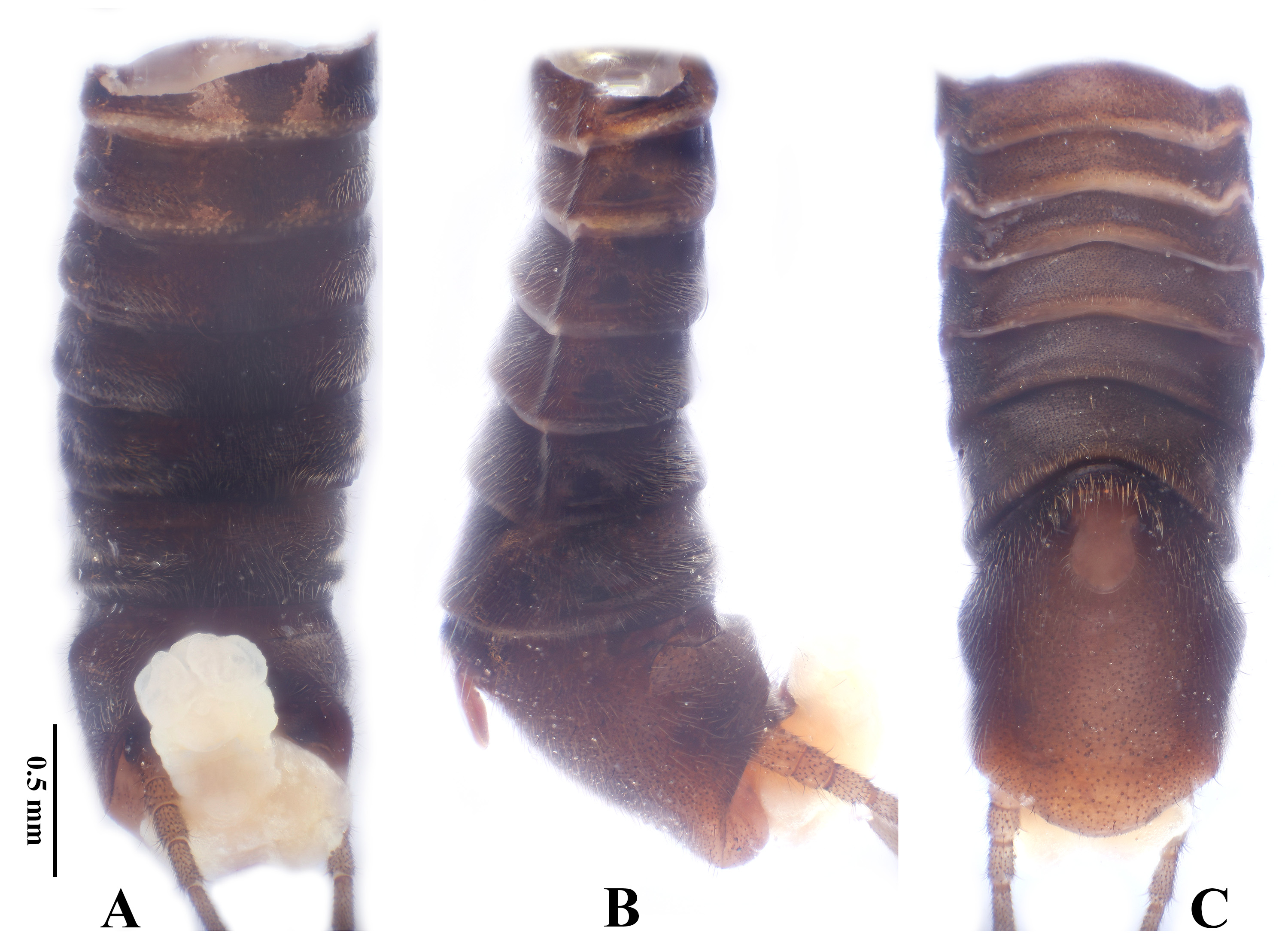
Abdominal segments mostly dark brown ( Fig. 6 View FIGURE 6 ); terga 1–3 with two pale lateral spots. Sternum 9 extended backwards, posterior margin rounded, posterolaterally with two curved impressions. Ventral lobe pale and elliptical, width longer than length, constricted near base, apical half margined with long lateral bristles. Posterior margin of tergum 9 with a shallow median notch ( Fig. 7A, B View FIGURE 7 ). Tergum 10 with a membranous median area; posteromedial margin sclerotized and curved upwards, subtriangular from caudal view ( Fig. 7A, B View FIGURE 7 ). Aedeagus membranous ( Fig. 7 View FIGURE 7 C–E), basally enlarged as a cushion, near the cushion with a small dorsal lobe, dorsoapically with three connected large lobes surrounding a tiny median lobe; from lateral view, anterior of the three large lobes with another lower large lobe. Cerci short, generally pale brown, with about 7 segments, basal segment unmodified.
Female. Body ( Fig. 8 View FIGURE 8 ) length ca. 7 mm (n = 17). Color pattern similar to males ( Figs. 8 View FIGURE 8 , 9A View FIGURE 9 ). Abdominal tergum 10 slightly projected backwards ( Fig. 9A View FIGURE 9 ). Subgenital plate large and broad, bilobed with a shallow posterior notch or truncate at posterior margin, covering half of sternum 9 ( Fig. 9B, C View FIGURE 9 ).
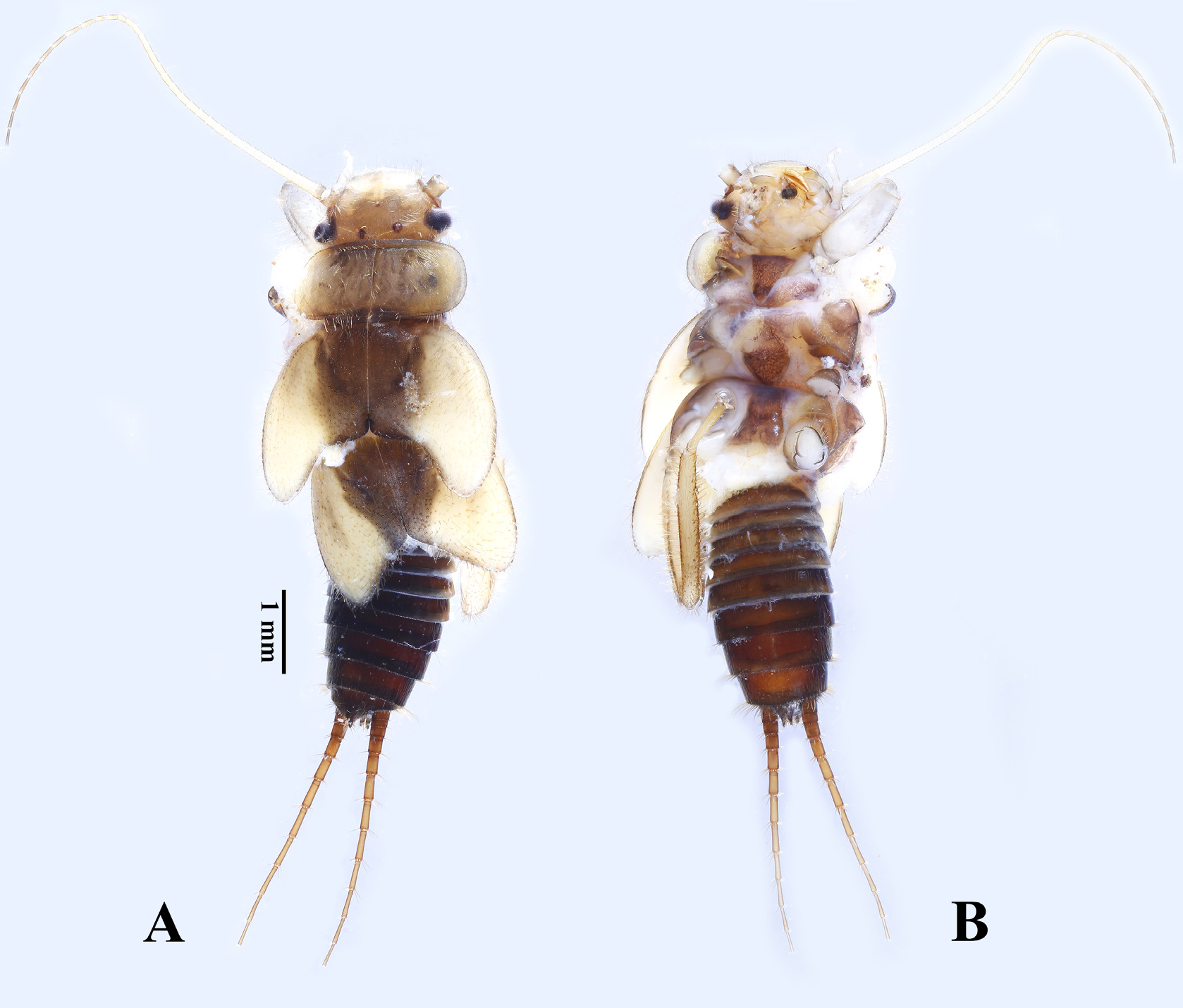
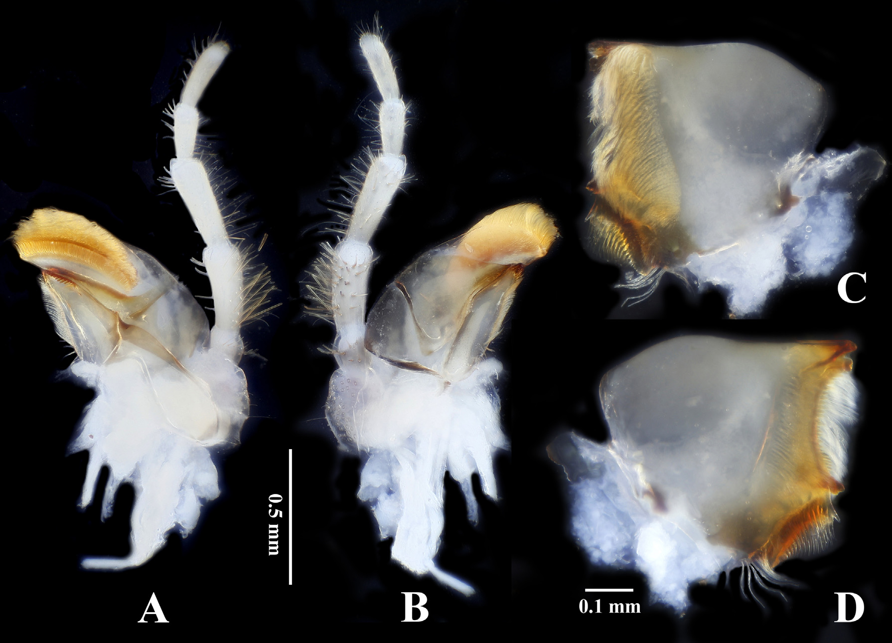
Larva. Body length ca. 6.5 mm ( Figs. 10 View FIGURE 10 , 11 View FIGURE 11 ). Head mostly pale, medially with a brown stigma covering ocelli ( Fig. 12A View FIGURE 12 ); compound eyes dark and glabrous; margins and submargins of head with long pale spines. Antennae pale and slender ( Fig. 12 View FIGURE 12 B–E), subequal to body length, each segment apically fringed with moderate cylindrical spines. Glossae of labium longer than paraglossae, similar to that in Huo & Du (2019). Stipes of maxilla with granules along outer margin ( Fig. 13A, B View FIGURE 13 ); galea broad, inner margin with a row of stout apical spines, apex with a huge crescent hairbrush on ventral aspect; lacinia long triangular, with several apical teeth and an inner comb of marginal spines. Mandible with three blunt main teeth ( Fig. 13C, D View FIGURE 13 ), ventral aspect with a huge hairbrush, dorsal aspect with a marginal comb of thick spines posterior to the main teeth; molar area pale.
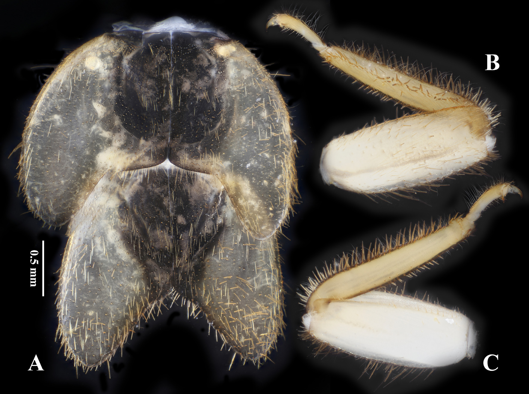
Pronotum wide and short with obtuse corners ( Fig. 14A View FIGURE 14 ), mostly pale, scattered with dark spots, covered and fringed with long cylindrical spines. Meso- and metanota dark, covered and fringed with long cylindrical spines which are easily detached. Wing pads pale, outer margins circular and fringed with long spines. Coxae and trochan- ters membranous and glabrous ventrally, dorsal and lateral aspects covered with dense moderate spines. Femora dorsally covered and fringed with long cylindrical spines, ventrally near glabrous except for the short spines along posterior margin; inner groove of femora covered with conspicuous granules; outer margin with several moderately long swimming hairs ( Fig. 14B, C View FIGURE 14 ). Tibiae dorsally with a longitudinal median row of long spines, inner margin fringed with moderate spines, outer margin with dense long spines and moderately long swimming hairs; ventral surface of tibiae near glabrous, with a sparse row of short spines near inner margin. Tarsal segments with short and long spines along inner margin, with long swimming hairs along outer margin. Claws sharp and glabrous.
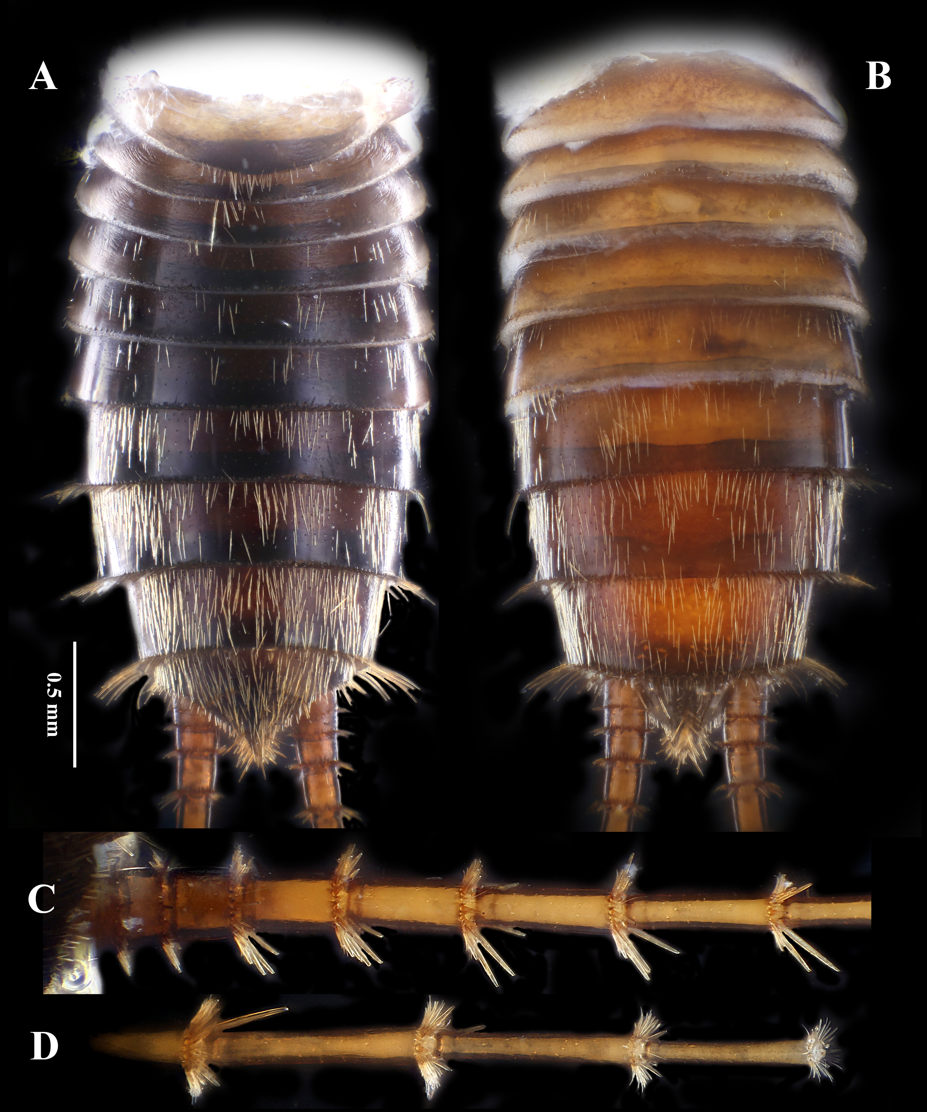
Abdominal segments generally dark brown ( Fig. 15A, B View FIGURE 15 ), each segment covered and posteriorly fringed with long spines which are easily detached; posterior half of each segment dark. Tergum 10 projected backwards, near triangular in shape. Paraprocts conical and sharp, covered by dense long spines. Cerci mostly brown ( Fig. 15C, D View FIGURE 15 ), subequal in length to abdomen, each segment with both long and short cylindrical apical spines, length of which does not exceed the segment length.
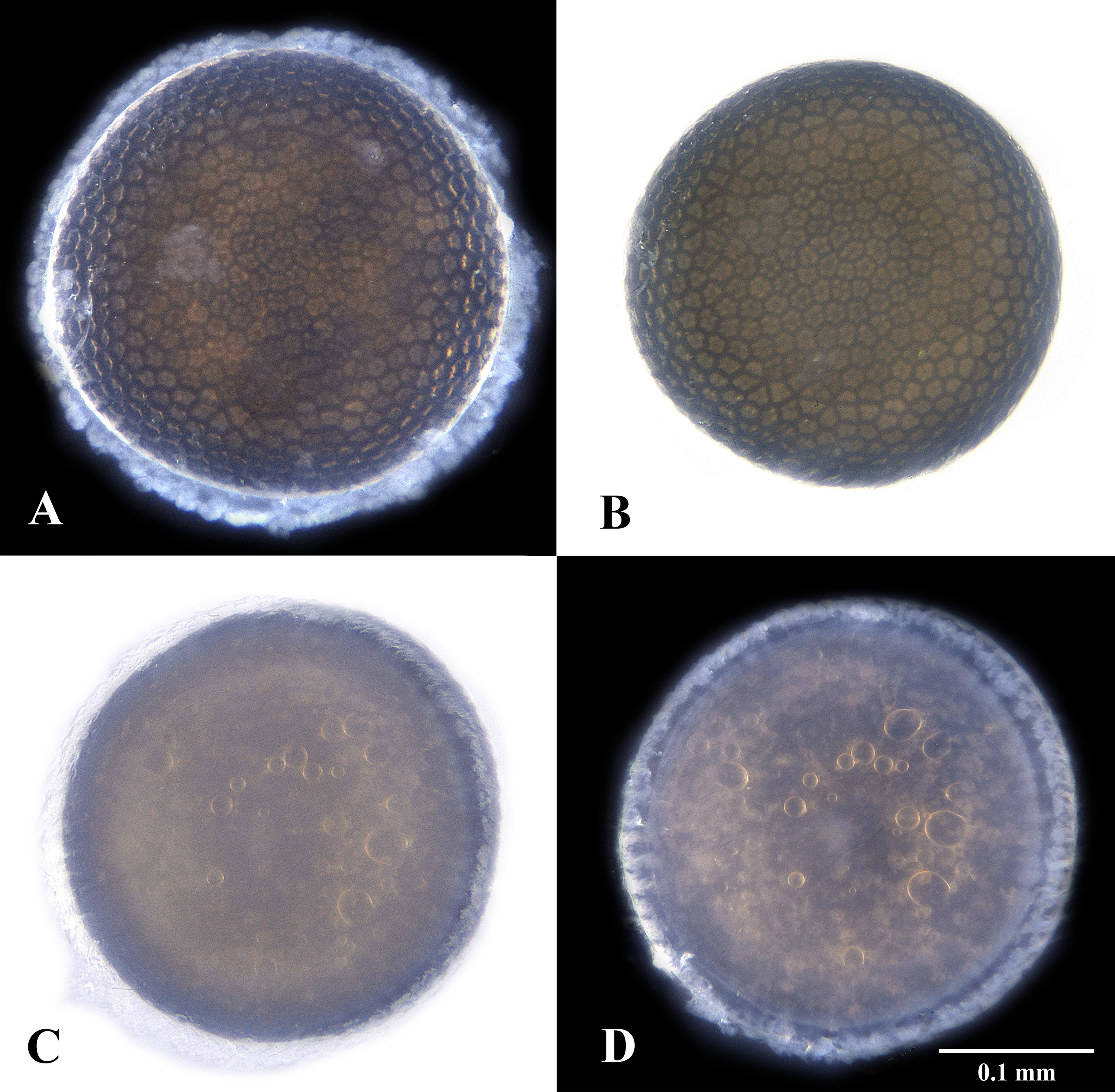
Egg. Shape typical for Peltoperlidae ( Fig. 16 View FIGURE 16 ), both dorsal and ventral aspects flat, diameter approximately 0.3 mm. Irregular follicle cell impressions present on chorionic surface except for the circular lid, inner area of which unmodified ( Fig. 16C, D View FIGURE 16 ).
Remarks. Chen & Song (2019) concluded that wing venation, shape and size of the ventral lobe of the male, shape and extent of the subgenital plate of the female and egg morphology are currently utilized characters for specific identification of Microperla . However, that study found the conspecific variations concerning the width of ventral lobe and shape of sternum 9 in the male; this study also revealed intraspecific variations on subgenital plate of the female and interspecific similarity of terminalia and wing venation. Fortunately, details of aedeagus and egg morphology are diagnostic among the currently named and completely described species.
The newly designated Neotype is generally identical to the original descriptions of Chu (1928), despite the variable width of male ventral lobe and shape of the female subgenital plate. Wing venation of the Neotype is almost identical with the original descriptions of M. geei except for the number of crossveins beyond Sc and between M and CuP, whereas it is similar to the venation of M. qinlinga . The abdominal tergum 9 of the Neotype is notched at posterior margin, but was shown as complete in Chu (1928) and in the supposed redescription in Stark & Sivec (2000). The aedeagus of M. geei is newly described, clearly different from that of M. qinlinga ( Chen & Song 2019, Huo & Du 2019). Larva of M. geei is apparently different from M. qinlinga by showing different body color and details of mouthparts ( Huo & Du 2019). Egg morphology of the newly studied material apparently differs from that provided by Stark & Sivec (2000) in showing no cell impressions on the circular lid. The Microperla species described by Stark & Sivec (2000) from Henan Province is not conspecific with M. geei .


No known copyright restrictions apply. See Agosti, D., Egloff, W., 2009. Taxonomic information exchange and copyright: the Plazi approach. BMC Research Notes 2009, 2:53 for further explanation.
|
Kingdom |
|
|
Phylum |
|
|
Class |
|
|
Order |
|
|
Family |
|
|
Genus |
Microperla geei Chu, 1928
| Chen, Zhi-Teng 2020 |
Microperla geei
| Chu 1928 |