
Gnathoncus sechuanus, Lackner, 2020
|
publication ID |
https://doi.org/ 10.37520/aemnp.2020.24 |
|
publication LSID |
lsid:zoobank.org:pub:AC387BAF-E7A8-40B2-9486-E5642074587D |
|
DOI |
https://doi.org/10.5281/zenodo.4549639 |
|
persistent identifier |
https://treatment.plazi.org/id/039A87F1-FFC5-FFBF-FEF3-B7F5FF5FF916 |
|
treatment provided by |
Felipe |
|
scientific name |
Gnathoncus sechuanus |
| status |
sp. nov. |
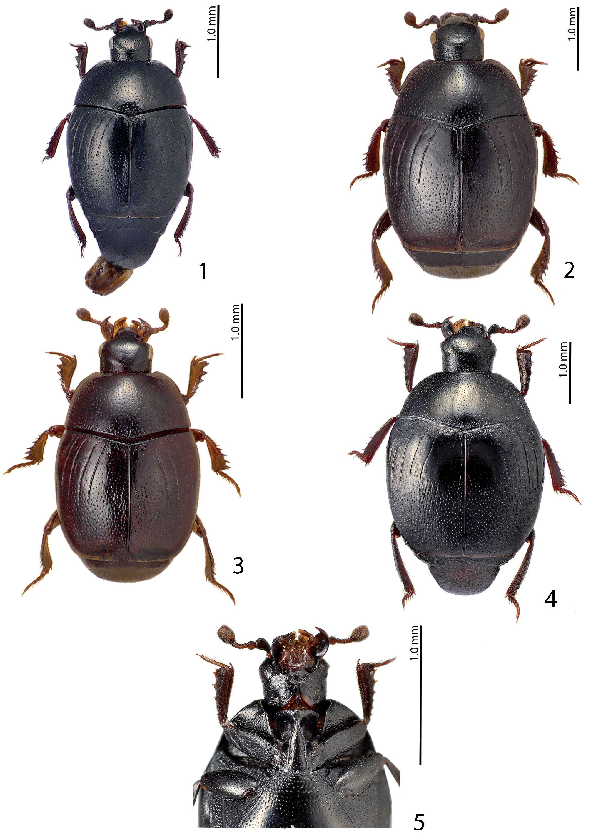
Gnathoncus sechuanus View in CoL sp. nov.
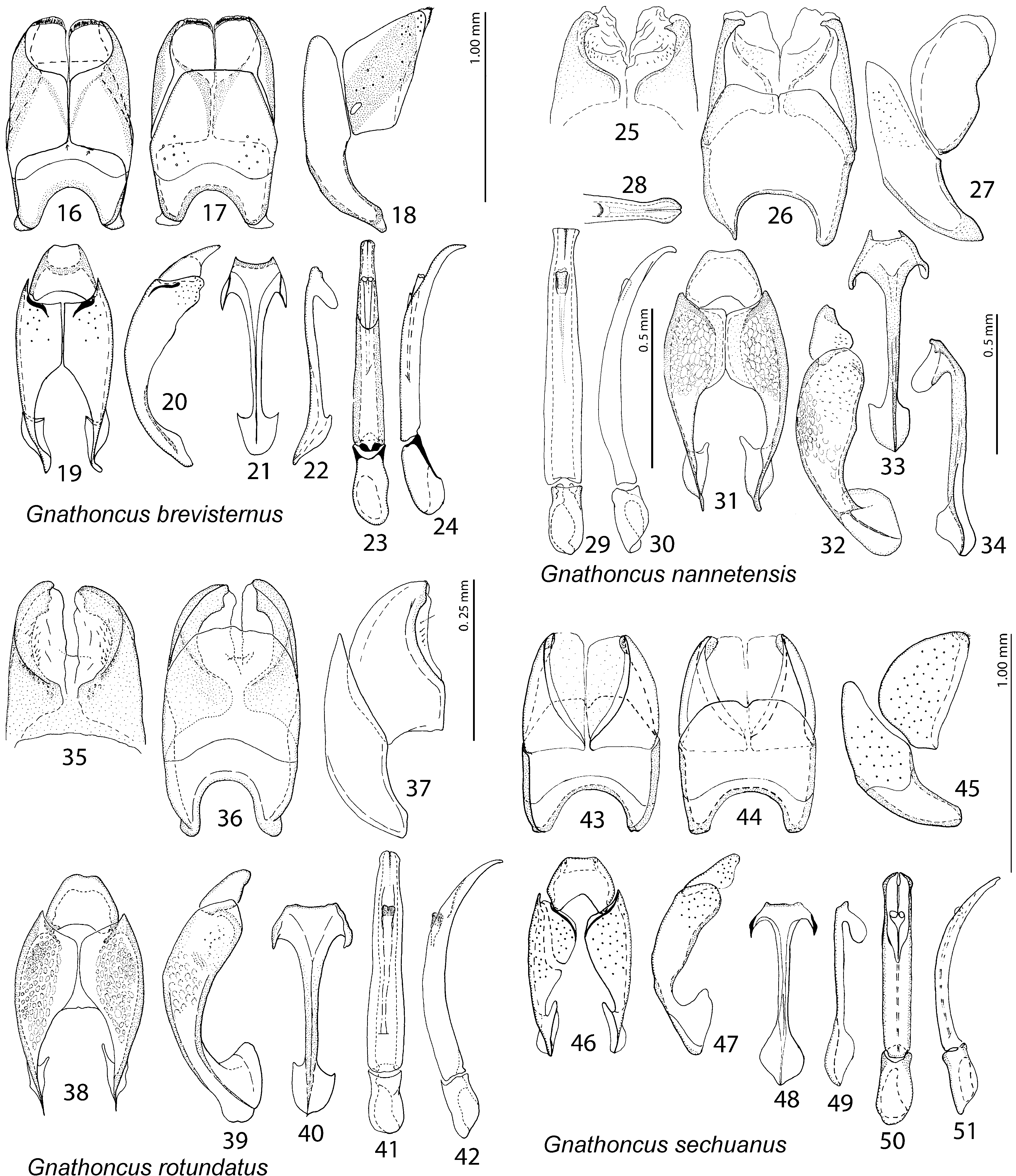
( Figs 4, 5 View Figs 1–5 , 43–51 View Figs 16–51 )
Type locality. China: Sichuan, Jiuzhaigou env., Zhongchacun.
Type material examined. HοΓοτΥΡΕ: ♁, side-mounted on a triangular mounting card, with the genitalia extracted and disarticulated, glued to the same triangular card as the specimen,‘ China, N Sichuan | Jiuzhaigou env. | Zhongchacun | 33°17’13”N 103°50’1”E | 9.-13.VII.2017, 2400- 3000m | lgt. Ondřej Konvička’ [printed] || ‘ Gnathoncus | spec. nov. | Det. T. Lackner 2018’ [printed-written] || ‘ S. Mazur | O.K.!’ [printed-written] || ‘ Gnathoncus | sechuanus sp. nov. | HOLOTYPE | det. T. Lackner 2019 ’ [red label, written] ( ZSM). GoogleMaps PΑRΑτΥΡΕ: ♁, with extracted genitalia glued to the same mounting card as the specimen, locality label identical to holotype ( OKZC).
Description. Body ( Fig. 4 View Figs 1–5 ): PEL: 2.40–2.50 mm; APW: 0.50–0.55 mm; PPW: 0.90–1.00 mm; EL: 1.70–1.80 mm; EW: 1.90–2.00 mm.
Body roundly oval, black, body appendages dark brown, antennal club lighter, reddish-brown. Head and clypeus evenly punctate, punctures separated by several times their diameter; antennal scape with microsetae; sensory structures of the antennal club not examined; other mouthparts not examined.
Pronotum ( Fig. 4 View Figs 1–5 ) narrowing anteriorly, marginal pronotal stria on basal half slightly distanced from pronotal margin, stopping short of pronotal base. Entire pronotal disc with punctation, laterally punctures very dense and almost confluent, becoming finer and sparser medially, where they are separated by several times their diameter. Pronotal hypomeron asetose; along pronotal base present double row of larger punctures; scutellum small, triangular.
Elytra widest in middle, elytral epipleuron with two rows of tiny punctures; marginal epipleural stria double, fine; marginal elytral stria thin, complete, continued as very fine and complete apical elytral stria. Humeral elytral stria almost obliterated under very dense aciculate punctures; dorsal elytral stria I significantly shortened basally, reaching apically approximately elytral mid-length; dorsal elytral striae II–III carinate, only slightly shortened basally, stopping short of elytral mid-length; dorsal elytral stria IV basally hooked inwardly, approaching elytral base, between that and sutural elytral stria present characteristic short hooked appendix; sutural elytral stria basally hooked inwardly, stopping short of elytral mid-length. Elytral intervals 1–2, and partly also 3 with scattered fine punctures and dense alutaceous microsculpture; basal half of third elytral interval and space among fourth dorsal elytral and sutural striae without such microsculpture creating ‘mirror’ covered only with scattered microscopic punctures. Otherwise entire elytral disc, with exception of elytral base and extreme apex with variously dense punctures and strong alutaceous microsculpture creating almost matte appearance; punctation becoming denser apically.
Propygidium and pygidium densely punctate, punctures of pygidium separated by less than their diameter, interspaces with microsculpture.
Prosternum. Prosternal process ( Fig. 5 View Figs 1–5 ) basally widened, strongly convergent apically, carinal prosternal striae on basal half sub-parallel, thence strongly convergent apically, united in tiny prosternal fovea; lateral prosternal striae carinate, very short, reaching middle of prosternal process.
Mesoventrite ( Fig. 5 View Figs 1–5 ) approximately 3.5 times as broad as long, with variously-sized punctures; meso-metaventral stria undulate, situated slightly anterad of meso-metaventral suture. Metaventrite along sides and base with large deep punctures, medially punctation becomes sparser and finer; lateral metaventral stria well developed, carinate, stopping short of metacoxa. Metepisternum with four rows of very dense elliptical punctures; intercoxal disc of first visible abdominal ventrite completely striate laterally, laterally with large dense punctures becoming sparser and finer medially.
Legs. Protibia on outer margin with 4–5 low teeth topped by tiny denticle, followed by several minute denticles; protibial stria carinate, complete; protarsal groove shallow; surface between outer protibial margin and protibial stria with microscopic strioles; setae of median row regular, short, situated on definite stria; protibial spur short, hooked, articulated near tarsal insertion. Posterior surface of protibia convex, covered with tiny strioles, posterior protibial stria complete; inner row of setae double; protibial apex with three tiny denticles. Mesotibia slender, on outer margin with seven thin denticles growing in size in proximal direction; mesotibial spur short, stout; metatibia similar to mesotibia, but on outer margin medially with two tiny denticles, another two longer denticles present apically, near tarsal insertion.
Male genitalia. Eighth sternite ( Figs 43–44 View Figs 16–51 ) widely separated medially, apex with large almost asetose velum; eighth tergite ( Fig. 44 View Figs 16–51 ) outwardly arcuate, medially with tiny “notch”; eight sternite and tergite not fused laterally ( Fig. 45 View Figs 16–51 ). Ninth tergite ( Figs 46–47 View Figs 16–51 ) separated medially, densely covered with pores and pseudopores; tenth tergite ( Fig. 46 View Figs 16–51 ) basally deeply inwardly arcuate. Ninth sternite (spiculum gastrale) with both “head” and “tail” developed, “tail” outwardly arcuate and pointed apically ( Figs 48–49 View Figs 16–51 ); “head” ( Fig. 48 View Figs 16–51 ) rather slender, heavily sclerotized laterally. Aedeagus ( Figs 50–51 View Figs 16–51 ) strongly sclerotized on flanks, almost parallel-sided, bluntly pointed apically. Parameres fused in basal two-thirds approximately.
Differential diagnosis. This species is easily distinguishable from other congeners by dense microsculpture of elytra.
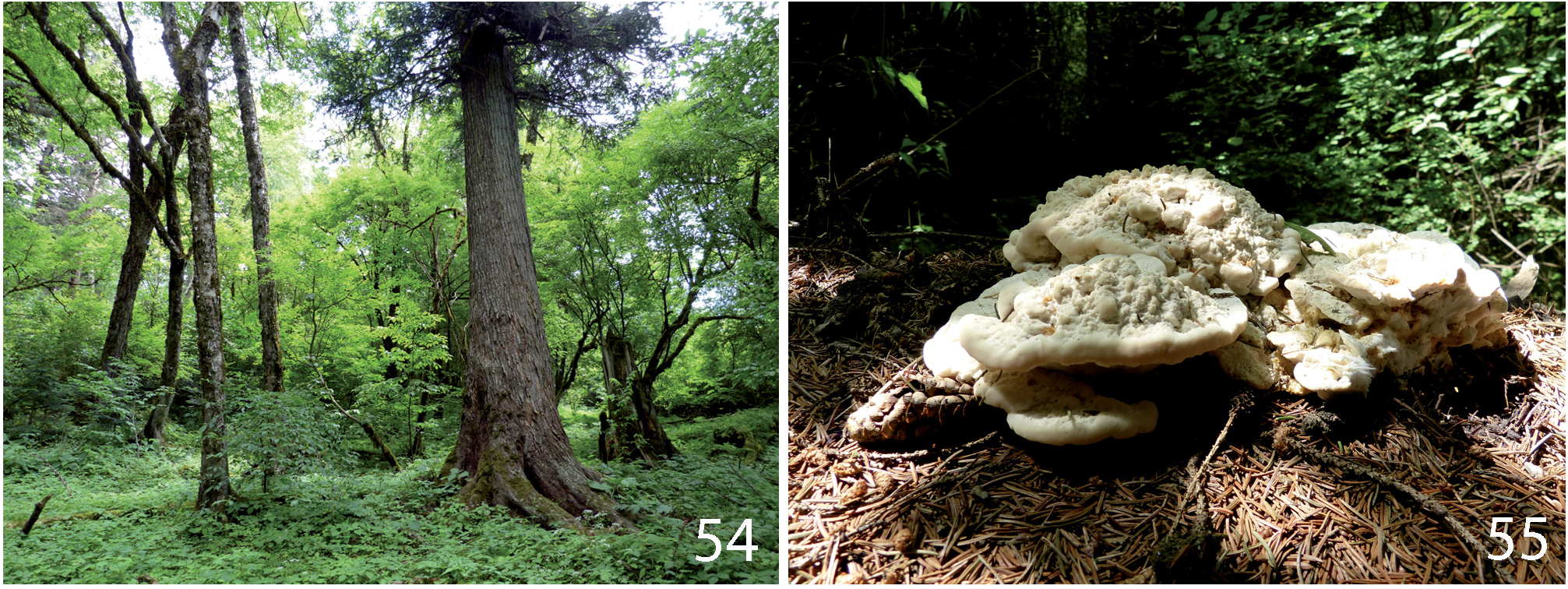
Biology. Found in a mixed forest on the underside of a mushroom ( Bondarzewia sp.?) growing on a tree trunk ( Figs 54–55 View Figs ).
Distribution. China: Sichuan; known only from the type locality.
| T |
Tavera, Department of Geology and Geophysics |
| ZSM |
Bavarian State Collection of Zoology |
No known copyright restrictions apply. See Agosti, D., Egloff, W., 2009. Taxonomic information exchange and copyright: the Plazi approach. BMC Research Notes 2009, 2:53 for further explanation.
|
Kingdom |
|
|
Phylum |
|
|
Class |
|
|
Order |
|
|
Family |
|
|
Genus |