
Lathahypossia Schweitzer, Artal, Van Bakel, Jagt & Karasawa, 2007
|
publication ID |
https://doi.org/ 10.11646/zootaxa.5351.2.4 |
|
publication LSID |
lsid:zoobank.org:pub:BFA5DC03-2B14-4CB1-9B55-C3E973C3FEE9 |
|
DOI |
https://doi.org/10.5281/zenodo.8399750 |
|
persistent identifier |
https://treatment.plazi.org/id/039AB01C-FFEA-FFAE-42E6-DC67AB675BCB |
|
treatment provided by |
Plazi |
|
scientific name |
Lathahypossia Schweitzer, Artal, Van Bakel, Jagt & Karasawa, 2007 |
| status |
|
Genus Lathahypossia Schweitzer, Artal, Van Bakel, Jagt & Karasawa, 2007
Type species. Titanocarcinus aculeatus Busulini, Tessier & Visentin, 1984 View in CoL , by monotypy. Gender feminine.
Species included. Lathahypossia aculeata ( Busulini, Tessier & Visentin, 1984) ; L. campolongensis ( Beschin, Busulini, Fornaciari, Papazzoni & Tessier, 2018) n. comb.
Remarks. When Schweitzer et al. (2007: 293) erected Lathahypossia , they noted that it differs from Hypothalassia mainly in the carapace being proportionally much wider than long with the surface covered with coarse granules (versus carapace proportionally narrower, with many sharp spines on the dorsal surface), and the frontal margin being armed with three spines on each side of the median notch (versus with two supplementary spines).
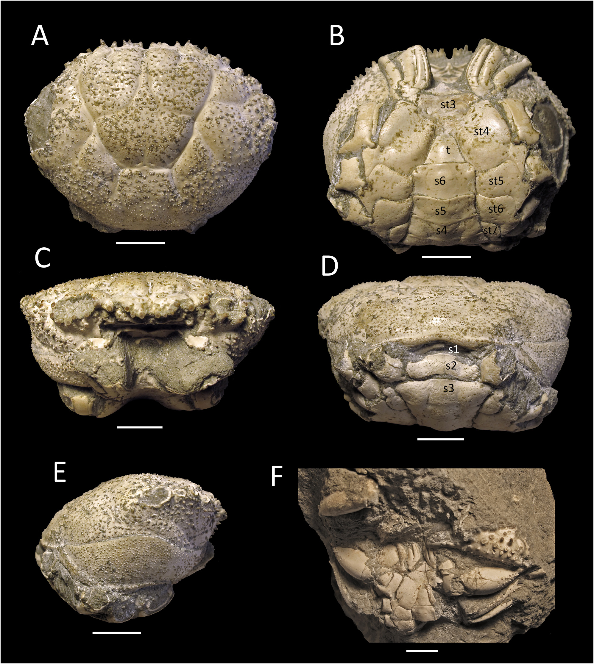
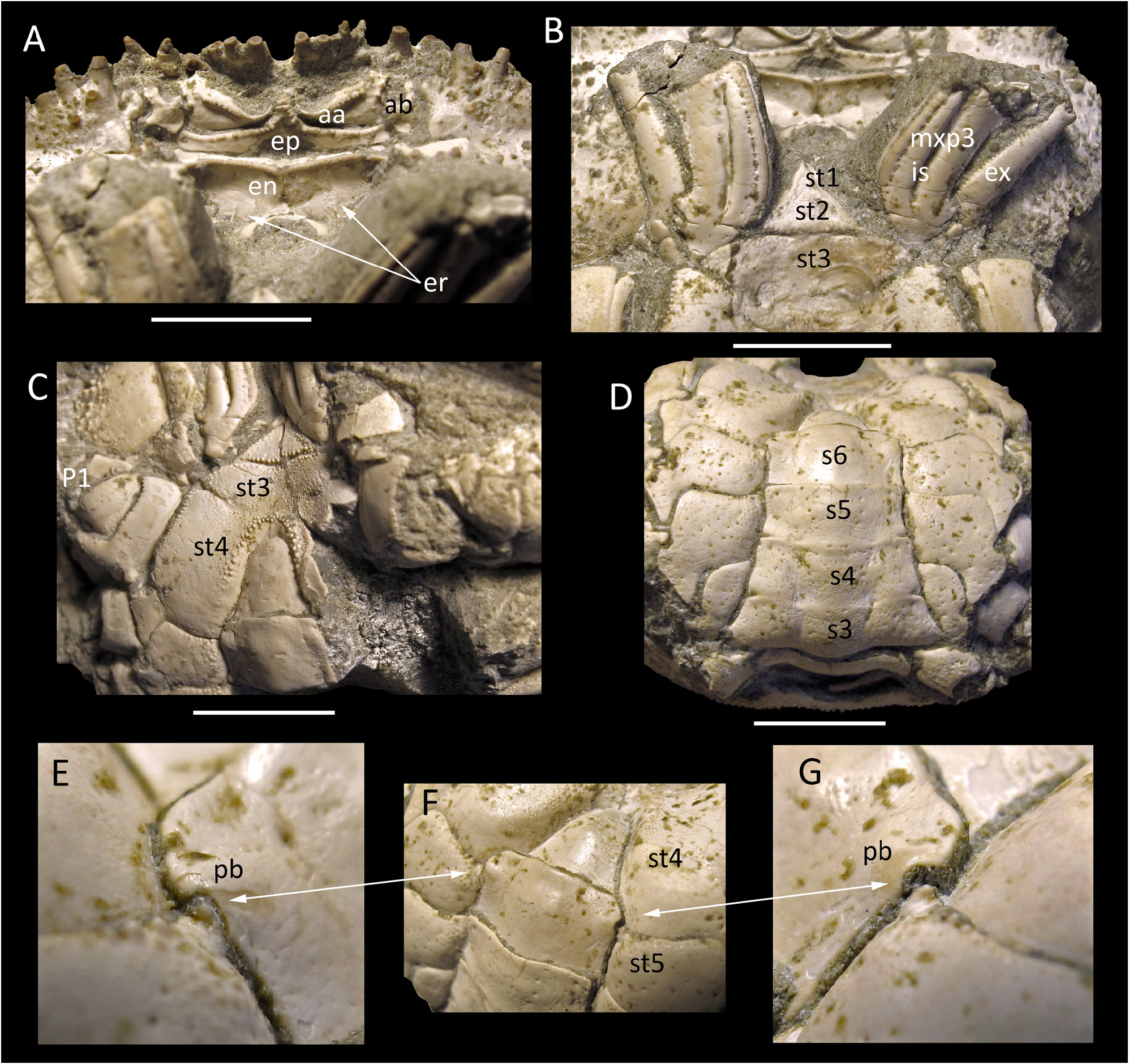
Whereas Lathahypossia has a dorsal appearance reminiscent of Hypothalassia , especially in the spinose ornamentation of the carapace and pereopods, these similarities are only superficial. A detailed study of the present material shows that there are significant differences between Lathahypossia and Hypothalassia that demonstrate they are not closely related: (i) the posterior carapace margin is smooth and entire in Lathahypossia ( Fig. 2D View FIGURE 2 ) (versus posterior carapace margin with a submarginal groove in Hypothalassia ; Ng & Davie 2020: fig. 3B); (ii) the sternopleonal cavity of Lathahypossia is deep and narrow, with the anterior lateral margins of the cavity angular, sloped, with edges granulated, reaching anteriorly beyond the level of the midlength of the P1 coxae ( Figs. 2B, F View FIGURE 2 , 3C View FIGURE 3 ) (versus sternopleonal cavity relatively shallower, smooth, barely reaching anteriorly to level of the anterior edges of the P1 coxae ( Koh & Ng 2000: figs. 4a, b, 7b; Ng & Davie 2020, fig. 6B, H); (iii) the thoracic suture 3/ 4 in Lathahypossia is only visible laterally and is interrupted medially ( Figs. 2B, F View FIGURE 2 , 3C View FIGURE 3 ) (suture distinct, deep and complete in Hypothalassia ; Ng & Davie 2020, fig. 6B); (iv) the sternal tubercle of the male pleonal locking mechanism is on the proximal edge of sternite 5, adjacent to suture with sternite 4 ( Figs. 3E–G View FIGURE 3 ) (versus tubercle on distal edge of sternite 5 adjacent to suture with sternite 6 in Hypothalassia ; Ng & Davie 2020: fig. 7B); (v) the proximal margin of the male telson is distinctly narrower than somite 6 ( Figs. 2B View FIGURE 2 , 3F View FIGURE 3 ) (versus male telson wider, with the proximal margin as wide as the distal margin of somite 6 in Hypothalassia ; Ng & Davie 2020: fig. 6H); and (vi) the male pleon of Lathahypossia has the somites 3–5 distinctly fused, with the sutures separating them shallow ( Figs. 2B View FIGURE 2 , 3D View FIGURE 3 ) (versus somites 3–5 completely free in Hypothalassia ; Davie et al. 2015b: 1079; Ng & Davie 2020, fig. 6H).
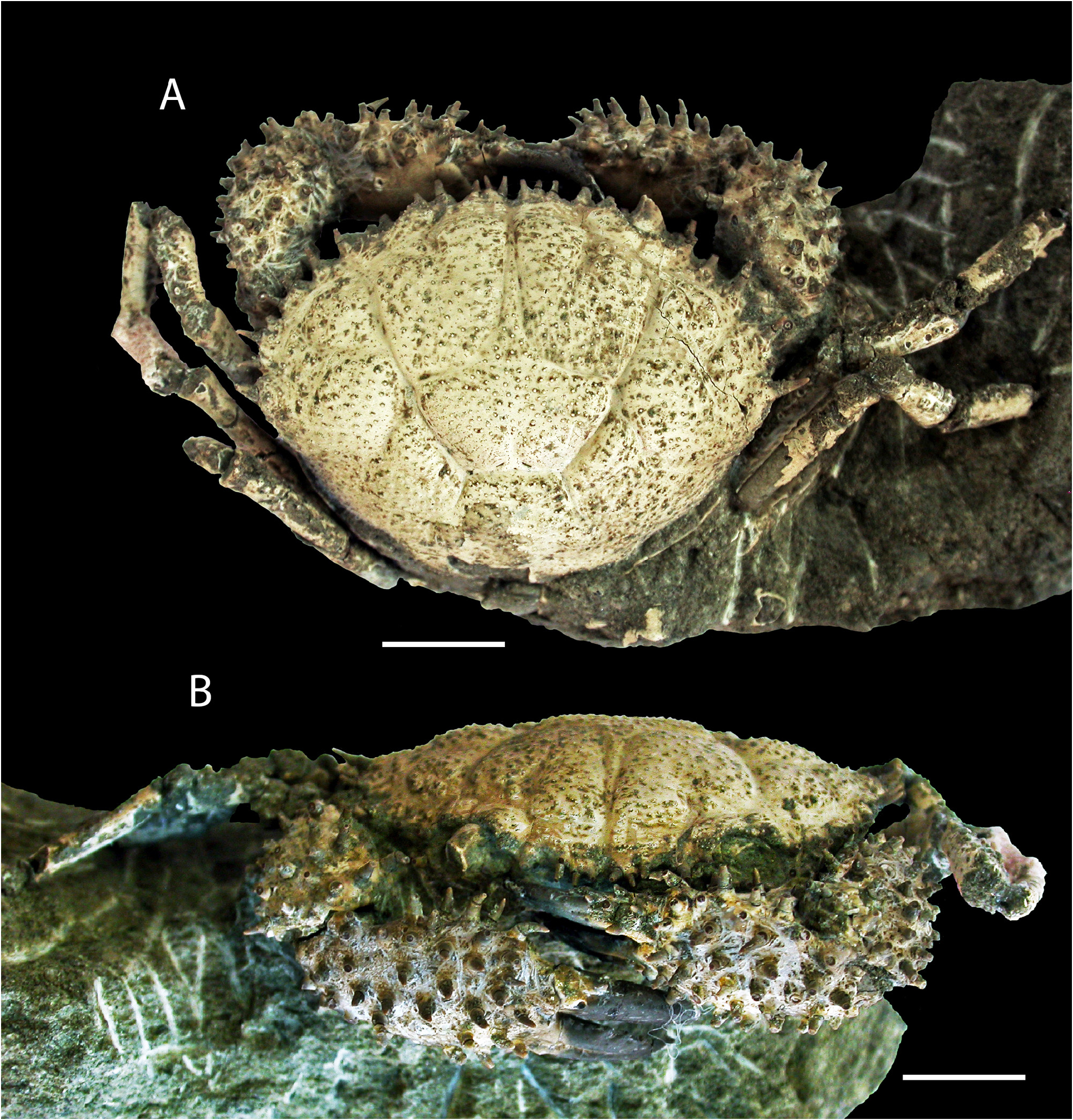
The anteriorly positioned sternopleonal cavity, which reaches well into sternite 4 to a level between the coxae of the chelipeds, is a character not typical of eriphioids and not observed in Hypothalassia and the Hypothalassiidae . All families of the Eriphioidea have the sternal tubercle of the male pleonal locking mechanism usually on the posterior part of sternite 5, adjacent to suture 5/6 (as in Hypothalassiidae ), with in some families on the middle of sternite 5 (cf. Ng & Davie 2020: table 1). This is in sharp contrast to that of Lathahypossia , which has the tubercle more anterior in position, adjacent to suture 4/5. Most significantly, male pleonal somites 3–5 are clearly fused, even though the sutures between them are shallow and just visible ( Fig. 3D View FIGURE 3 ). This is almost identical to the condition observed in xanthid genera such as Neoxanthias Ward, 1932 , Ladomedaeus Števčič, 2005 , Glyptocarcinus Takeda, 1973 , Antrocarcinus Ng & Chia, 1994 and Cyrtocarcinus Ng & Chia, 1994 in which the sutures demarcating somites 3–5 are visible to varying degrees but the somites are nevertheless immovably fused (see Ng & Chia 1994; Manuel-Santos & Ng 2007). Therefore, Lathahypossia cannot be an eriphioid, a group in which all the male pleonal somites are freely articulating. The lateral walls of the carapace are not well preserved, but Lathahypossia does not appear to have any pleurites ( Figs. 3D, E View FIGURE 3 ), and that again is the usual condition for xanthoids. In addition, in Lathahypossia , the carapace is distinctly more ovate ( Figs. 1A View FIGURE 1 , 2A View FIGURE 2 ) (versus more hexagonal in Hypothalassia ; Koh & Ng, 2000: figs. 1a, b, 2a, b, 3, 6a, b), and the cervical grooves reach more posteriorly to below the middle part of the carapace in Lathahypossia , with the branchiocardiac grooves deep and distinct ( Figs. 1A View FIGURE 1 , 2A View FIGURE 2 ) (versus cervical grooves only reach to the median part of the carapace, with the branchiocardiac grooves very shallow in Hypothalassia ; Koh & Ng 2000: figs. 1–3, 6).
In conclusion, all the characters point to Lathahypossia being a member of the Xanthidae sensu stricto, and not Hypothalassiidae . As for the two species of Lathahypossia currently recognized, L. aculeata is superficially similar to some species of Pilumnoidea, which has already been pointed out by Beschin & De Angeli (2004: 20, 21): the carapace is dorsally vaulted with spinose frontal and anterolateral margins, and strongly spinose chelae, features that resemble extant pilumnid species such as Pilumnus dofleini Balss, 1933 , P. acanthosoma Ng, 2000 and P. armatus Komai & Motoh, 2012 from the Indo-Pacific, and American taxa such as P. townsendi Rathbun, 1923 , P. spinohirsutus ( Lockington, 1877) , P. spinosissimus Rathbun, 1898 , and P. sayi Rathbun, 1897 (cf. Rathbun 1930: text-fig. 79, pls. 201 figs. 4–7, 203, 204 figs. 1, 2; Ng 2000: figs. 3, 4a; Komai & Motoh 2012: figs. 1, 8). These pilumnids, however, are all quite small, with a carapace width less than 30 mm. More significantly, all pilumnoids have male pleonal somites 3–5 completely free. The eriphiids, Eriphia verrucosa ( Forskål, 1775) (from the Mediterranean Sea) and Eriphides hispida ( Stimpson, 1860) (North America), are also similarly spinose, but the spines on their chelae are not as long and their carapace shape is distinctly more trapezoidal (cf. Rathbun 1930: pls. 225, 226; Koh & Ng 2008: fig. 1). The very spinose carapace and chelipeds of Lathahypossia also resemble the poorly known Pilumnus palmeri Garth, 1986 , from Ecuador, for which Števčić (2011) established a new genus, Garthopilumnus , and new family Garthopilumnidae , even though he did not examine the specimens. Ng et al. (2008: 142) and Poore & Ahyong (2023) opted to tentatively retain it in Pilumnidae . The second author examined the material in the Los Angeles County Museum of Natural History and the types are juveniles and in poor condition. The figures of the species by Garth (1986) are somewhat schematic and most of the sternal elements are damaged. Nevertheless, in general, the carapace shape of Pilumnus palmeri is distinctly trapezoidal, the frontal margin is trilobed, the gastric regions are prominently raised and the ambulatory legs are unarmed. Garth (1986: fig. 4) figured the male pleon as having all somites free but as his specimen is a juvenile male, the adult character state and the significance of this cannot be ascertained.
Hypothalassia campolongensis described from the Upper Eocene of Campolongo (Vicenza, northeastern Italy), is here referred to Lathahypossia for the first time. The species possesses a subovate, vaulted dorsal carapace surface with a spiny bilobed front that is notched medially, and spinose anterolateral margins: all features of Lathahypossia . It also has the same morphology of the carapace regions, which are demarcated by very shallow grooves, and the shape of the gastric process is the same. These features clearly differ from those of Hypothalassia , as stated above, and excludes it from that genus. It is actually better placed in Lathahypossia and the Xanthidae (see Beschin et al. 2018: 196–198, figs. 132, 133a, b). Unfortunately, the ventral surfaces and male pleonal condition of this species are not known.
Considering the extant xanthoid fauna with focus on “spiny crabs”, there are few taxa quite like Lathahypossia , or Hypothalassia for that matter. For Xanthoidea s. str., in which male pleonal somites 3–5 are functionally fused, the closest perhaps are the three species now in the American xanthid genus Heteractaea Lockington, 1877 but they are also relatively small crabs with a carapace width less than 30 mm (cf. Rathbun 1930: pl. 212 fig. 6, pl. 213).
No known copyright restrictions apply. See Agosti, D., Egloff, W., 2009. Taxonomic information exchange and copyright: the Plazi approach. BMC Research Notes 2009, 2:53 for further explanation.