
Anatosuchus minor Sereno et al., 2003
|
publication ID |
https://doi.org/ 10.3897/zookeys.28.325 |
|
publication LSID |
lsid:zoobank.org:pub:A979ECDE-871F-4AFC-9ABA-63A0FD6DC323 |
|
DOI |
https://doi.org/10.5281/zenodo.3790365 |
|
persistent identifier |
https://treatment.plazi.org/id/039B2B68-FF80-9B76-A5FA-B27FFDAC50CD |
|
treatment provided by |
Plazi |
|
scientific name |
Anatosuchus minor Sereno et al., 2003 |
| status |
|
Anatosuchus minor Sereno et al., 2003
Figs. 4 View Figure 4 –10, 12, 13
Tables 2–6
Sereno et al. (2001, figs. 1, 2)
Holotype. MNN GAD603 ; nearly complete skull with lower jaws of a subadult individual; margins of the skull are eroded away. The holotype was previously catalogued as “ GDF603 ” ( Sereno et al. 2003).
Type locality. Gadoufaoua, Agadez District, Niger Republic (N 16° 46’, E 9° 22’) (Fig. 1A, C).
Horizon. Elrhaz Formation, Tegama Series; Lower Cretaceous (Aptian-Albian), ca. 110 Mya ( Taquet 1976). In association with a diverse dinosaurian fauna ( Taquet 1976; Sereno et al. 1998, 1999, 2007; Taquet and Russell 1999; Sereno and Brusatte 2008) and the crocodyliforms Sarcosuchus imperator ( Broin and Taquet 1966; Sereno et al. 2001), Araripesuchus wegeneri (Buffetaut and Taquet 1979) , and Stolokrosuchus lapparenti ( Larsson and Gado 2000) . At a single field locality (G109), specimens were recovered that are referable to Anatosuchus minor ( MNN GAD 18) and Araripesuchus wegeneri ( MNN GAD 19).
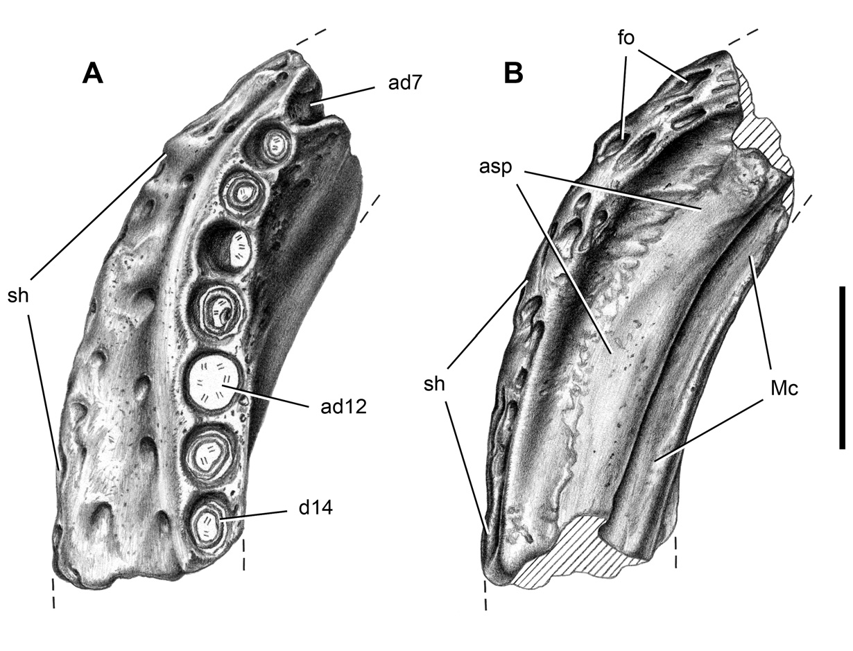
Referred material. MNN GAD 17 ( Figs. 4–8 View Figure 4 View Figure 5 View Figure 6 View Figure 7 View Figure 8 , 12, 13), nearly complete skull with lower jaws lacking only the anterolateral corner of the snout in articulation with a postcranial skeleton lacking the right pectoral girdle and forelimb, most of both hind limbs, sacrum, and tail; MNN GAD 18 ( Fig. 9 View Figure 9 ), mid-section of the left dentary preserving alveoli 7–14 and the anterior tip of the left splenial.
Revised diagnosis. Small-bodied metasuchian (<1.0 m) with low transversely expanded snout that forms the broadest portion of the cranium, broad-based anteriorly projecting pointed internarial bar, lenticular-shaped external nares, elevated narial bridge which expands transversely behind the external nares, prominent median edentulous dentary margin, laterally projecting vascularized dentary shelf on parasagittal portion of dentary ramus, enlarged neurovascular foramina located along the anterior snout margin, anterior snout margin smooth, vertical and sharply defined on the premaxilla and maxilla, oval splenial fenestra on the anterior transverse portion of the lower jaw, six premaxillary teeth, premaxillary and anterior maxillary tooth row that angles ventrolaterally toward the corner of the snout at approximately 25°, largest upper and lower teeth positioned along the bend in the L-shaped tooth row (m4, d12), three pairs of cervical osteoderms that decrease in size posteriorly, large manus (30% skull length), elongate poorly recurved manual unguals on digits I-III, and manual digit IV with six phalanges.
The initial description was based on an immature skull embedded in a hematitic concretion ( MNN GAD 603). The concretion was discovered on the surface with prominent edges of the skull, such as the anterior end of the snout, trimmed by erosion ( Sereno et al. 2003). Th e likeness drawn between Anatosuchus and the South American genus Comahuesuchus was based on a few seemingly unique features, such as a diastema between the premaxillary tooth rows, which we can now say arose in the immature skull of Anatosuchus as an artifact of erosion. Th e revised diagnosis is based mainly on a referred adult skull and partial articulated postcranium ( MNN GAD 17) that preserves an intact portion of the paravertebral shield ( Fig. 4 View Figure 4 ). Th is well preserved skull was found embedded in sandstone, the right corner of the snout, right limbs, sacrum and tail lost to erosion. Th e additional information available for both Anatosuchus and Comahuesuchus confirms Martinelli’s (2003) view that these genera are not closest relatives among known notosuchians.
Dorsal skull roof. In A. minor the snout becomes relatively broader and longer during growth. In the juvenile holotype specimen MNN GAD603 , the width of the skull across the rounded anterior corner of the snout is subequal to that across the suborbital ramus of the jugal ( Sereno et al. 2003). Preorbital length, in addition, is subequal to that of the remainder of the skull. In mature individuals, in contrast, the anterior snout corner is the broadest region of the skull, and preorbital length is approximately 20% greater than the posterior portion of the skull ( MNN GAD17 ; Figs. 5 View Figure 5 , 6 View Figure 6 ; Tables 2, 3). Th e following description is based primarily on this specimen .
The premaxilla is a broad bone housing six recurved teeth. Th e base of the internarial process is broad, unlike that in Araripesuchus , but similar in this regard to Simosuchus ( Buckley et al. 2000) . It extends anteriorly at approximately 30° above the horizontal, and tapers to a point, where it joins at a sharp angle the nearly horizontal internarial process of the nasal ( Figs. 5 View Figure 5 , 6 View Figure 6 ). Th e external nares, as a result, are dorsoventrally compressed and appear as a narrow slit in lateral view. In dorsal view, the external nares are elliptical, the floor of the narial passage broadly exposed to each side of the tapering internarial process of the nasal. The floor of the narial passage, which is formed by the premaxilla, is raised and slightly extended anterolaterally by a short tongue-shaped flange ( Figs. 5B View Figure 5 , 6B View Figure 6 , 7A View Figure 7 ). The anterior half of the external nares projects beyond the first premaxillary tooth, a narial structure that projects anteriorly more prominently than in any other crocodyliform.
The narial fossa is clearly demarcated as a smooth subtriangular surface located lateral to the external nares and restricted to the premaxilla. In glancing light, a subtle division of the surface is visible. A teardrop-shaped fossa within the narial fossa is the largest surface, its tip emerging from under the lip of the rim of the external naris. In ventral view, the anterior projection is smooth and incorporates into the narial fossa the alveolar margin dorsal to premaxillary teeth 1–3. The lateral margin of the narial fossa is delimited by a shallow trough from the smooth, highly vascularized, vertical alveolar margin, which extends laterally toward the premaxilla-maxilla suture. No other crocodyliform known thus far closely approaches the form and orientation of the external nares in A. minor .
The remainder of the external surface of the premaxilla can be divided into the alveolar margin and the ramus that tapers between the nasal and maxilla. The alveolar
are measured on left side except as indicated.
margin faces primarily anteriorly, has a vertical orientation, and is gently transversely convex ( Fig. 7A, B View Figure 7 ). As in Araripesuchus wegeneri , two large neurovascular foramina are situated between the narial fossa and the premaxilla-maxilla foramen. The ventral margin is scalloped to match the position of the lateral three premaxillary teeth ( Fig. 7B View Figure 7 ) as occurs in Simosuchus , but unlike the straight margin in Araripesuchus . The dorsal margin meets the dorsal surface of the snout at nearly a right angle along a rugose edge. Small foramina and grooves for impressed vessels are visible on the dorsal surface of the snout near the narial fossa and alveolar margin. Th at texture becomes deeply pitted as the premaxilla tapers to a point on the lateral aspect of the nasal bridge.
In ventral view, the premaxilla is divided between the transversely convex surface of the internarial bar, the raised edges of the alveoli that scallop the alveolar margin, and the flat palatal surface, which is only partially exposed ( Figs. 5C View Figure 5 , 6C View Figure 6 ).
The maxilla is the most expansive bone in the skull and forms most of the snout. Its external surface is composed of a narrow alveolar margin and broader posterodorsal and posteroventral rami that extend above and below the antorbital opening, respectively. Like the premaxilla, the alveolar surface is vertical ( Fig. 7B View Figure 7 ). It faces anterolaterally, borders the premaxilla-maxilla foramen, and gives passage to one additional large neurovascular foramen. Th e dorsal edge protrudes over this foramen before curving posteroventrally to join the scalloped ventral margin near the overhanging corner of the snout adjacent to the fourth maxillary tooth. Several large foramina are present just above this edge on the corner of the snout ( Fig. 7B View Figure 7 ).
The dorsal surface of the maxilla remains lightly textured along a band near the sharp anterior margin of the snout from the narial fossa to the anterolateral corner. This same low texture is present across the posteroventral ramus lateral to the antorbital depression, a muted textural pattern that resembles that seen in Simosuchus . In both taxa most of the maxilla below the antorbital opening is only lightly textured. In Araripesuchus , by contrast, the comparable region of the maxilla above m3 and m4 is more deeply sculpted with pits (Figs. 14A, 15A). As in most crocodyliforms, in A. minor a row of neurovascular foramina runs above the alveolar margin along the posteroventral ramus, although these are smaller than those at the anterior end of the snout. The maxilla forms the smooth and elongate anterior wall of the antorbital fossa, which is pierced by a foramen ( Fig. 7C View Figure 7 ). Th e posterodorsal ramus of the maxilla is deeply pitted and slightly elevated as it passes over the antorbital depression to join the lacrimal and prefrontal.
The nasal extends from the tip of the internarial bar anteriorly to a subquadrate process posteriorly. Th e texture is reduced on the nasals immediately posterior to the external nares. Nonetheless, shallow sculpting is present, and the nasals do not contribute to the smooth narial fossa, which is isolated on the premaxilla as in Araripesuchus (Fig. 16A), Simosuchus ( Buckley et al. 2000) and other crocodyliforms. The elevated nasal bridge is narrowest in width at mid-length along the snout, after which it broadens slightly to equal interorbital width ( Figs. 5B View Figure 5 , 6B View Figure 6 ). A narrow median trough is present from mid-snout to the subrectangular interdigitating ends of the nasals.
The L-shaped lacrimal has anterior and ventral rami, which join near a laterally prominent process for articulation with a missing anterior palpebral ( Fig. 7C View Figure 7 ). The lacrimal foramen is tucked under this process within the orbit. Th e anterior ramus is deeply pitted and joins the maxilla along a subrectangular suture. Th e ventral ramus is smooth and divided into an orbital margin and medially inset posterior margin of the antorbital fossa.
The palpebrals are disarticulated in both known skulls. In the adult skull, however, they have fallen into orbital and temporal spaces, where they are partially exposed. A pair of articular fossae, the anterior on the lacrimal and prefrontal and the posterior on the postorbital, supported anterior and posterior palpebrals, respectively, as in many crocodyliforms ( Fig. 7C, D View Figure 7 ). Th e prefrontal-frontal suture courses anteriorly, extending parallel to the inset of the fossa for the anterior palpebral. Th e prefrontal narrows in mid-section, where it contacts the lacrimal, and then extends anteriorly to contact the maxilla, effectively separating the nasal and lacrimal. Th e prefrontal pillar angles ventromedially and slightly posteriorly, tapering strongly from the skull roof to the palate.
The frontal and parietal are fused to their opposites and joined to each other by an interdigitating frontoparietal suture in both the adult and subadult skulls. The deeply pitted frontals have a median crest. The flat skull table formed by the parietals is also deeply pitted and separates the supratemporal fossae to a greater degree than in Simosuchus ( Buckley et al. 2000) . During growth in A. minor , interorbital width expands relative to the width of the skull table, such that the two measurements are subequal in a subadult ( Sereno et al. 2003) whereas the former is nearly twice the latter in an adult ( Figs. 5B View Figure 5 , 6B View Figure 6 ).
In the adult skull the frontal forms the anteromedial rim and distinctive corner of the supratemporal fossa, which is not the case in the subadult skull. Th at corner, in addition, is invaded by diverticulae from the supratemporal fossa. Although there is a similar corner in the rim of the fossa in Araripesuchus wegeneri , the rim is not undercut by pneumatic invagination. Simosuchus , on the other hand, has diverticulae resembling the condition in A. minor that undercut the anterior rim of the supratemporal fossa, a condition that has arisen a few times among crocodyliforms.
The frontal contributes to the rim of the supratemporal fossa and reaches the fossa in dorsal view. Frontal participation in these supratemporal structures seems to occur with maturity, given the exclusion of the frontal in a subadult skull ( Sereno et al. 2003). Th e posterior margin of the skull table is scalloped to each side of a short posteromedian projection formed by the supraoccipital, which joins the parietals along a shallow V-shaped suture. Simosuchus , in contrast, is shown with a nearly straight posteromedian margin. In this case, notching of the posterior margin of the parietals by the supraoccipital may have been obliterated by coossification.
The right side of the skull has rotated slightly posterolaterally, an asymmetry best seen in dorsal view ( Figs. 5B View Figure 5 , 6B View Figure 6 ). Because there is no pattern of postmortem distortion of the skull, this asymmetry appears to be pathological rather than preservational in origin. Th e articular notch for the posterior palpebral on the right side is shifted posterolaterally, altering the shape of the supratemporal fossa. Th e right fossa has a convex lateral margin and its maximum parasagittal length is about 10% longer than the left side.
The postorbital is notched by an articular facet for a small posterior palpebral. The surface of the postorbital between the facet and the supratemporal fossa varies, remaining textured with pits in some species, such as A. gomesii ( Price 1959) and A. tsangatsangana ( Turner 2006) , and smooth in others such as A. patagonicus ( Ortega et al. 2000) . In A. wegeneri that surface between the palpebral facet and supratemporal fossa is smooth and convex (Figs. 14B, 15B).
The squamosal is distinctly triradiate in dorsal view, the anterior process that contacts the postorbital the most slender. The dorsal surface of the anterior process is deeply pitted and depressed to form a shallow arcuate fossa ( Figs. 5B View Figure 5 , 6B View Figure 6 ). The posterior process is offset below the skull table and has a more subdued texture.
The jugal approaches, but does not contact, the posteroventral corner of the antorbital fossa ( Fig. 7C View Figure 7 ). Th e anterior ramus is moderately expanded dorsoventrally toward its anterior end and is deeply pitted, with an oval fossa located beneath the orbit ( Fig. 7D View Figure 7 ). The relatively slender postorbital process is inset at its base, the location for a very small siphonal opening. Th e posterior ramus is also relatively slender under the laterotemporal fenestra, where it terminates in a shallow inset articulation on the quadratojugal.
The L-shaped quadratojugal is partially fused to the quadrate near the quadrate condyle, where it approaches, but does not contribute to, the jaw articulation. The suture with the quadrate shaft is relatively straight, and surface texture is low and limited to the anterior portion of the bone.
Palate. The configuration of palatal sutures, shape and position of the suborbital fenestra, form of the mandibular rami of the pterygoid and ectopterygoid, position of the choanae, and form of the choanal septum ( Figs. 5C View Figure 5 , 6C View Figure 6 ) correspond well with those of Araripesuchus ( Price 1959) (Figs. 14C, 15C) and differ markedly from the palatal configuration described in Simosuchus ( Buckley et al. 2000) . In these regards, A. minor is less derived than Simosuchus .
The premaxillary portion of the palate is restricted to a broad-based triangle near the anterior margin. Th e premaxilla-maxilla suture, however, is exposed only near the alveolar margin. Th e premaxilla-maxilla foramen may communicate with the palate as in A. wegeneri ; a foramen is present at the anterior margin of the maxilla just posterior to the premaxilla-maxilla suture, as is the case on one side of a cranium of A. wegeneri (Figs. 14C, 15C). Furthermore, as in another skull of that species ( Fig. 20B View Figure 20 ), this palatal foramen appears to be associated with the tip of the fourth dentary crown ( MNN GAD 17, GAD 603)
The maxilla and palatine form the majority of the palate in A. minor ( Figs. 5C View Figure 5 , 6C View Figure 6 ). Th e median one-third appears to preserve its natural arching toward the midline, whereas the lateral one-third on each side lies closer to the horizontal. Neither the vomer nor pterygoid are exposed in the midline as in Simosuchus ( Buckley et al. 2000) . A slit-shaped foramen opens on the maxilla. Canted along an anterolateral-posteromedial axis, opening anterolaterally, and associated with a small palatal fossa, the foramen is far from the alveolar margin and may not correspond to maxillary foramina associated with the alveolar margin in other notosuchians.
The pterygoid and ectopterygoid form the posterior portion of the palate, including the posteroventrally projecting mandibular rami. Th e distal end of this process is modestly expanded as in Araripesuchus and lies in its natural position adjacent to the adductor fossa of the lower jaw. The ectopterygoid overlaps the ventral aspect of the pterygoid on the lateral edge of the palate.
The suborbital fenestra, which is best exposed in the subadult skull ( Sereno et al. 2003), is subequal in size to the paired choanae and located farther anteriorly. The palatine-pterygoid suture, preserved on the right side, courses across a broad palatal border lateral to the choanae. In the midline of the adult skull, the posterior one-half of the very thin choanal septum is exposed, the remainder covered from view by extraneous bone pieces. Th e choanae are located as far posterior on the pterygoids as possible, butting against a posterior palatal ridge formed by the pterygoids. During growth the sigmoid curve of the posterior palatal ridge in the subadult becomes a broad arch in the adult ( Figs. 5C View Figure 5 , 6C View Figure 6 ). Unlike in some other species of Araripesuchus ( A. gomesii , A. wegeneri ), there is no development of a pair of parasagittal flanges extending from the posterior palatal ridge.
The quadrate angles posteroventrally from the recessed otic region toward the quadrate condyles. In the otic region, a large opening constitutes the fenestra ovalis and confluent cranioquadrate passage. Anterior to this opening is the preotic siphonium, ventral to which is a circular fossa ( Fig. 7D View Figure 7 ) as in Araripesuchus wegeneri .
A sharp vertical crest on the quadrate contributes to the posterior skull margin, joining the paroccipital process with the rim of the medial condyle. In posterior view, a foramen aërum opens on the posterior aspect of the quadrate shaft just above the medial condyle ( Fig. 8 View Figure 8 ). In lateral view, the posterior margin of the quadrate angles anteroventrally as in Simosuchus ( Buckley et al. 2000) rather than posteroventrally as in nearly all other crocodyliforms. Th e quadrate condyles are relatively flat and separated by a marked V-shaped cleft ( Fig. 7D View Figure 7 ).
Braincase. The braincase is well preserved and exposed in the holotype and referred skulls ( Figs. 5C View Figure 5 , 6C View Figure 6 ). The supraoccipital forms a small median pitted triangle on the dorsal skull roof. On the occiput, the supraoccipital forms a short vertical nuchal keel with broad flanges extending to either side, more closely resembling that in Simosuchus than in Araripesuchus . Th e proatlantal elements are fused together forming an inverted chevron that is preserved in articulation with the protruding dorsal rim of the foramen magnum ( Figs. 5B View Figure 5 , 6B View Figure 6 ). Th e paroccipital processes project to each side, arching ventrolaterally to a sharp edge that connects the squamosal above and quadrate condyles below ( Fig. 8 View Figure 8 ). Th e ends of the paroccipital processes are marked by a series of striations or ridges as in Araripesuchus and extant crocodylians.
The ventrally deflected occipital condyle is formed almost exclusively by the basioccipital. Th e remainder of the bone angles anteroventrally at approximately 45° and forms most of the braincase floor posterior to the palate. A small posterior Eustachian foramen is located in the midline just anterior to the occipital condyle. Farther anteriorly, a median crest rises (larger in the subadult skull), followed by a large anterior Eustachian foramen. Th is circular foramen opens posterodorsally between the basioccipital and basisphenoid. Th e lateral edges of the basioccipital curl against the medial edge of low basal tubera formed by the anterior extremity of the exoccipital.
A large lateral Eustachian foramen opens posterodorsally on the anterior side of each basal tuber between the otoccipital (exoccipital + opisthotic) and basisphenoid. As in Araripesuchus , four foramina are present adjacent to the occipital condyle, the largest an anteroventrally opening foramen for the internal carotid. Along the lateral edge of the braincase, a pair of low crests is present running anteromedially from the quadrate to the pterygoid. In lateral view, the otoccipital extends from the very large cranioquadrate passage anteriorly to the paroccipital process posteriorly, just separating the squamosal and quadrate ( Figs. 5A View Figure 5 , 6A View Figure 6 ). Th e basisphenoid has only a narrow, V-shaped ventral exposure. It floors a narrow depression between the pair of lateral crests and a small median patch between the basioccipital and the posterior margin of the palate.
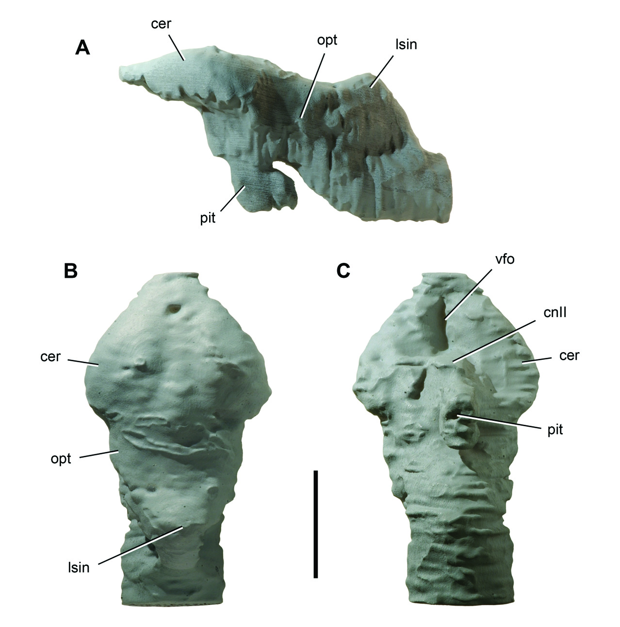
Endocast. An endocast, generated from the computed-tomographic scan of cranium MNN GAD 17 (Fig. 10), closely resembles that for Araripesuchus ( Fig. 22 View Figure 22 ). In both the cerebral hemispheres are spade-shaped as seen in dorsal view and measure ap-
Figure |0. Endocast of the crocodyliform Anatosuchus minor . Endocast ( UCRC PVC 2) prototyped from a computed-tomography scan of skull MNN GAD 18. Th e endocast lacks a portion of the pituitary fossa and right and left labyrinths. A Lateral view. B Dorsal view. C Ventral view. Scale bar equals 2 cm. Abbreviations: cer, cerebrum; lsin, longitudinal sinus; opt, optic lobe.
proximately one-half of total endocast length. In general the forebrain in the endocast compares more closely with that reported for Sebecus ( Hopson 1979) than the more rounded, symmetrical cerebral hemispheres in Alligator (Fig. 11) or Caiman ( Hopson 1979). A sagittal venous sinus flanked by shallow longitudinal depressions outlines the medial aspect of each hemisphere. In lateral view, the cerebral hemispheres are compressed dorsoventrally. In A. minor the posterior portion of the hemisphere is a little deeper than in Araripesuchus wegeneri . In ventral view, the absence in A. minor of the ventromedian fossa between the hemispheres observed in A. wegeneri may be an artifact of the quality of the scan. Swellings for optic lobes are visible posterior to the cerebral hemispheres. Although not well preserved in A. minor , the dorsal surface of the cerebellar region is near the height of the cerebral hemispheres.
Dentition. There are 6 premaxillary teeth, 19 maxillary teeth, and 21 dentary teeth, as established on the basis of the exposed teeth and a computed-tomographic scan of skull MNN GAD 17. In a subadult skull ( MNN GAD 603), there are 6 premaxillary teeth,
Figure ||. Endocast of Alligator mississippiensis . Endocast ( UCRC PVC 6) prototyped from a computed-tomography scan of a recent skull ( TMM M-983). A Lateral view. B Dorsal view. C Ventral view. Scale bar equals 1 cm. Abbreviations: asc, anterior semicircular canal; cer, cerebrum; lsc, lateral semicircular canal; lsin, longitudinal sinus; opt, optic lobe; pit, pituitary fossa; psc, posterior semicircular canal.
15 maxillary teeth, and an unknown number of dentary teeth. Sereno et al. (2003) originally reported 5 premaxillary teeth in the subadult skull, although it is now clear that the first premaxillary tooth was broken away on both sides based on comparison with the adult skull. Premaxillary tooth number thus appears to be stable in the final 30% of growth in the skull, while maxillary and probably dentary tooth counts increase by a comparable percentage. Th e lower jaws and tooth rows become much more U-shaped during maturation. The diagnostic breadth of the snout and transverse orientation of the anterior ends of each dentary emerge late in post-hatching growth. On the other hand, the characteristic inclination of the anterior dentition from the midline to the corner of the snout changes very little; the tooth row in anterior view of both subadult and adult skulls is angled at approximately 25° from the horizontal.
Upper and lower crowns are subconical with the base of the crown very slightly expanded from the root. Th e crowns curve lingually. There is no distinct neck or marked constriction between root and crown. All but the first premaxillary crown have unornamented mesial and distal carinae and very fine interweaving striae, which can be seen under strong magnification on the labial side of premaxillary and maxillary crowns. Tooth wear is not nearly as pronounced as in Araripesuchus . There are no wear facets and only a few crown tips with thinned enamel or exposed dentine from apical abrasion.
Six premaxillary teeth are one or two more than common among crocodyliforms. Pm1–3 project ventrally unopposed by dentary teeth, the first of which projects between pm3 and pm4. Th e tip of d4 projects dorsally into a fossa between pm6 and m1 ( MNN GAD 17, GAD 603), a typical dental configuration among crocodyliforms. If the teeth at the junction of premaxilla, maxilla and dentary teeth are regarded as homologous with those in other crocodyliforms, additional premaxillary teeth must have been added to the original plesiomorphic tooth count of four or five teeth, beginning at the medial end of the tooth row.
The crown of pm1 is approximately 20% smaller than the crowns of pm2–6, lacks carinae, and is positioned lateral to the midline. Th e alveolar margins of opposing premaxillae are separated in the midline by a subtriangular gap, such that the opposing first premaxillary crowns are separated by a median diastema approximately twice that between ipsilateral premaxillary crowns.
Premaxillary teeth 2–6 are very similar in size and crown detail. Th e alveoli of all premaxillary teeth are raised as rugose cylinders. Th e inner set of alveoli (pm1–3) are separated by concave intercrown festoons, whereas the raised rim of the alveolus in the outer set (pm4–6) are linked together by a rugose alveolar ridge. Th e festooning of the inner set, thus, is the result of the concave margin between alveoli; festooning in the outer set and in the maxillary series, by contrast, is the result of the dorsally concave labial rim of the alveoli ( Fig. 7B View Figure 7 ).
The mesial premaxillary crowns (pm1–3) are functionally distinctive. They oppose a prominent edentulous edge of the dentary, which is 9 mm in transverse width in the adult skull. As confirmed by computed tomography, the first dentary tooth is positioned 11 mm from the dentary symphysis. Th at tooth (d1) projects toward the base of the fourth premaxillary alveolus. Successive dentary crowns (d2–4) project toward small circular fossae between pm5 and pm6 and into a large palatal opening, respectively. The palatal opening is visible on both available skulls and possibly connected with the nearby premaxilla-maxilla foramen. Given that a similarly positioned fossa in Araripesuchus receives the tip of the caniniform fourth dentary tooth, the dental and palatal relationships in A. minor appear to be modified from that observed in other notosuchians.
The maxillary teeth have crowns that are more closely spaced than the premaxillary teeth with alveoli that begin to coalesce toward the distal end of the tooth row. The first maxillary crown is approximately 20% larger than the sixth premaxillary crown. Crown size reaches its maximum in m4 at the depressed corner of the snout, distal to which it gradually decreases (m5–20). A caniniform crown is not differentiated. All maxillary crowns curve lingually with carinae that are shifted lingually. Were the crown to be split by a plane through the carinae, the labial portion would comprise most of crown volume.
The dentary teeth are more poorly exposed. Crown shape seems similar to that in the maxilla and they equal opposing maxillary crowns in size. Crown size reaches its maximum in d11–13 at the depressed corner of the snout ( Fig. 8A View Figure 8 ), distal to which it gradually decreases (d14–21). A caniniform crown is not differentiated, and the dentary series ends mesial to the maxillary series; tooth d21 opposes m14 or m15, leaving at least m16–20 free of opposing dentary crowns. The differential between upper and lower tooth rows in A. minor is greater than that in Araripesuchus .
Lower jaw. The lower jaw broadens significantly during growth, gaining its distinctive U-shape with maturity. Th is shape is similar to that in the lower jaws of mature individuals of Simosuchus as seen in dorsal view ( Buckley et al. 2000). Th e lower jaw in A. minor , however, is anteroposteriorly nearly twice as long as its maximum width; in Simosuchus jaw length and width are subequal. The profile of the lower jaw differs from that in either Simosuchus or Araripesuchus . With jaws abducted, the anterior portion of the lower jaws fits within the snout and is obscured in lateral view ( Figs. 5A View Figure 5 , 6A View Figure 6 ). Th e lateral ramus of the dentary gradually increases in depth to a point ventral to the postorbital bar and dorsal to the external mandibular fenestra, after which it tapers rapidly to an elongate, narrow retroarticular process.
The dentary has an immobile interdigitating symphysis with its opposite in the midline. The medial 9 mm of the dentary projects anterodorsally at about 45° with an articular edge for the premaxillary palate that protrudes to the height of adjacent dentary crowns. In ventral view, the process has a gently convex articular edge in contact with the premaxillary palate. In cross-sectional views derived from the computed- tomographic scan, the edentulous margin appears to narrow to a sharp cutting edge. This masticatory structure has no parallel among other crocodyliforms ( Figs. 5C View Figure 5 , 6C View Figure 6 ).
Lateral to the median process, the dentary decreases in width and twists into a subhorizontal plane as it approaches the corner of the snout. As it turns the corner, it becomes broader transversely than deep, a very unusual proportion and quite different from Simosuchus ( Buckley et al. 2000) . Much of the additional width is due to the highly vascularized dentary shelf, which extends lateral to the scalloped alveolar margin ( Fig. 9 View Figure 9 ). In ventral view, Meckel’s canal lies in a groove along the medial edge, lateral to which is a broad articular surface for the splenial ( Fig. 9B View Figure 9 ).
The dentary extends posteriorly, its deep posterodorsal ramus forming the anterior portion of the coronoid process and anterodorsal margin of the external mandibular fenestra. There is a small triangular posteroventral ramus that terminates on the angular ventral to the external mandibular ramus, as evident in several species of Araripesuchus ( Price 1959) .
The splenial contributes to the median symphysis anteriorly ( Figs. 5A View Figure 5 , 6A View Figure 6 ). Its posterior margin at the symphysis is damaged in the adult skull. In the subadult skull there is some development of a posteromedian thickening; it seems likely there was a posteromedian splenial “peg” in the adult as in many other notosuchians. In Simosuchus the posteromedian eminence is formed by the dentary, as the splenial approaches but fails to reach the symphysis. Th e splenial extends laterally from the symphysis as a thin sheet of bone with a near horizontal orientation, similar to that of the dentary. That orientation is maintained around the corner of the lower jaw, after which a vertical ramus expands across the medial side of the dentary. A large oval foramen opens on the transverse ramus of the splenial and continues as a groove medially toward the posterior margin of the symphysis.
The surangular extends from the jaw articulation anterodorsally along the top of the coronoid process, a ramus that is swollen laterally with pitted ornamentation except where it bounds the external mandibular fenestra ( Fig. 7D View Figure 7 ). It appears to form the lateralmost portion of the jaw articulation, after which it continues as a slender unornamented process between the articular and angular to the tip of the long retroarticular process ( Fig. 7D View Figure 7 ). The angular also has raised pitted ornamentation except for the portion contributing to the margin of the external mandibular fenestra ( Fig. 7D View Figure 7 ). It extends as a slender unornamented process to the tip of the retroarticular process.
The articular forms the saddle-shaped glenoid for the quadrate condyles ( Fig. 8 View Figure 8 ). The surface is transversely convex to accommodate the cleft between the condyles and gently concave anteroposteriorly, the medial socket situated farther ventrally than the lateral socket. There is no anterior or posterior lip to the glenoid. Th e shape of the quadrate condyles and accommodating surface on the articular is similar to that in Araripesuchus . In posterior view, there is a prominent attachment crest ventral to the jaw joint. Th e articular extends to the tip of the slender, dorsoventrally flattened retroarticular process, which is twisted to face dorsomedially.
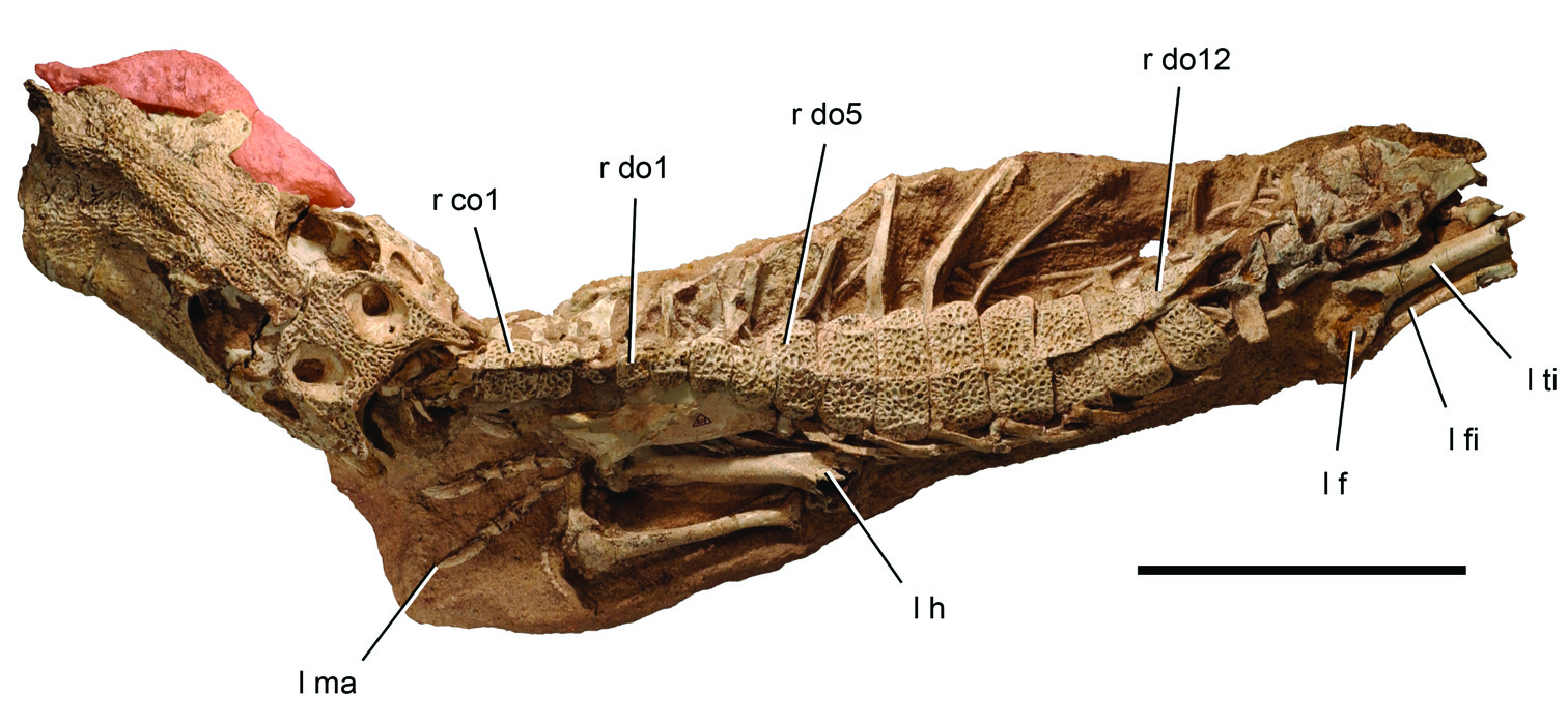
Axial skeleton. The axial skeleton is preserved in articulation from the proatlas to the fifteenth dorsal vertebra. Th is is one of the most complete presacral series available for any notosuchian. Th e axial column is well exposed immediately posterior to the skull and partially exposed, mainly in right lateral view, more posteriorly. Because this is one of the rare specimens that also shows the relationship between the osteoderms and vertebrae, we left all bones in place during preparation and obtained a computed-tomographic scan to observe details hidden from view. A subadult specimen of Araripesuchus gomesii is the other notable basal metasuchian preserving a complete cervicodorsal column ( Hecht 1991).
Extant crocodylians have a proatlas, 8 cervical vertebrae and 16 dorsal vertebrae ( Mook 1921). Th e ribs for C3–7 are short, overlapping, and parallel the vertebral column. The rib for C8 angles posteroventrally and is transitional to longer, broader-shafted dorsal ribs. There are typically 16 dorsal vertebrae in extant crocodylians ( Chiasson 1962). Hecht (1991: 346) suggested there were “about seven cervicals” and 17 dorsal vertebrae (thoracic and lumbar) in the subadult specimen of Araripesuchus gomesii . Th e vertebra that would be the eighth cervical, however, is partially covered by the scapula. Its rib is transitional in form between the short cervical and long dorsal rib, which is typical of the eighth cervical rib in extant crocodylians ( Mook 1921). A similar vertebral formula and transitional rib has recently been reported in Araripesuchus tsangatsangan a ( Turner 2006). Th e axial column in A. minor also appears to have 8 cervical vertebrae and probably 16 dorsal vertebrae. Only 15 dorsal vertebrae are preserved, but a sixteenth may be inferred from the position of the sacral vertebrae, which is based on the position of the associated hind limb ( Fig. 4 View Figure 4 ). Cervical centra are amphiplaytan and lack hypapophyses. Dorsal centra become amphicoelous.
This vertebral formula differs from that described recently in the notosuchian Notosuchus . Th is genus may posses as many as 10 cervical vertebrae, 19 dorsal vertebrae, and 3 sacral vertebrae (Pol 2005; Fiorelli and Calvo 2008). Th e cervicodorsal column, thus, has 29 rather than 24 vertebrae and the sacrum 3 rather than 2 vertebrae.
A proatlas is preserved in articulation with the occiput in A. minor . It is an inverted V-shaped median element with a dorsal keel similar to that in extant crocodylians ( Mook 1921). Th e proatlas in A. minor appears to be somewhat larger relative to the atlas, which is composed of separate, paired neural arches and an intercentrum. The transverse width of the proatlas is greater than that of the atlantal neural arches.
The axis has a low subrectangular neural spine that projects only slightly posterior to the centrum as in extant crocodylians ( Mook 1921; Chiasson 1962). Cervical vertebrae three through eight have tall anteriorly tilted neural arches and vertical neural spines as described in the Notosuchus (Pol 2005) . Th e neural spine in C3 is subrectangular, about twice as tall as long. The neural spine in C7 is considerably taller and narrower, about five times as tall as long. Tall neural arches may characterize notosuchians (Pol 2005).
The dorsal vertebrae are somewhat longer relative to their width in A. minor than in Araripesuchus gomesii ( AMNH 24450; Hecht 1991). Th e broadest width in both taxa occurs in the posterior dorsal vertebrae, which have long transverse processes ( Fig. 4 View Figure 4 ). In A. minor maximum width across the transverse processes is approximately twice centrum length, whereas in A. gomesii maximum width is about three times centrum length. In both genera, the parapophysis migrates out onto the transverse process anterior to the diapophysis (D9–11), eventually coalescing to form a single rib articulation (D12), as in extant crocodylians. Similar elevation and fusion of the parapophysis does not appear to occur in Notosuchus (Pol 2005; Fiorelli and Calvo 2008).
The straight rib s of the atlas and axis are preserved on the left side (Fig. 12). The shorter triradiate ribs of C3–8 are preserved on the right side in articulation with each other. After they clear the paravertebral shield, the shafts of the anterior dorsal ribs bend ventrally and expand slightly to form a flange along their anterior margin as in A. gomesii ( Hecht 1991) and A. tsangatsangan a ( Turner 2006). In the posterior dorsal ribs, the capitulum and tuberculum lie in the same plane and eventually coalesce into a single head. Gastralia are preserved ventrally between the girdles ( Fig. 4 View Figure 4 ). Th ere do not appear to be any ventral osteoderms in A. minor .
Parasagittal rows of osteoderms are preserved above the cervicodorsal column, with each pair joining its opposite in the midline along an interdigitating suture (Fig. 12; Table 5). Articulation between successive rows of osteoderms is limited to overlap by the posterior edge of a given osteoderm with the anterior edge of the successive ipsilateral osteoderm. As in Araripesuchus ( Hecht 1991; Turner 2006), there is no development of anteromedial processes as is common among basal crocodylomorphs, and the
Figure |2. Pectoral girdle and forelimb of the crocodyliform Anatosuchus minor . Left pectoral girdle, forelimb and anterior portion of the paravertebral shield ( MNN GAD 17) in dorsal view. Scale bar equals 5 cm. Abbreviations: C2, axis; co1, 3, 4, cervical osteoderm 1, 3, 4; do1, 5, dorsal osteoderm 1, 5; h, humerus; l, left; r, right; ra, radius; rC1, atlantal rib; rC2, axial rib; sc, scapula; ul, ulna.
overlap within each parasagittal column of osteoderms is a narrow smooth articulation limited to the edges of the dorsal series.
No osteoderms are positioned over the proatlas, atlas or axis (Fig. 12). Four paired cervical osteoderms are associated with C3–8 and 12 osteoderms are positioned over D1–12. Osteoderms distal to the twelfth were weathered away. The first cervical osteoderm is the largest of the cervical series and articulates over the neural spines of C3–5. It has a trapezoidal shape with a broader anterior end and a low keel that is most promi- nent on the posterior one-half of the osteoderm. As in the other cervical osteoderm rows, there is some asymmetry in the paired plates. Th e keel in the first cervical osteoderm row is laterally displaced on the left but centered on the right side. Th e second cervical osteoderm is smaller and articulates with the neural spine of C6. Its shape is similar to the first cervical osteoderm, the keel now reduced to a swelling along the rounded posterolateral corner on the left side or centered on the right side. Th e third cervical osteoderm is the smallest among all preserved and articulates with the neural spine of C7. It is subtriangular on the left and subquadrate on the right and does not have a keel. The fourth and final cervical osteoderm is slightly larger than the third cervical osteoderm and has a shape reminiscent of many of the succeeding dorsal osteoderms. The laterally displaced keel is low and set back from the anterior margin of the plate. The lateral corners of the plate are rounded, the anterolateral corner more so than the posterolateral corner. Th ere is no overlap between the last cervical and first dorsal osteoderm. The cervical osteoderms would allow considerable lateral and dorsoventral flexibility of the cervical series as may have been needed during foraging on land or subaquatic feeding.
The dorsal osteoderms have a one-to-one relationship with underlying dorsal vertebrae as described in extant crocodylians ( Ross and Mayer 1984) ( Figs. 4 View Figure 4 , 12). Each dorsal osteoderm contacts the neural spine of its respective vertebrae, extends posteriorly across the interspinous gap, and rests on the anterior portion of the successive neural spine. This is well exposed in the middle of the dorsal series, where the right column of osteoderms is displaced ventrally against the transverse processes, exposing the natural articulation between the neural spines and the left column of osteoderms. The junction between the osteoderms appears to be positioned so as to coincide functionally with the joints between the centra to enhance mobility of the trunk ( Salisbury et al. 2006).
The first dorsal osteoderm closely resembles the last cervical osteoderm but is slightly larger and extends over the leading edge of the successive osteoderm. Each dorsal osteoderm has a smooth beveled leading edge approximately 1.75 mm broad for articulation with the next anterior osteoderm. Th e sculpted pitting is reduced in a narrow parallel band of slightly greater width adjacent to the leading articular surface. Dorsal osteoderms 2–12 are more flexed than more anterior osteoderms, the portion of the plate lateral to the keel deflected ventrally. Th e keel remains parallel to the midline across the series. Osteoderm length gradually increases until about the middle of the series ( Table 5). Osteoderm shape remains very similar throughout the series, the rounding of the anterolateral corner somewhat less in posterior dorsal osteoderms.
Appendicular skeleton. The left pectoral girdle and forelimb and portions of the left tibia, fibula and pedal phalanges are preserved in association with the adult skull (Figs. 3, 11; Table 5). The left scapula has broad proportions comparable to those in Araripesuchus gomesii ( Hecht 1991) . Th e blade does not appear to flare as strongly distally as in A. tsangatsangana ( Turner 2006) . Th e distal end of the blade is tucked under the edge of the anterior dorsal osteoderms as in extant crocodylians (Fig. 12). The elongate coracoid is exposed distally near its contact with the interclavicle.
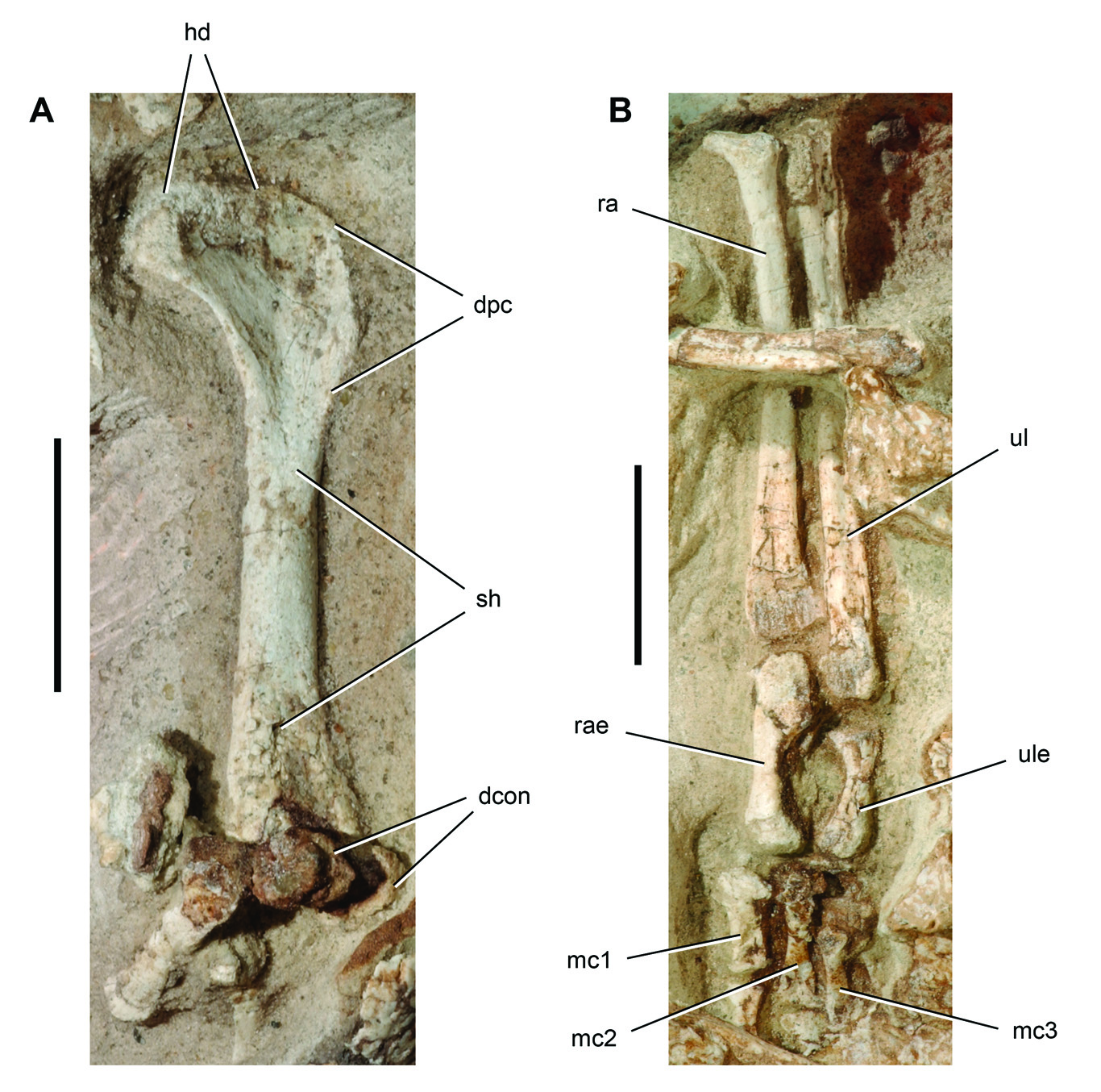
The humerus has a straight shaft and gracile proportions, with shaft diameter less than 10% of its length ( Turner 2006) ( Table 5). The deltopectoral crest is directed anteriorly, and the fossa for the olecranon process is well developed distally as in Araripesuchus ( Hecht 1991; Turner 2006). Th e proximal end of the radius is strongly flared, measuring more than twice mid-shaft diameter. Flaring of the proximal end of the radius to this degree is also present in Araripesuchus ( Fig. 25B View Figure 25 ) and Notosuchus (Pol 2005) . Th e radius is shorter than the ulna, because the ulna extends along the lateral side of the radiale. Th e ulna in A. minor is only partially exposed, its shaft noticeably curved. The differential in length between the radius and ulna is about 10%, as preserved in articulation in Araripesuchus ( Fig. 25B View Figure 25 ). The radiale is a very robust bone in A. minor , its shaft just slightly less robust than the mid-shaft of the radius (Fig. 13A). The broad lateral facet for the ulna on the proximal end confirms the offset in the joint between the forearm bones (radius, ulna) and the proximal carpals (radiale, ulnare). From the radiale, it is clear that this offset is also present in A. tsangatsangana ( Turner 2006) and Notosuchus terrestris (Pol 2005) . Very little of the ulnare is not exposed in A. minor , but the bone would have been considerably smaller than the radiale. The offset at the forelimb-carpus joint, the general robustness of the radiale, and the differential in robustness between the radiale and ulnare are primitive for Crocodylomorpha, given their presence in Terrestrisuchus ( Crush 1984) , Hesperoschus ( Clark et al. 2000), Dibothrosuchus (Wu and Chaterjee 1993) , Junggarsuchus ( Clark et al. 2004) , and Protosuchus ( Colbert and Mook 1951) , although often muted in extant crocodylians ( Mook 1921).
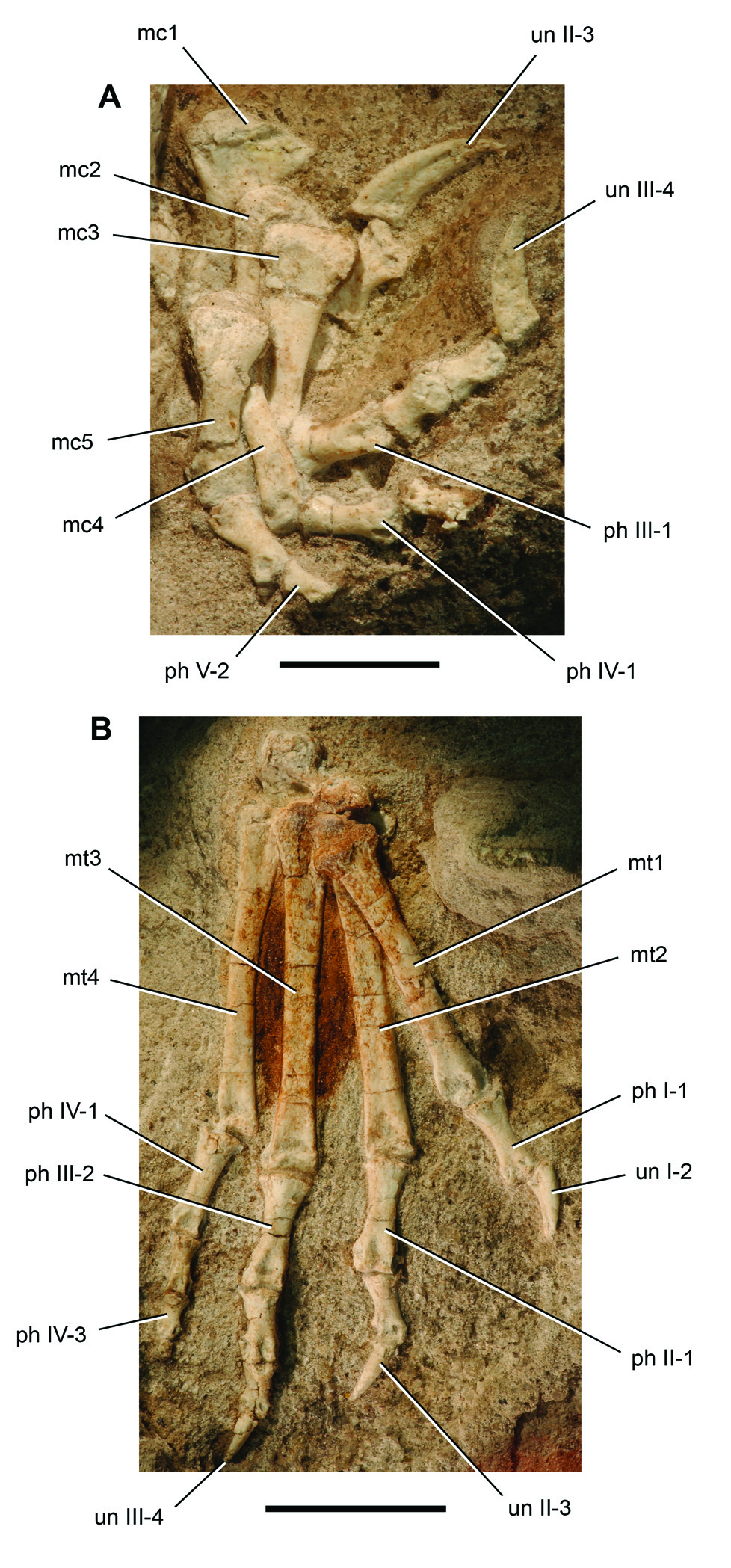
The manus is well preserved and exposed (Fig. 13A). As in A. wegeneri ( Fig. 26A View Figure 26 ), the metacarpals and phalanges have well developed distal condyles marked by dorsal extensor pits. Th e manus is very large relative to the forearm. Digit three is approximately 80% the length of the radius, whereas in other terrestrial crocodylomorphs that percentage is between 50 to 60% ( Mook 1921; Colbert and Mook 1951; Crush 1984; Wu and Chaterjee 1993; Clark et al. 2000; Clark et al. 2004). Besides its size, two other features of the manus are unusual. Digit IV has six phalanges, two more than is usual among crocodylomorphs (Fig. 13B). Total length of the phalanges of digit IV is approximately 80% the length of the phalanges of digit III, a typical crocodylian proportion. Much of the length of the phalanges of digits I-III is due to elongate unguals. Th e phalanges of digit IV are longer than the nonungual phalanges of digit III. Th e unguals of the inner digits are unusually long. The unguals have a narrow attachment groove that extends toward from the base to the tip (Fig. 13B). This groove converges with the dorsal margin of the ungual. The ventral margin is arched proximally and straight distally toward the tip. These unusual features, which are absent in the more typical manus in Araripesuchus wegeneri ( Fig. 26A View Figure 26 ), are indicative of specialized function.
Figure |3. Manus of the crocodyliform Anatosuchus minor . Left carpus and manus ( MNN GAD 17). A Left carpus and manus in dorsal view. B Left manual digits III and IV in dorsomedial view. Scale bars equal 2 cm. Abbreviations: I-IV, digits I-IV; ph, phalanx; ra, radius; rae, radiale; un, ungual.
Referred species. A. gomesii ( Price 1959) , A. wegeneri (Buffetaut and Taquet 1979) , A. patagonicus ( Ortega et al. 2000) , A. buitreraensis ( Pol and Apesteguia 2005) , A. tsangatsangana ( Turner 2006) .
Revised diagnosis. Small-bodied metasuchians with autapomorphies including (1) trapezoidal snout cross-section just anterior to the orbit in which the lacrimal is split between dorsal and lateral rami; (2) premaxilla external surface smooth with ornamentation limited to the distal end of the ascending ramus; (3) presence of one or two neurovascular foramina opening anterolaterally or anteroventrally just posterior to the narial fossa; (4) premaxillary teeth 1–4 aligned in a straight row; (5) maxillary postcaniniform alveolar margin dorsally arched; (6) smooth buccal emargination on lateral maxillary and dentary alveolar margins adjacent to postcaniniform teeth; (7) confluent alveoli for postcaniniform maxillary and mid- and posterior postcaniniform dentary teeth; (8) medial alveolar wall absent along mid- and posterior postcaniniform dentary teeth with root crypts enclosed medially by the splenial.
Discussion. The monophyly of the genus Araripesuchus has been controversial. Some features that were initially thought to be diagnostic for the genus were discovered to have broader distributions among notosuchians such as Uruguaysuchus . The generic assignment of one species in particular, A. wegeneri , has been questioned ( Ortega et al. 2000). Comparison among species has been diffi cult due to incomplete specimens and descriptions. Th e dentition, for example, is critical for evaluation of species and generic distinction, but the morphology of a relatively fresh (unworn) dentition is not available for most species within Araripesuchus or immediate outgroups (e.g., Uruguaysuchus ). Here we describe derived features that may unite some or all of the species in the genus Araripesuchus .
The geometric shape of the cross-section at the base of the snout ( Ortega et al. 2000) involves a distinct flexure in the body of the lacrimal that gives the snout a trapezoidal cross section just anterior to the orbit. The vertical portion of the lacrimal is not broadly exposed in dorsal view of the skull (Figs. 14B, 15B). Th e lacrimal is gently arched and broadly visible in dorsal view in most short-snouted notosuchians, such as Mariliasuchus ( Zaher et al. 2006) , or long-snouted neosuchians, such as Hamadasuchus ( Larsson and Sues 2007). The lacrimal in Uberabasuchus ( Carvalho et al. 2004) and Stolokrosuchus ( Larsson and Gado 2000) are closest in form to that in Araripesuchus .
Most of the premaxilla is smooth and lacks the rugose texture and small foramina typical of other regions of the snout in the vast majority of crocodyliforms. Only the tip of the ascending ramus is textured, as it curves onto the dorsal aspect of the snout tapering between similarly textured surfaces of the nasal and maxilla (Fig. 16A). The body of the premaxilla is also smooth in A. gomesii ( Price 1959: pl. 1) and A. tsangatsangana ( Turner 2006: fig. 20), whereas the condition in A. patagonicus ( Ortega et al. 2000) and A. buitreraensis ( Pol and Apesteguia 2005) remains poorly known.
Two large neurovascular foramina open on the lateral surface of the premaxilla on a smooth surface just posterior to a depression (narial fossa) and just anterior to the premaxilla-maxilla foramen (Fig. 15A). Th e same pair are present in the same position in A. gomesii (AMNH 24450; Hecht 1991), although there appears to be only a single large foramen in the smaller species A. tsangatsangana ( Turner 2006: figs. 19, 20). In other genera, such as Hamadasuchus ( Larsson and Sues 2007: fig. 3) or Stolokrosuchus ( Larsson and Gado 2000) , small foramina are often present but are not relatively as large, isolated, or located on a smooth surface related to the margins of the narial fossa.
The straight, rather than labially convex, arrangement of alveoli 1–4 in the premaxillary tooth row is unusual. The external profile of the alveolar margin of the premaxilla, likewise, is also straight or even slightly concave in ventral view (Figs. 13C, 14C). Th is feature is currently known in A. wegeneri , A. gomesii ( Price 1959; Turner 2006; AMNH 24450], and A. tsangatsangana ( Turner 2006) . A similar premaxillary margin was very likely present in a new species of Araripesuchus described below, given the opposing straight, anteromedially oriented margin at the anterior end of the dentary ( Figs. 27C View Figure 27 , 28 View Figure 28 ). In A. tsangatsangana the alveolar margin of the premaxilla is gently concave ( Turner 2006: fig. 49A), and the corresponding anteriormost dentary teeth also have a straight, rather than curved, alignment [ Turner 2006: fig. 41A]. This unusual feature may eventually be shown to characterize other closely related notosuchians, such as Libycosuchus , which shows a similar condition ( Stromer 1914). In Uruguaysuchus the premaxillary margin is not well described but has been shown as gently convex ( Rusconi 1933; Price 1959). Anatosuchus ( Figs. 5 View Figure 5 , 6 View Figure 6 ), Uberabasuchus ( Carvalho et al. 2004) , Hamadasuchus ( Larsson and Sues 2007) and most other crocodyliforms show the plesiomorphic condition; a line drawn through the centroids of the premaxillary crowns arches from the midline to the lateral aspect of the snout.
The postcaniniform alveolar margin on the maxilla is dorsally arched, above which is a smooth buccal emargination (Figs. 14–16). Both features characterize Araripesuchus . Although in some other crocodylomorphs the alveolar margin of the maxilla is sinuous, the portion distal to the caniniform that is dorsally convex is limited to several crowns and followed by a margin that is ventrally convex, as in Hamadasuchus ( Larsson and Sues 2007). Araripesuchus is distinctive because the entire postcaniniform series has a dorsally convex margin (Figs. 14A, 15A). This appears to be related to the enlargement of the opposing dentary teeth ( Fig. 20A View Figure 20 ); when the enlargement of opposing crowns is more limited, the arching of the maxillary series is more subtle, as in A. patagonicus ( Ortega et al. 2000) and A. tsangatsangana ( Turner 2006) . In Uruguaysuchus the postcaniniform series also appears to be very gently arched and may ultimately share this feature with Araripesuchus . Th e buccal emargination is also present on the dentary dorsal to a row of neurovascular foramina (Figs. 18A, 31A). As discussed below, there may have been a fleshy cheek margin functioning for temporary storage during mastication parallel to that in basal ornithischian and sauropodomorph dinosaurs ( Taquet 1976).
As discussed most notably by Pol and Apesteguia (2005), the alveoli are confluent for postcaniniform maxillary and for mid- and posterior postcaniniform dentary teeth in Araripesuchus . In other words, the posterior two-thirds of both upper and lower dentitions, have incompletely divided alveoli. This is well preserved in the upper and lower jaws of A. wegeneri (Figs. 14C, 15C, 16C, 19B, 20A, 21B, 27C). In the maxilla, medial and lateral walls of the alveoli extend ventrally to an equal degree, so the incomplete septa separating the alveoli are best seen in ventral view (Figs. 14C, 15C, 16C). A similar condition may be present in the reduced postcaniniform series in Libycosuchus ( Stromer 1914) as well as some other basal metasuchians, although more comparative detail is needed. In Notosuchus the alveolar septa are incomplete along the entire upper tooth row ( Lecuona and Pol 2008).
In the dentary, the lateral alveolar margin is much taller than the medial margin, so the incomplete septa separating the alveoli are broadly visible in medial view of a disarticulated dentary (Figs. 18B, 21B, 27B). Th e lack of a medial wall enclosing these alveoli is a remarkable feature. Th e crypts for the roots of the mid- and posterior postcaniniform teeth in the dentary are actually enclosed medially by the splenial in Araripesuchus ( Pol and Apesteguia 2005) . Th is condition does not appear to be present in the stout mandibular rami of Libycosuchus ( Stromer 1914) .
Several features used previously to distinguish Araripesuchus ( Ortega et al. 2000; Pol and Apestiguia 2005; Turner 2006) clearly have a broader distribution among genera that may be closely related within Notosuchia. Th ese include teeth showing marked differentiation of tooth type into anterior incisiforms with bulbous subconical crowns, caniniforms, and squat postcaniniforms; a sharp transition in tooth form between the upper caniniform tooth (m3) and smaller and similar sized, squat-crowned, denticulate postcaniniforms; the presence of a basal constriction between crown and root in most teeth; and inclined denticles along the carinae of many upper and lower teeth. All of these features are present, for example, in Uruguaysuchus ( Rusconi 1933) and Uberabasuchus ( Carvalho et al. 2004) , both of which may fall within Notosuchia.
The lateral bulge at the anterior end of the maxilla ( Pol and Apesteguia 2005) is filled by the root of the maxillary caniniform (m3), as seen in a computed-tomographic scan of the cranium (Fig. 17C). Th us the degree of bulging in the maxilla of Araripesuchus is related to the relative size of the caniniform, as it is in many crocodyliforms. Interpreted in this manner, this feature is not restricted to Araripesuchus but has a much broader distribution. Th e corresponding bulge in Hamadasuchus , for example, occurs somewhat farther posteriorly, corresponding to the more posterior position of the caniniform ( Larsson and Sues 2007).
The jugal ascending ramus diverges at a point posterior to the midpoint of the ventral rami in Araripesuchus ( Pol and Apesteguia 2005) , a feature also present in Anatosuchus ( Figs. 5A View Figure 5 , 6A View Figure 6 ) and Uberabasuchus ( Carvalho et al. 2004) . The ascending ramus, in contrast, is positioned at the midpoint of the ventral rami in Uruguaysuchus ( Rusconi 1933) and many other basal crocodyliforms. Th e interpretation of this feature as a synapomorphy uniting species of Araripesuchus ( Pol and Apesteguia 2005) thus is not clear.
Several features have an uncertain distribution or polarity to function as unambiguous synapomorphies uniting species of Araripesuchus . A. wegeneri , A. gomesii ( Price 1959) and A. tsangatsangana ( Turner 2006) have five premaxillary teeth whereas A. patagonicus ( Ortega et al. 2000) has four, a more common condition among crocodyliforms. A prominent wedge-shaped posteroventral (quadrate) process on the pterygoid characterizes A. wegeneri (Figs. 14C, 15C, 16C, 17C) and A. gomesii ( Price 1959) but is absent in A. patagonicus ( Ortega et al. 2000) and A. tsangatsangana ( Turner 2006) . The polarity of this character is uncertain. Th e choanal septum has a flat ventral surface and T-shaped cross-section in A. patagonicus , but the condition in other species of Araripesuchus seems variable; the septum is flattened to a lesser degree in A. buitreraensis and a subadult specimen of A. gomesii ( Pol and Apesteguia 2005) and is present as a narrow strut with a rounded ventral edge in A. wegeneri (Figs. 14C, 15C) and a mature specimen of A. gomesii ( Price 1959) .


No known copyright restrictions apply. See Agosti, D., Egloff, W., 2009. Taxonomic information exchange and copyright: the Plazi approach. BMC Research Notes 2009, 2:53 for further explanation.