
Araripesuchus rattoides, Sereno & Larsson, 2009
|
publication ID |
https://doi.org/ 10.3897/zookeys.28.325 |
|
publication LSID |
lsid:zoobank.org:pub:A979ECDE-871F-4AFC-9ABA-63A0FD6DC323 |
|
DOI |
https://doi.org/10.5281/zenodo.3790375 |
|
persistent identifier |
https://treatment.plazi.org/id/CF171699-D3FD-4B5C-909A-21C4412BCB0E |
|
taxon LSID |
lsid:zoobank.org:act:CF171699-D3FD-4B5C-909A-21C4412BCB0E |
|
treatment provided by |
Plazi |
|
scientific name |
Araripesuchus rattoides |
| status |
sp. nov. |
Araripesuchus rattoides sp. n.
urn:lsid:zoobank.org:act:CF171699-D3FD-4B5C-909A-21C4412BCB0E
Figs. 27–30 View Figure 27 View Figure 28 View Figure 29 View Figure 30
Table 9
Etymology. Rattus (Latin) ; -oides, likeness (Latin). Named for the enlarged, procumbent first dentary tooth, which is reminiscent of the condition in many rodents.
Holotype. CMN 41893; right dentary preserving alveoli 1–14.
Referred material. UCRC PV3; anterior portion of left dentary preserving alveoli 1–8.
Type locality. Er Rachidia District (exact locality unknown), eastern Morocco (Fig. 1A, B). A referred specimen ( UCRC PV 3) was surface collected in 1990 in a small wash at Darelkarib (south of Erfoud).
Horizon. Kem Kem Beds; Upper Cretaceous (Cenomanian), ca. 95 Mya ( Sereno et al. 1996). Th e referred specimen ( UCRC PV 3) appears to have come from the lower member (pers. commun. D. Dutheil).
Diagnosis. Small-bodied metasuchian (<1 m) with an enlarged procumbent first dentary tooth that is set immediately adjacent to the midline; smaller procumbent second dentary tooth; a caniniform fourth dentary tooth that is particularly large (twice the basal dimensions of adjacent crowns); and a smooth anterior surface on the dentary symphyses with an oval fenestra opening into the first alveolus.
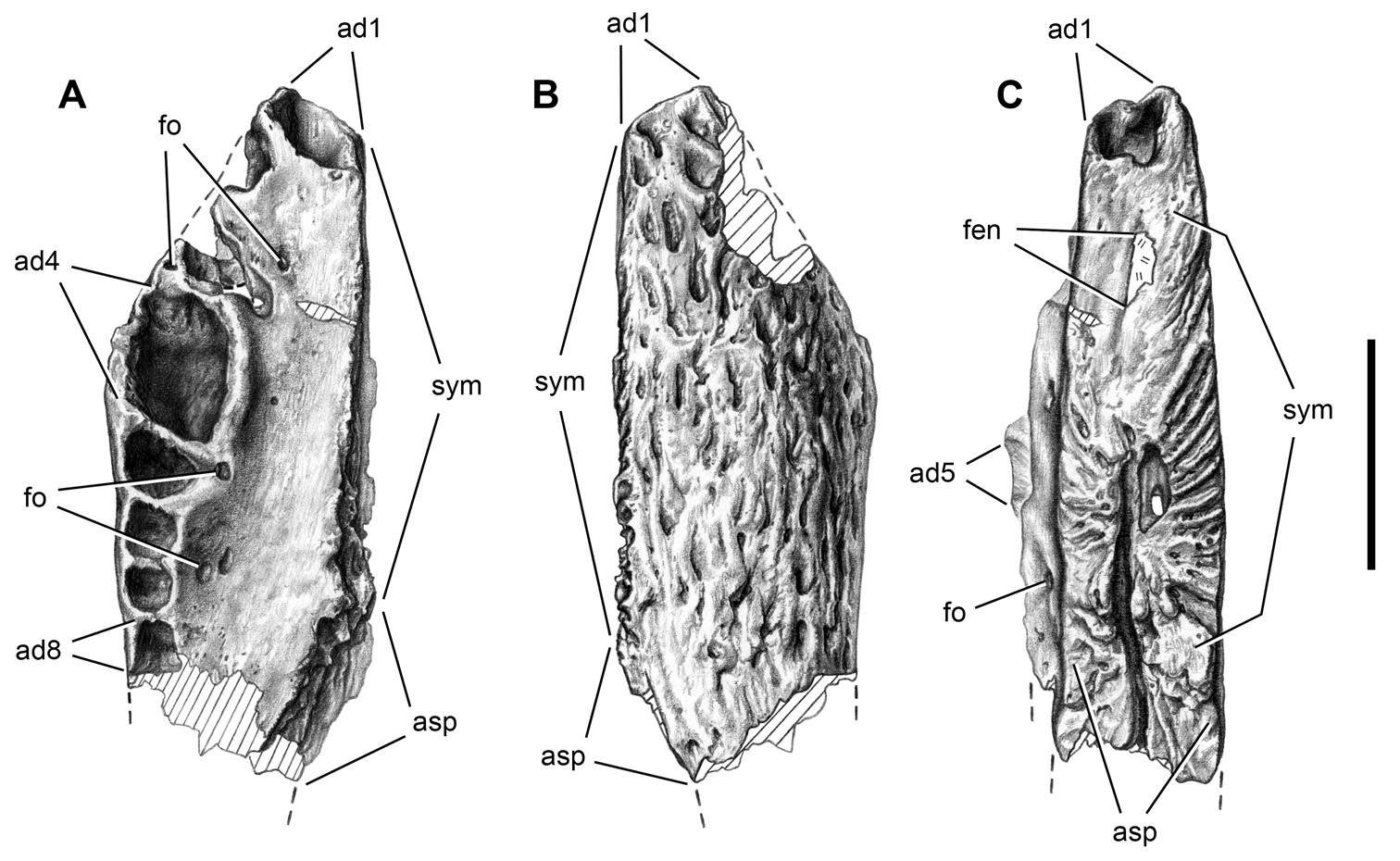
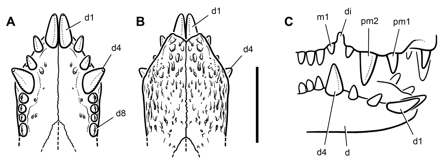
Dentary. The dentary of A. rattoides show a series of features that distinguishes it from the previously named species A. wegeneri and from a contemporary unnamed species from Cenomanian beds in Niger that closely resembles A. tsangatsangana ( Turner 2006) . Th e skull in A. rattoides appears to be proportionately narrower than in A wegeneri , based on the angle of divergence of the dentary tooth row from the midline. In A. wegeneri , the tooth row diverges at an angle between 20 and 25° from the midline (Fig. 18C), an angle matching the divergence of the upper tooth row (Figs. 14C, 15C). In A rattoides , by contrast, the angle of divergence is approximately 10° ( Fig. 27C View Figure 27 ), or less than half that in A. wegeneri . Th e anterior end of the dentary in A. rattoides is proportionately deeper than in A. wegeneri and other species of Araripesuchus . This difference is visible in both anterior and lateral view (Figs. 18A, D, 27A, D).
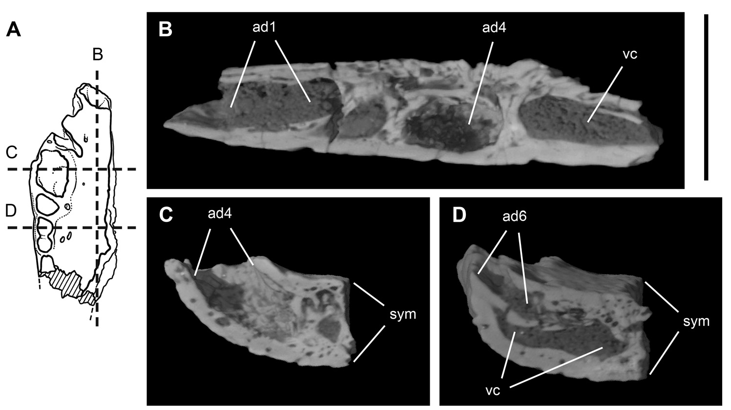
The orientation of the alveoli for teeth d1–11 is more procumbent in A. rattoides . The first and second alveoli project more strongly anteriorly than dorsally, a difference best appreciated in anterior view (Figs. 18D, 27D). Succeeding alveoli, including the caniniform (d4) and d5–11, are visible in lateral view ( Fig. 27A View Figure 27 ), whereas they are hidden by the dorsal edge of the alveolar margin in A. wegener i (Fig. 18A). Despite the more pronounced anterior projection of the anteriormost pair of teeth, the symphyseal region below these teeth ( Fig. 27A View Figure 27 ) is deeper than in A. wegeneri (Fig. 18A) and in a larger contemporary of A. wegeneri (Fig. 31A). Moreover, unlike these other species, the symphyseal articular surface of the dentary is not uniformly rugose in A. rattoides as it is in A. wegeneri and its larger contemporary (Figs. 18B, 31B). Th e anterior portion is smooth and fenestrated, as seen in two specimens ( Figs. 27B View Figure 27 , 28C View Figure 28 ).
Tooth size is also distinctive in A. rattoides ( Table 9). The first tooth is 75% the average diameter of the caniniform tooth (d4), which is already twice the diameter of adjacent crowns. In A. wegeneri the first dentary tooth is small ( Fig. 27C View Figure 27 ), and the caniniform is considerably less than twice as large as adjacent crowns ( Fig. 20B View Figure 20 ).
Tooth number may have been slightly greater in A. rattoides . In A. wegeneri , the largest postcaniniform teeth are d11 and d12 (Figs. 18B, 20A). In A. rattoides the largest postcaniniform dentary teeth are d12 and d13 ( Fig. 27 View Figure 27 , Table 9).
Other features in A. rattoides confirm its status as a species of Araripesuchus . Both A. rattoides and A. wegeneri have an unusual anterior extension of the articular scar for the splenial located dorsal to the symphysis on the subhorizontal palatal surface. This articular extension of the splenial, which is located medial to the alveoli for d4–6 ( Fig. 27C View Figure 27 ), is continuous posteriorly with the more typical vertical splenial attachment scar dorsal to Meckel’s canal. A. wegeneri shows a similar articular extension of the splenial (Fig. 18C). The alveoli posterior to d11, in addition, are open medially with alveolar septa poorly developed as low rounded ridges ( Fig. 27B, C View Figure 27 ). A similar condition is present in A. wegeneri (Fig. 18B) and some other species (Fig. 31C) ( Pol and Apesteguia 2005).


No known copyright restrictions apply. See Agosti, D., Egloff, W., 2009. Taxonomic information exchange and copyright: the Plazi approach. BMC Research Notes 2009, 2:53 for further explanation.