
Laganosuchus thaumastos, Sereno & Larsson, 2009
|
publication ID |
https://doi.org/ 10.3897/zookeys.28.325 |
|
publication LSID |
lsid:zoobank.org:pub:A979ECDE-871F-4AFC-9ABA-63A0FD6DC323 |
|
DOI |
https://doi.org/10.5281/zenodo.3790377 |
|
persistent identifier |
https://treatment.plazi.org/id/B9B7ACB2-A32A-4190-810E-F93ADE61C245 |
|
taxon LSID |
lsid:zoobank.org:act:B9B7ACB2-A32A-4190-810E-F93ADE61C245 |
|
treatment provided by |
Plazi |
|
scientific name |
Laganosuchus thaumastos |
| status |
sp. nov. |
Laganosuchus thaumastos sp. n.
urn:lsid:zoobank.org:act:B9B7ACB2-A32A-4190-810E-F93ADE61C245
Figs. 37 View Figure 37 –41
Tables 12, 13
Etymology. Thaumastos, astonishing (Greek). Named for the remarkably slender depth of its lower jaws and its straight spike-shaped teeth.
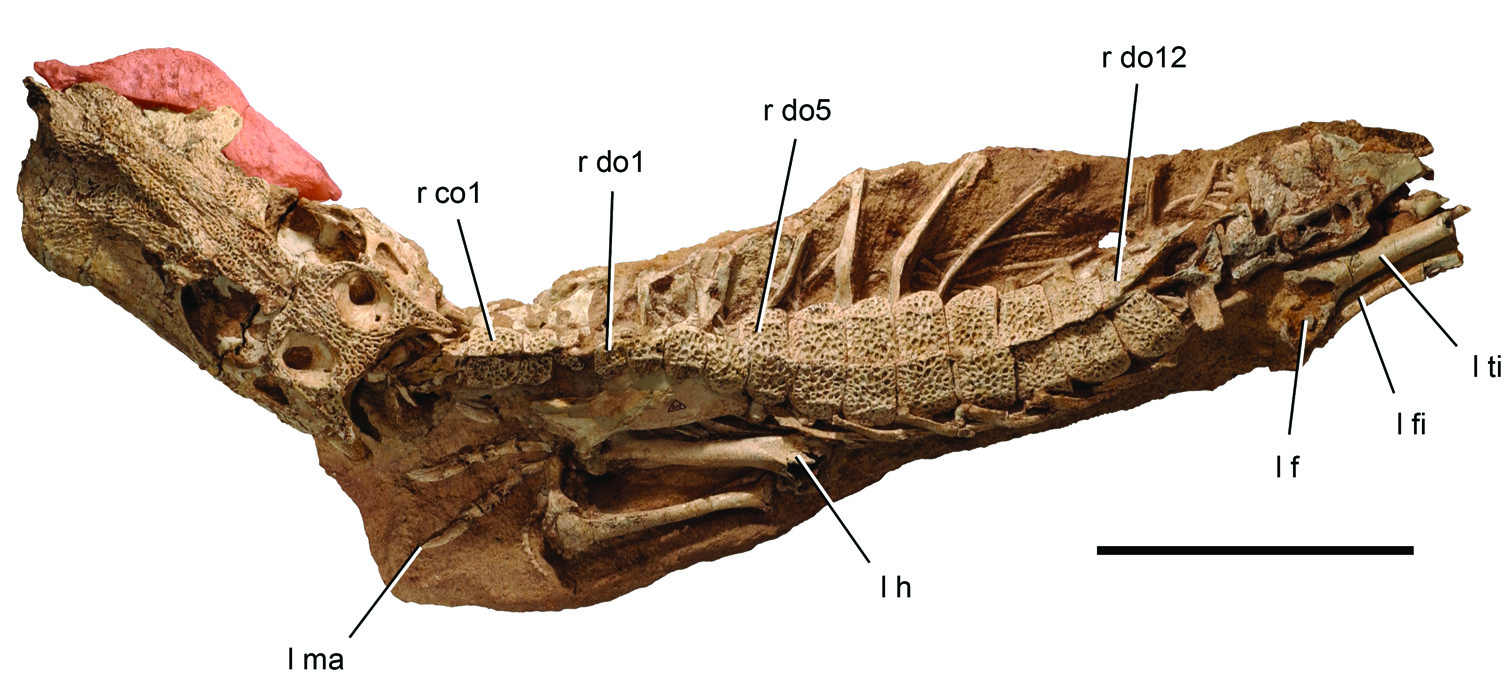
Holotype. MNN IGU13 ; nearly complete lower jaws missing only the left retroarticular process ( Fig. 37 View Figure 37 ).
Type locality. Iguidi (west of In Abangharit), Agadez District, Niger Republic (N 17° 56’, E 5° 38’) (Fig. 1A).
Horizon. Echkar Formation, Tegama Series; Upper Cretaceous (Cenomanian), ca. 95 Mya ( Taquet 1976). Found in association with the crocodyliform Kaprosuchus saharicus , abelisaurid Rugops primus , spinosaurid Spinosaurus sp., carcharodontosaurid Carcharodontosaurus iguidensis , an unnamed rebbachisaurid, and titanosaurian sauropods.
Diagnosis. Metasuchian characterized by alveoli for dentary teeth 1–10 with a depressed labial rim that exposes the upper portion of the alveolus in labial view; slightly procumbent d1 and d2 teeth; two pairs of twinned dentary teeth with conjoined alveolar margins among postcaniniforms; and splenial anterior end split into a pair of short flanges.
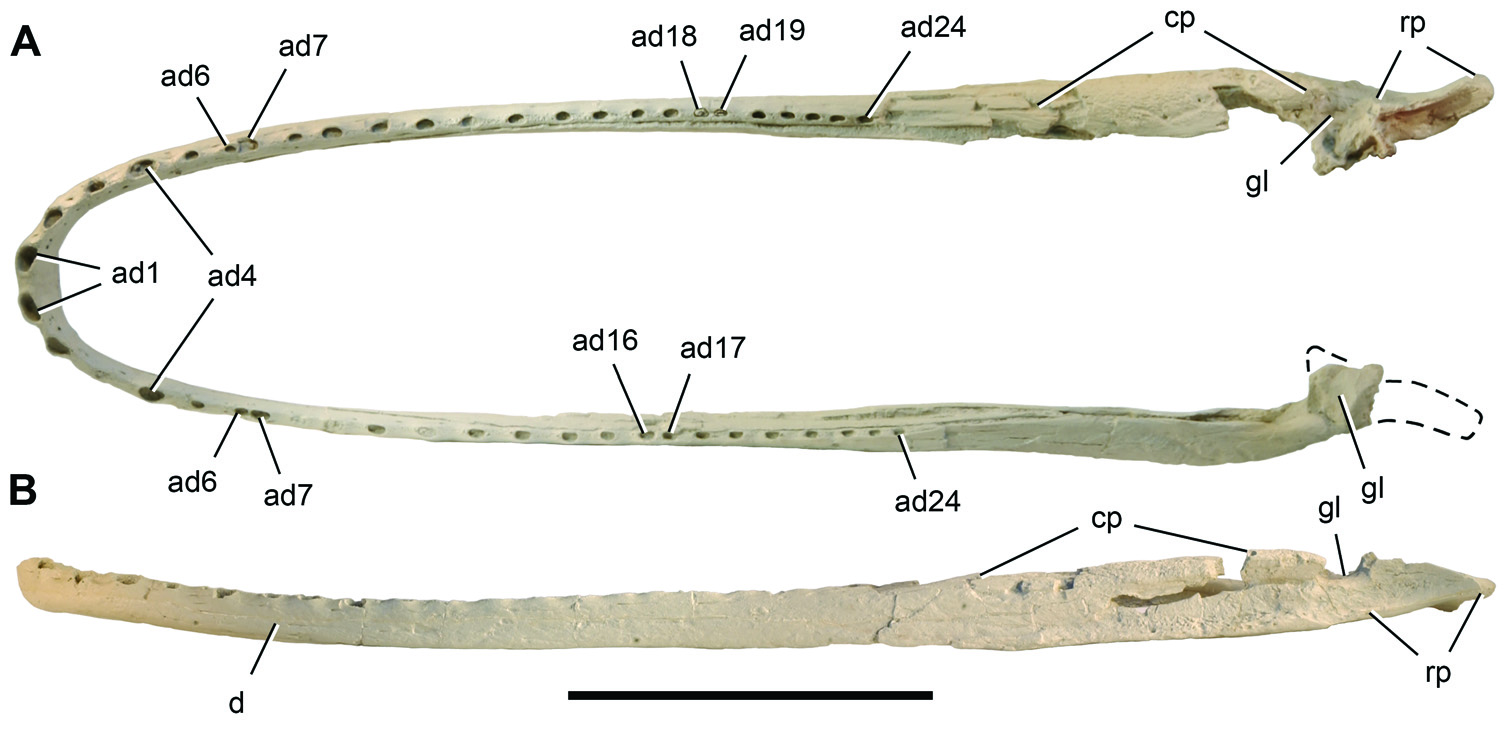
Lower jaw. The lower jaws of Laganosuchus thauma and Stomatosuchus inermis are remarkably slender and elongate and the symphysis extremely reduced compared to any extant crocodylian ( Fig. 37 View Figure 37 ). The lower jaws of Laganosuchus measure 0.84 m in length (Table 12) and probably pertain to a crocodyliform four-to-six meters in body length. Th e jaws of Stomatosuchus are 250% that of Laganosuchus , or approximately 2.1 m long. Th is is comparable to the length of the strongly built, robustly joined lower jaws in the largest individuals of Sarcosuchus ( Sereno et al. 2001) , the largest well documented crocodylomorph.
In dorsal view the mandible in Laganosuchus is U-shaped ( Fig. 37A View Figure 37 ). Each side is gently bowed, with curvature toward the symphysis increasing at about the seventh alveolus. In lateral view the ventral margin of the lower jaw is also gently curved as in Stomatosuchus ( Fig. 37B View Figure 37 ).
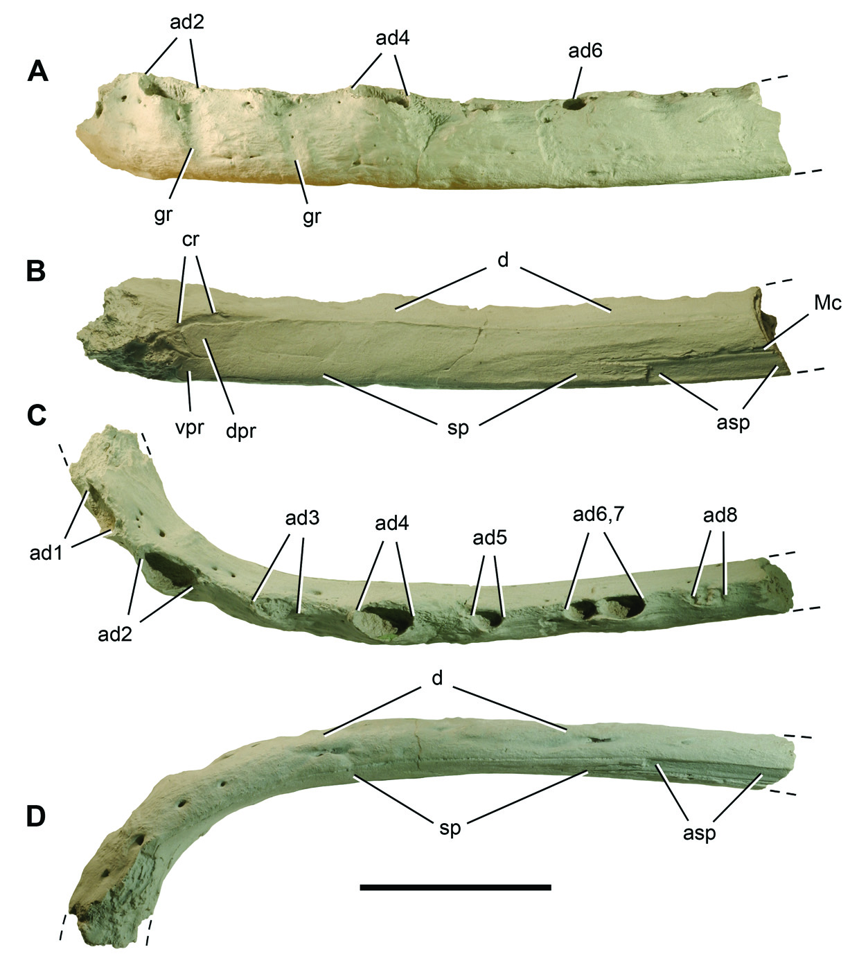
The dentary is most slender in the region of alveolus five and six (Table 12). At the symphysis, the dentary joins its opposite, an articulation that appears to have been fused in the holotype. Th e broken ventral margin at the symphysis appears to have been thickened dorsoventrally, forming a low chin ( Fig. 38D View Figure 38 ). The internal (labial) aspect of the dentary near the symphysis is convex with a discrete crest running along the dorsal edge of the splenial ( Fig. 38B View Figure 38 ).
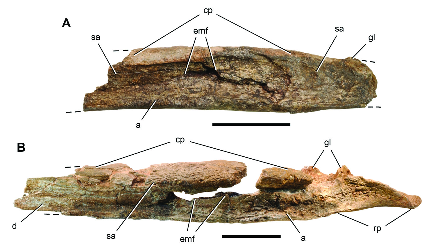
At mid-length the dentary has an elliptical cross-section. Th e dorsal, festooned, alveolar margin is transversely broader than the ventral margin. In medial view, a very narrow neurovascular groove is exposed where the splenial has broken away ( Fig. 38B View Figure 38 ). In lateral view the dentary splits into two posterior rami below alveoli 22 and 23. The dorsal ramus, which is the longer of the pair, twists onto the dorsal side of the coronoid process. Th ere it extends posteriorly as a tongue-shaped process that overlaps the surangular. Th is relation is unusual compared to extant crocodylians, as the surangular typically extends anteriorly, overlapping the dentary and approaching the posteriormost tooth. Th e subtriangular ventral ramus is short, the angular lapping it medially and extending anteriorly between the dentary and splenial.
The splenial is a very thin sheet of bone that extends toward, but does not participate in, the symphysis ( Fig. 38B View Figure 38 ). Th e distal end of the splenial is bifurcated, with Meckel’s canal terminating in the notch between the processes. In the anterior one-half of the dentary, Meckel’s canal is developed as a narrow incised groove lapped medially by the splenial ( Fig. 38B View Figure 38 ). Externally, the symphyseal ramus of the dentary is marked by two rows of neurovascular foramina, one extending near the ventral margin in lateral view ( Fig. 38A View Figure 38 ) and the other visible only in ventral view ( Fig. 38D View Figure 38 ).
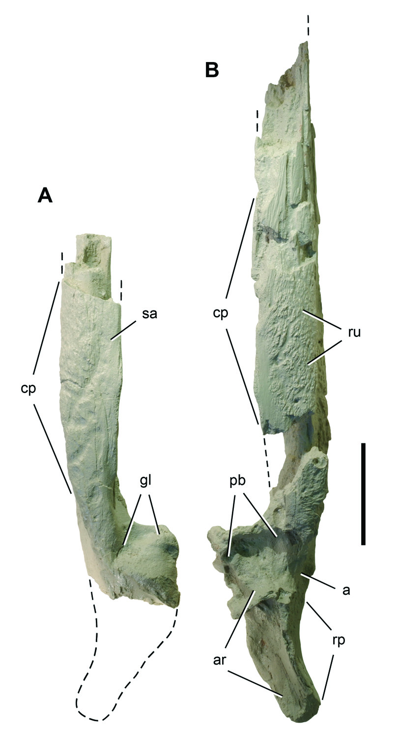
The posterior end of the lower jaw is characterized by a rugose, low, and transversely broad coronoid process, below which is a strongly reduced, slit-shaped external mandibular fenestra ( Figs. 39 View Figure 39 , 40 View Figure 40 ). In medial view, the remarkably small adductor fossa is located immediately anterior to the glenoid. As seen on the left side ( Fig. 40A View Figure 40 ), the articular surface of the glenoid is saddle-shaped, convex along an anterolateral-posteromedial axis and concave along an anteromedial-posterolateral axis ( Fig. 40A View Figure 40 ). The right side is concave with irregular edges and shows signs of bone pathology.
The retroarticular process, preserved only on the right side ( Figs. 39B View Figure 39 , 40B View Figure 40 ), has a triangular cross-section with sides that are concave. Th in posterior rami of the angular and prearticular completely overlap the articular on lateral and medial sides. The articular forms all of the dorsomedial face of the process, which is canted at an angle of approximately 45° ( Fig. 40B View Figure 40 ).
Dentition. There are 24 alveoli in each dentary with some variation in the position of two pairs of twinned alveoli. On both sides, the alveoli for tooth d6 and d7 are joined, the former is the smaller of the pair ( Fig. 37A View Figure 37 ). A similar twinning, although less complete and involving alveoli of comparable size, occurs between alveoli of d16 and d17 on the left side and d17 and d18 on the right side.
The alveolus for d1 is the largest in the tooth row and slightly larger than d4, commonly enlarged as a caniniform among crocodyliforms, and d2 (Table 13). The alveoli of d1 and d2 are canted labially and probably projected anterior to the rim of the opposing premaxilla. Th e alveolus for d3 is small ( Fig. 38C View Figure 38 ). In lateral view, this alveolus is flanked mesially and distally by canted troughs that accommodated crowns of the opposing maxillary series ( Fig. 38A View Figure 38 ). The dorsal margin between alveoli is developed as a ridge that becomes rounded posterior to d7. Festooning of the alveolar margin involves elevation of the rim of each alveolus with concave embayment of the lateral aspect of the interalveolar margin. Th e resulting undulating alveolar margin doubtless accommodated the interdigitation of opposing crowns.
Figure 4 View Figure 4 |. Tooth of the crocodyliform Laganosuchus thaumastos gen. n. sp. n. Medial view of the replacement crown in the eleventh alveolus of the right dentary (MNN IGU13). Scale bar equals 5 mm. Abbreviations: ca, carina; d, dentary; sp, splenial.
Several broken crowns remain in place, their crown bases tightly fitted to their respective alveoli. In cross-section, these crowns are oval with a large central lumen. We exposed replacement teeth in several crypts (Fig. 41). Th e crowns are spikeshaped in lateral view, lacking recurvature or any apparent asymmetry. Th ey are oval in cross-section at their base, above which they become transversely compressed with sharp, unornamented mesial and distal carinae. Th ere is no ornamentation of the crown surface.
The spike-shaped crowns remove any doubt that Laganosuchus was an active predator (Fig. 41). Because the spaced, oval alveoli resemble in size and shape those described in the anterior half of the maxilla of Stomatosuchus , it is possible that the latter genus had maxillary crowns of similar form ( Stromer 1925). Th e alveolar margin of the dentary in Stomatosuchus was depicted as smooth, lacking large alveoli or a festooned margin ( Fig. 2B, C View Figure 2 ), although Stromer (1925) questioned its state of preservation.
Both genera would have fed on fish in a very different manner than extant crocodylians, given the mechanical limitations of such a slender, hoop-shaped mandible, unexpanded cross- section at the symphysis, posteriorly positioned coronoid process, and short span available between the coronoid process and supratemporal region for the adductor musculature. Bite forces would have been limited. Stomatosuchids may best be interpreted as sit-and-wait predators in shallow water, closing their interdigitating spike-shaped dentition on unsuspecting prey that wandered within the U-shaped perimeter of their long jaws.

| MNN |
Musee National du Niger |
No known copyright restrictions apply. See Agosti, D., Egloff, W., 2009. Taxonomic information exchange and copyright: the Plazi approach. BMC Research Notes 2009, 2:53 for further explanation.