
Subrasaca flavolineata (Signoret, 1855)
|
publication ID |
https://doi.org/10.11646/zootaxa.3637.4.4 |
|
publication LSID |
lsid:zoobank.org:pub:1D85C538-793B-4CB1-B100-4415E03126C2 |
|
DOI |
https://doi.org/10.5281/zenodo.6160809 |
|
persistent identifier |
https://treatment.plazi.org/id/039B8793-FFB4-FFE6-37A8-2BDEFB60F9D7 |
|
treatment provided by |
Plazi |
|
scientific name |
Subrasaca flavolineata (Signoret, 1855) |
| status |
|
Subrasaca flavolineata (Signoret, 1855) View in CoL
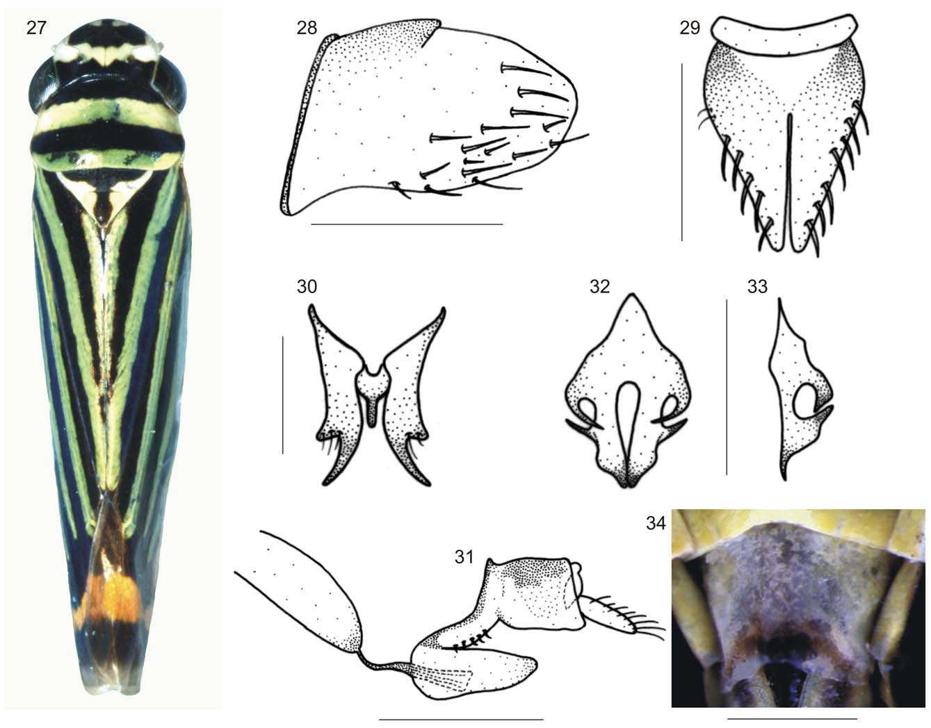
( Figs 27–34 View FIGURES 27 – 34 , 44 View FIGURES 42 – 45 )
Tettigonia flavolineata Signoret, 1855: 791 , pl. 24, fig. 11. Lectotype designated by Young (1964), female (Museum für Naturkunde der Humboldt-Universität, Berlin); photograph examined.
Length. Males, 5.4-5.5 mm (n = 2); females, 6.1-6.2 mm (n = 2).
Head and thorax. Structural features of head and thorax much as described above for S. constricta sp. nov., except for anterior margin of crown more broadly rounded, ocelli located slightly behind line between anterior eye angles, and forewing membrane restricted to small transverse area at apex of apical cells.
Color. Anterior dorsum ( Fig. 27 View FIGURES 27 – 34 ) dark brown to black; crown with pair of apical yellow maculae (originated from frons) and transverse yellow stripe located at level of antennal ledges and anterior eye portions; pronotum with broad transverse yellow-green stripe on anterior half and transverse yellow-green stripe along posterior margin; mesonotum with lateral portions yellow, delimiting conspicuous, central T-shaped dark brown to black area. Forewings ( Figs 27 View FIGURES 27 – 34 , 44 View FIGURES 42 – 45 ) yellowish-green with four longitudinal dark brown to black stripes, one on clavus, one on claval sulcus and two on corium, posterior portions of outermost three stripes connected by dark brown area followed by conspicuous yellow or orange transverse stripe; basal portion of costal margin dark brown to black. Face yellow; frons with pair of dark brown to black longitudinal stripes originated from anterior portion of crown and joining each other medioventrally to form stripe that reaches apex of clypeus, median superior portion of frons with smaller dark brown to black longitudinal stripe originated from crown; genae with posterior dark brown area below eyes. Lateral and ventral portions of thorax yellow, mesothorax with anepisternum and katepisternum mostly dark brown; legs brownish-yellow.
Male genitalia. Pygofer ( Fig. 28 View FIGURES 27 – 34 ), in lateral view, moderately produced posteriorly; posterior margin broadly rounded; without processes; macrosetae distributed mostly on distal third of disk and more anteriorly on ventral portion. Subgenital plates ( Fig. 29 View FIGURES 27 – 34 ), in ventral view, subtriangular, basal third expanded laterally, then tapering gradually toward apex; connected to each other basally by large, membranous triangular area; with uniseriate macrosetae. Connective ( Fig. 30 View FIGURES 27 – 34 ), in dorsal view, very short, Y-shaped, stalk narrow. Styles ( Fig. 30 View FIGURES 27 – 34 ), in dorsal view, extending much farther posteriorly than apex of connective; preapical lobe distinct, conspicuous, with few setae; apical portion elongate, directed outward; apex obliquely truncate. Aedeagus ( Fig. 31 View FIGURES 27 – 34 ) short; in lateral view, with conspicuous lobe directed posteriorly; without processes; gonoduct distinct, sclerotized. Paraphyses ( Figs 32, 33 View FIGURES 27 – 34 ) with two rami, each one with two lateral spiniform processes; anterior spiniform process strongly curved.
Female genitalia. Abdominal sternite VII ( Fig. 34 View FIGURES 27 – 34 ), in ventral view, narrowed toward apex; apical area more sclerotized than remainder of surface; posterior margin broadly shallowly concave. Internal sternite VIII, in dorsal view, with pair of small sclerites connected to each other medially. First valvifers, in lateral view, not distinctly expanded toward posterior portion, posterior margin truncate; in dorsal view, posterior portion curved medially. Pygofer, ovipositor valvulae, and gonoplacs much as described above for S. constricta sp. nov. Second valvulae, in lateral view, with dorsal dentate apical portion distinctly smaller than ventral one.
Material examined. SE. Brazil, State of Rio de Janeiro, Serra dos Órgãos mountain range. Eight males, two females, “ BRASIL: RJ, \ Nova Friburgo \ Vale dos Pinheiros \ 16.VIII.1992 \ G. Mejdalani leg.” (DZRJ, DZUP, MNRJ); two males, one female, “ Brasil, RJ \ Nova Friburgo \ 16-V-1992 \ G. Mejdalani” (DZRJ); two females, “RJ - Nova Friburgo - \ Estrada Mury-Lumiar \ Km 6 18-21/IV/2008 \ Gonçalves, A.C. & \ Aguiar, R.B. col.” (MNRJ); one male, “BR RJ Nova Friburgo \ Macaé de Cima \ 21-22/VI/2003 \ P. Ceotto col.” (MNRJ); one female, “ BRASIL - RJ \ Macaé de Cima \ 20-21/I/2001 \ Ceotto & Roquete” (MNRJ); one male, two females, “Teresópolis \ RJ/BR \ 30/III/2002 \ Ceotto, P. col.” (MNRJ); one female, “ Brasil, RJ, Teresópolis \ Vale da Revolta \ 19-VIII- 1996 \ Felix e Mejdalani cols” (DZRJ); one male, “BR-RJ-Teresópolis \ Vale da Revolta \ 29-V-1998 \ G. Mejdalani col.” (MNRJ); one male, “TERESÓPOLIS - RJ \ 20/IX/1992 \ G. MEJDALANI col.” (DZRJ).

No known copyright restrictions apply. See Agosti, D., Egloff, W., 2009. Taxonomic information exchange and copyright: the Plazi approach. BMC Research Notes 2009, 2:53 for further explanation.
|
Kingdom |
|
|
Phylum |
|
|
Class |
|
|
Order |
|
|
Family |
|
|
Tribe |
Cicadellini |
|
Genus |