
Victorgorgia nyahae, Moore & Alderslade & Miller, 2017
|
publication ID |
https://doi.org/ 10.11646/zootaxa.4304.1.1 |
|
publication LSID |
lsid:zoobank.org:pub:3D557C94-0783-4C39-80C3-9C321DA94800 |
|
DOI |
https://doi.org/10.5281/zenodo.6015397 |
|
persistent identifier |
https://treatment.plazi.org/id/401F301E-C5CD-43D0-83F4-F663CB47ECD2 |
|
taxon LSID |
lsid:zoobank.org:act:401F301E-C5CD-43D0-83F4-F663CB47ECD2 |
|
treatment provided by |
Plazi |
|
scientific name |
Victorgorgia nyahae |
| status |
sp. nov. |
Victorgorgia nyahae sp. nov. Moore, Alderslade & Miller
http://zoobank.org/401F301E-C5CD-43D0-83F4-F663CB47ECD2 ( Figs. 139–146 View FIGURE 139 View FIGURE 140 View FIGURE 141 View FIGURE 142 View FIGURE 143 View FIGURE 144 View FIGURE 145 View FIGURE 146 )
Material examined. Holotype: TMAG K3988, Cascade Seamount, Huon Commonwealth Marine Reserve (CMR), SW Tasman Sea, Australia, CSIRO RV Southern Surveyor, stn. 75, sample 32, (SS 200702 /075-032) 43.92– 43.934°S, 150.463 –150.479°E, depth 590–660 m, 10th April 2007.
Other material: TMAG K3989, Cascade Seamount, Huon CMR, SW Tasman Sea, Australia, CSIRO RV Southern Surveyor, stn. 77, sample 0 0 3, (SS 200702 /077-003), 43.915°S, 150.46°E, depth 590–660 m, 10th April 2007. GoogleMaps
Description:
Colony form: The holotype is a complete colony, now broken into three fragments ( Fig. 139 View FIGURE 139 A). When whole, the colony had three main branches emanating from a small holdfast, encrusting a piece of coral rubble. The largest of these branches is approximately 29 mm in length with a diameter ranging between 1.8–2.9 mm, and has a small group of polyps branching off close to the base. The other colony fragments are approximately 25–29 mm long with slightly narrower diameters (1.8–2.1 mm) and no secondary branching. The branches are occasionally bent and twisted, and often not circular in cross-section being more likely to be irregularly narrowed and distorted. No anastomoses are present. The colony holdfast is an encrusting membrane with a few scattered polyps. The colony is in good condition—preservation was directly into 70% alcohol.
Calyces and polyps are distributed along and all around the branches. They are particularly crowded towards the branch tip and so form terminal polyp bunches ( Fig. 139 View FIGURE 139 B, C). All calyces and polyps project substantially from the colony, mostly at right angles to the branches, with large, prominent calyces and rounded polyp heads. There is little space between calyces and only a few are isolated.
Colour: The photograph taken soon after collection shows the polyps and the distal half of the calyces are lilac, the retracted tentacles are purple and the rest of the colony is light cream. The colony is now light beige in alcohol.
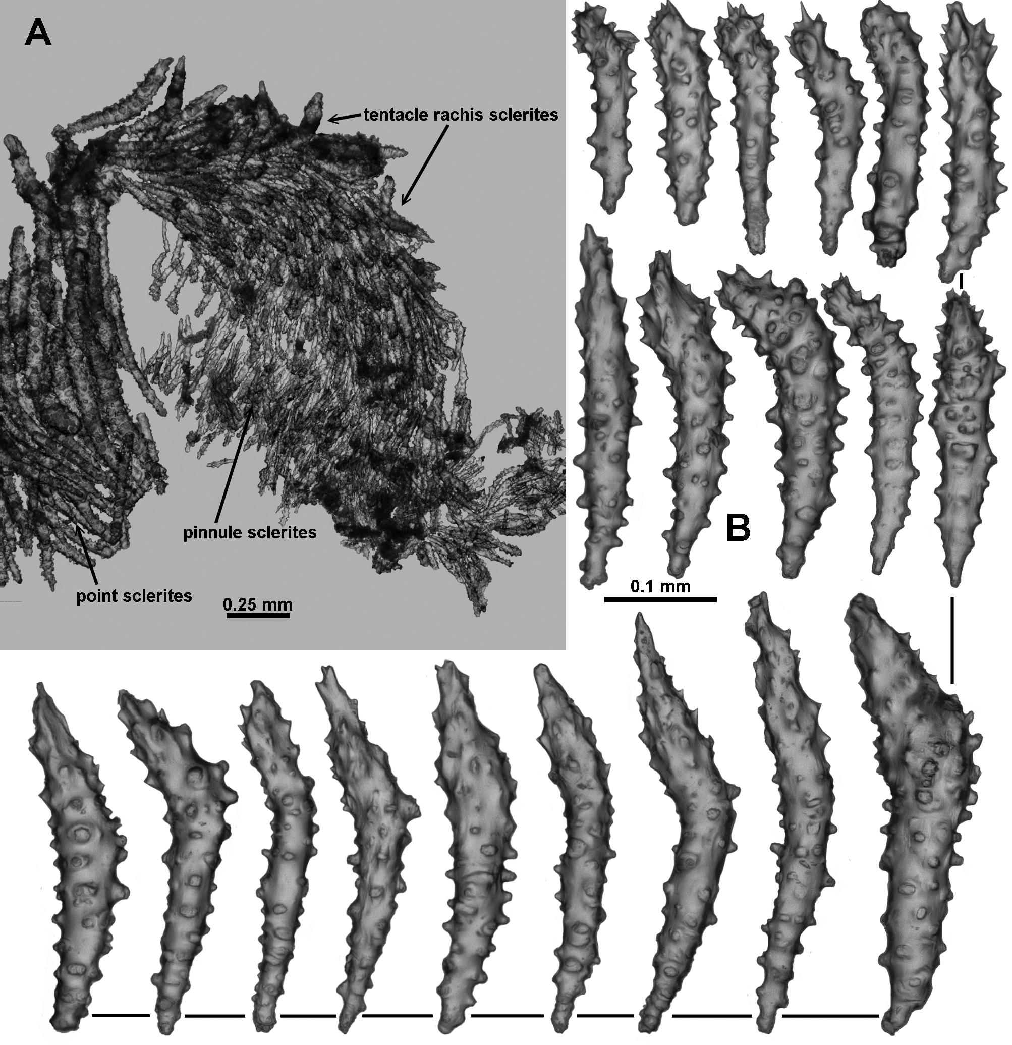
Calyces and polyps: The straight-sided calyces are large relative to the branch diameter, and range from 2.4– 3.9 mm tall and 1.4–2.6 mm wide. There are no polyps with the head fully retracted into the calyx although some are slightly retracted such that the polyp head is sitting on the lip of the calyx. Occasionally a partially retracted polyp head causes the calyx lip to flare out while the polyp head flattens ( Fig. 139 View FIGURE 139 Ca) making the whole polyp and calyx quite short and squat, however the majority of the polyps are exsert. Exsert polyps extended between 2–2.8 mm above the lip of the calyx, and the polyp heads have diameters of approximately 2–2.75 mm ( Fig. 139 View FIGURE 139 D). Both polyps and calyces are covered in a dense layer of crowded sclerites. Most polyps have the tentacles tightly folded over the polyp mouth so the polyp heads are rounded mounds with eight segments ( Fig. 140 View FIGURE 140 A). There are approximately 8–10 pinnules arranged in a single row along each side of the tentacles.
Medulla and Cortex: The branches of the scleraxonian colony are composed of a central medulla, made up of tightly packed longitudinal sclerites, surrounded by a thin cortex. The cortex and medulla are separated by a crowded series of longitudinal canals which anastomose to form an encircling boundary space allowing the cortex to be easily separated from the medulla. At the breakage point of the largest piece of colony, it can be seen that the medulla is extensively penetrated by longitudinal canals ( Fig. 140 View FIGURE 140 B, C). Many are large and encircled by a thin transparent layer of mesogloea, as mentioned by López-González and Briand, 2002 for V. josephinae . However, not all the canals have this layer and many are small and indistinct. For polyps which are arranged along and perpendicular to the branches, gastric cavities terminate at the medulla. For polyps which form the polyp clumps at the branch tip, their body cavity can extend internally down the branch for a short distance, eventually merging into the coelenteric canals in the central medulla.
Sclerites: The polyps and calyces are strongly protected with a dense covering of sclerites, predominately tuberculate sticks and spindles. On the polyp head, sclerites are arranged as a collaret and eight spiky points, with the sclerites bunched and crowded ( Figs. 139 View FIGURE 139 D; 140A). Around the head, approximately 10–15 transverse rows of sclerites form the collaret. Sclerites then angle en chevron up into the points, and eventually are longitudinally arranged at the tip of the points where the tentacle rachis begins. The collaret and point sclerites are mostly composed of tuberculate or warty sticks and spindles that are slightly curved or straight ( Fig. 141 View FIGURE 141 ). The points also have protruding, often large, warty spindles and clubs which are arranged with their modified spear-tip sticking out from the polyp head at the top of the points ( Figs. 140 View FIGURE 140 A; 141a). These sclerites give the polyp head a distinctive spiky appearance. Simple sclerites from the points and collaret range from 0.32–0.65 mm long while the more complex sclerites usually range from 0.5–0.7 mm long with occasional longer sclerites up to 0.91 mm. The sclerites from the polyp neck are simple tuberculate sticks and spindles, arranged obliquely, becoming transverse in the slightly contracted polyps.
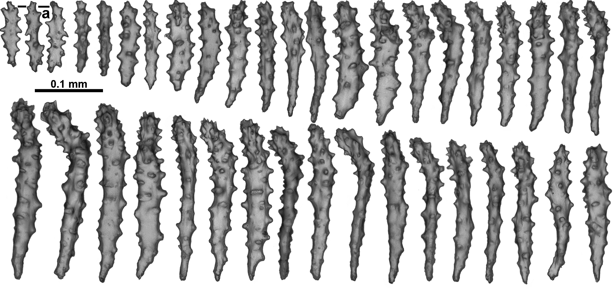
Sclerites continue longitudinally along the aboral side of the tentacles from the points ( Fig. 142 View FIGURE 142 A). These sclerites are sharply pointed thorn clubs with a tuberculate handle and a bent, spiny or thorny tip ( Fig. 142 View FIGURE 142 B). The thorny tip of the sclerites is distad in the tentacle and the bent tips project up from the tentacle rachis making it very bristly. The sclerites decrease in size distally along the tentacle rachis, grading from approximately 0.5 to 0.18 mm. Amongst these thorn clubs are josephinae clubs ( Fig. 143 View FIGURE 143 ) with a long, mostly smooth handle and a rounded spiny, bent tip. These tend to occur along the sides of the tentacles and extend into the pinnules while the thorn clubs are mostly restricted to the middle proximal ridge of the rachis ( Fig. 142 View FIGURE 142 A).
Sclerites, 0.1–0.27 mm long, are crowded longitudinally in the pinnules, reaching approximately half way down. They are mostly sparsely tuberculate, flat rods, straight clubs and josephinae clubs with the clubbed tips pointing distad ( Fig. 143 View FIGURE 143 ). Some of the larger sclerites have a reasonably well-developed clubbed tip, blurring the distinction between pinnule and tentacle rachis sclerites. There are also very small, flat rods with jagged edges ( Fig. 143 View FIGURE 143 a)—these appear to crowd around the distal end of the tentacle and can appear to be arranged transversely although this may simply be an artefact of contraction of the tentacles ( Fig. 142 View FIGURE 142 A).
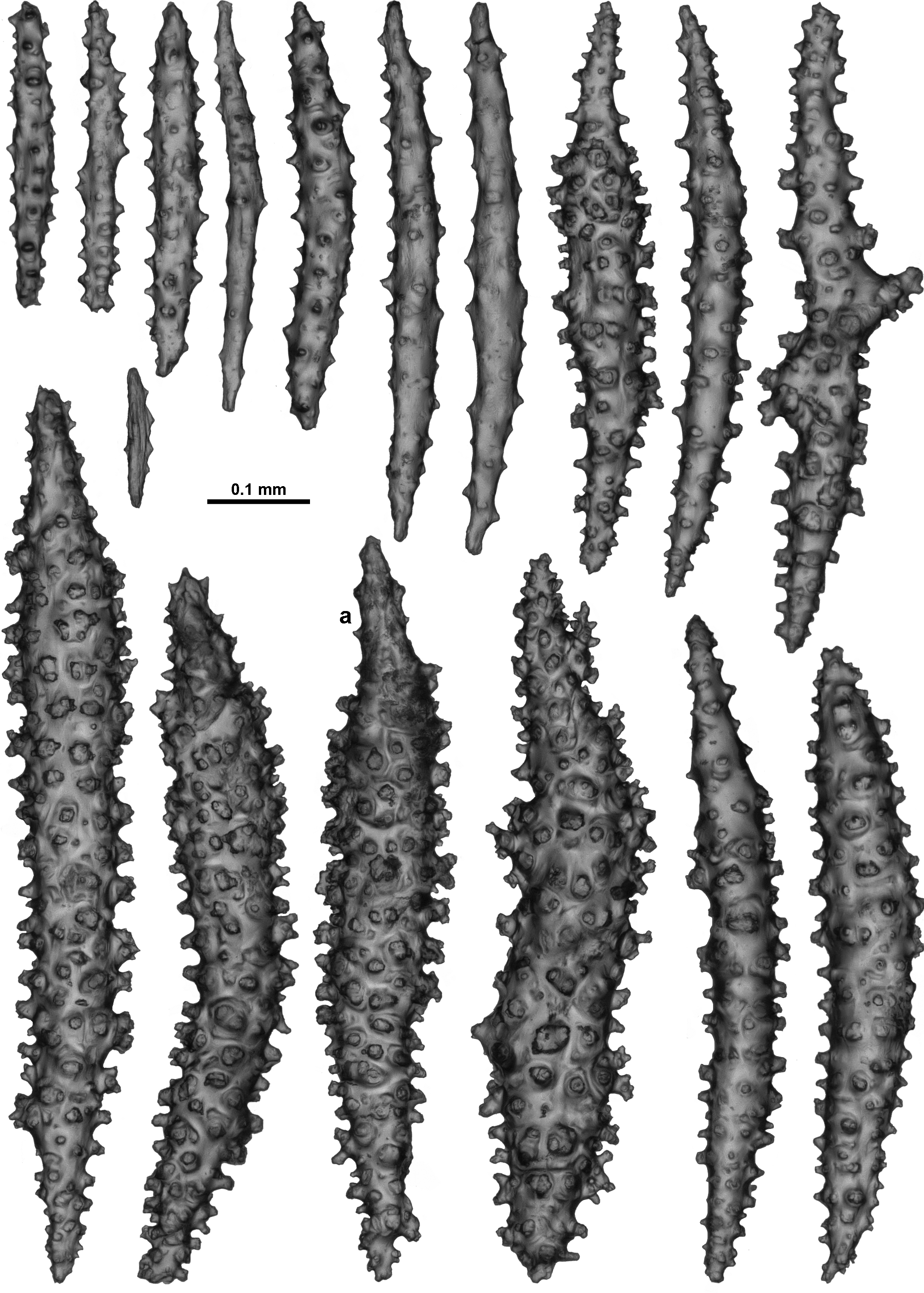
Calyces are covered in tightly packed sclerites, almost all sticks and spindles with tubercles varying from simple to complex branched warts ( Fig. 144 View FIGURE 144 ). Sclerites can range from 0.13–0.88 mm long but most are between approximately 0.36–0.78 mm. Bulky spindles with complex warts can have a spiny tip ( Fig. 144 View FIGURE 144 a) although these are not as developed as those present in the points and tentacles. Most sclerites are positioned longitudinally on the calyx, and tend to group into columns so the calyx has eight vaguely defined longitudinal ridges ending at the lip as eight indistinct mounds. The warty spindles are all orientated with the thorny tip projecting out from the calyx, sometimes giving the calyx a slightly prickly appearance particularly near the lip.
No sclerites were found in the pharynx.
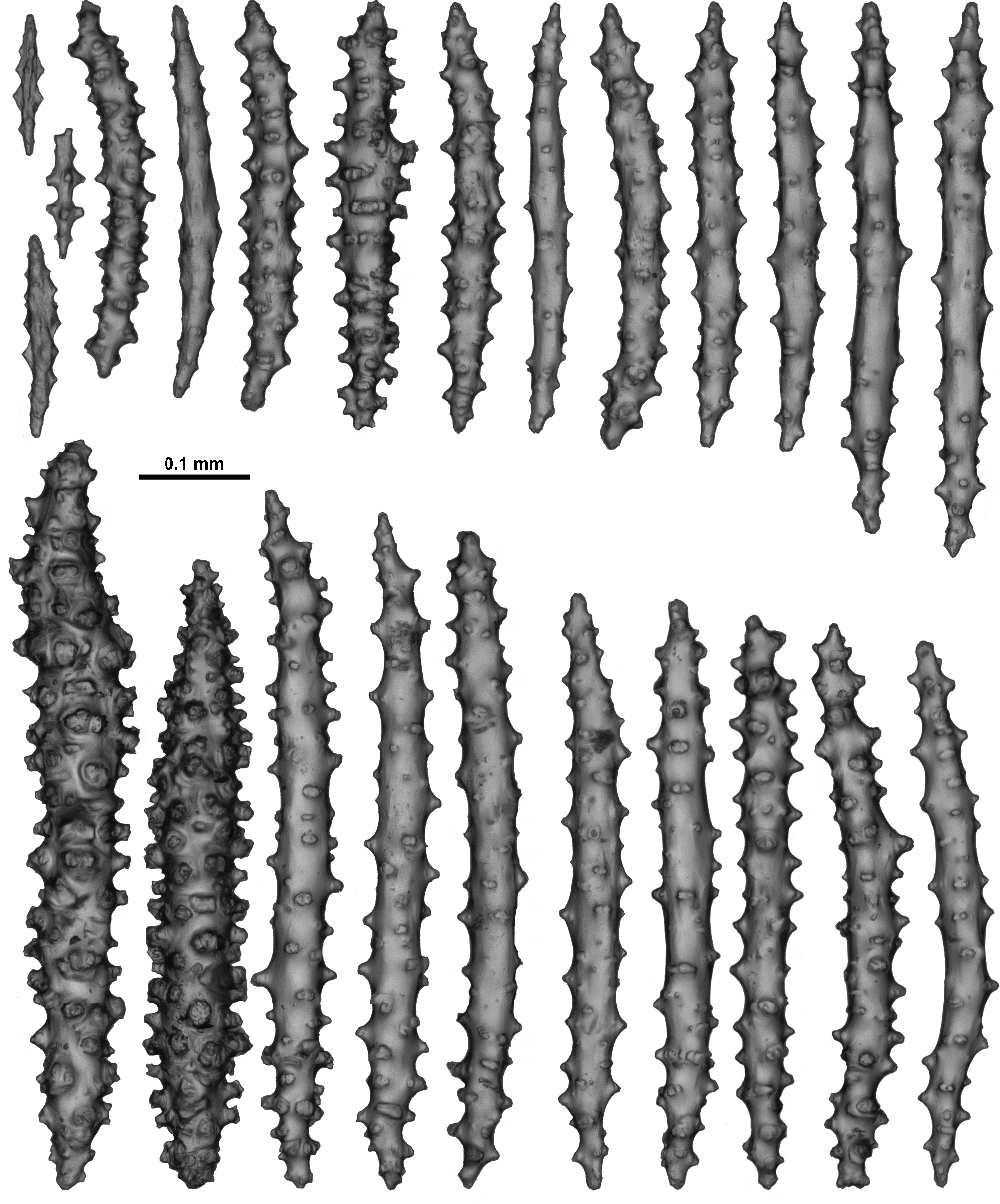
The thin cortex of the colony is composed of a dense layer of longitudinally arranged sticks and spindles from 0.23–0.71 mm long ( Fig. 145 View FIGURE 145 ). Most sclerites have mainly simple tubercles but there are some spindles with a dense covering of complex warts.
The medulla is composed of tightly packed longitudinal sclerites—mostly tuberculate and warty sticks and spindles ( Fig. 146 View FIGURE 146 ). Occasionally there are large sticks and spindles with only sparse warts, often with branches, forks and fused areas. Bulky spindles with complex warts are also present but only infrequently. Most sclerites are from 0.30–0.76 mm long although longer sclerites up to 1 mm were observed. It was difficult to ensure these long sclerites remained undamaged during sampling so the prevalence of these cannot be estimated. Similar to the calyx and surface, small, mostly smooth spindles, from 0.12–0.22 mm in length, occur amongst the bigger sclerites.
Sclerites are uniformly transparent under transmitted light.
Variability: The specimen TMAG K3989 is a small fragment closely resembling the holotype. It is a piece of a narrow branch with polyps which have a similarly prickly appearance, obvious large, straight-sided calyces and polyps which are mostly extended. No polyps are fully retracted. The sclerites are similar to those in the holotype. Unfortunately there is no live photograph of this colony. However, there is a significant genetic difference between the sequences from this specimen when compared with those from the holotype (see discussion below) thus it is not included as a paratype.
Distribution: Southern Tasmanian seamounts.
Depth: 590–660 metres.
Remarks: V. nyahae n. sp. is differentiated from other Victorgorgia species by the presence of sharply pointed thorn clubs and spear-tipped spindles in the tentacles, points and top of the calyx. See molecular section for details of the divergent sequences obtained from the two specimens.
Etymology: The species is named after the first author’s daughter, Nyah, as a small recompense for a childhood overshadowed by a PhD.


No known copyright restrictions apply. See Agosti, D., Egloff, W., 2009. Taxonomic information exchange and copyright: the Plazi approach. BMC Research Notes 2009, 2:53 for further explanation.
|
Kingdom |
|
|
Phylum |
|
|
Class |
|
|
Order |
|
|
Family |
|
|
Genus |