
Eschrichtioides gastaldii, Bisconti, 2008
|
publication ID |
https://doi.org/ 10.1111/j.1096-3642.2008.00374.x |
|
persistent identifier |
https://treatment.plazi.org/id/039BEA09-110A-FD6C-FF13-FE17380EFF43 |
|
treatment provided by |
Felipe |
|
scientific name |
Eschrichtioides gastaldii |
| status |
comb. nov. |
ESCHRICHTIOIDES GASTALDII COMB. NOV.
Cetotherium cortesii – Brandt, 1873: pls 21, 22; p. 153. Cetotherium gastaldii: Strobel, 1875: p. 8 .
Plesiocetus cortesii: Van Beneden, 1875 ; p. 755, 756 Cetotherium gastaldii: Strobel, 1881 : pls 1, 2, 5; p. 13. Balaenoptera gastaldii: Portis, 1883: p. 20 .
Balaenoptera gastaldii: Portis, 1885 : pls 1, 2; p. 17.
Balaenoptera acutorostrata: Caretto, 1970: p. 57 View in CoL .
‘ Balaenoptera View in CoL ’ gastaldii: Deméré et al., 2005: p. 105 , 119
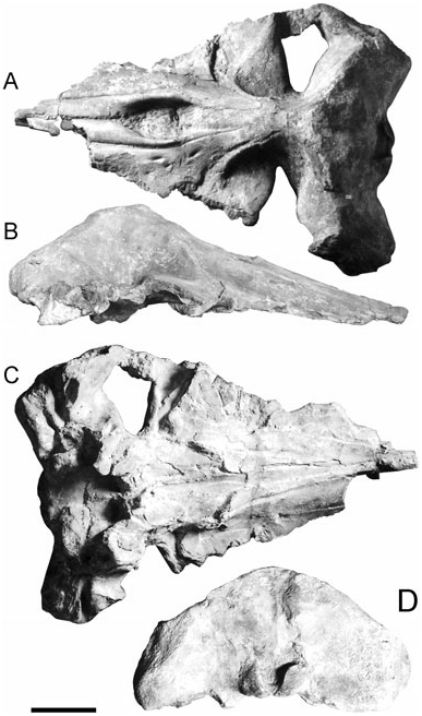
Formation and age: Sabbie d’Asti Formation. The Sabbie d’Asti Formation (Asti Sands Fm.) is developed at the top of the Argille Azzurre Formation (Blue Clays Fm.) ( Ferrero & Pavia, 1996). Foraminifers sampled at the top of the Argille Azzurre Formation suggested that deposition of this formation ended during the latest Early Pliocene. The Sabbie d’Asti Formation is characterized by a high density of mollusc shells whose occurrence in the Mediterranean Basin has been constrained within a range of a few million years between the start of the Pliocene (5.3 Ma) and 3.0 Ma by studies of Raffi and co-workers ( Raffi, Stanley & Marasti, 1985; Monegatti & Raffi, 2001). Interpretation of mollusc occurrence and extinction in the Sabbie d’Asti Formation by Ferrero & Pavia (1996, and references therein) suggests that the age of this formation can be con- Holotype: MRSN 13802 including skull, right dentary (13802/3), left dentary (13802/4 PU), atlas (13802/8), axis (13802/6), fourth cervical vertebra (13802/7), a lumbar vertebra (13802/18), four caudal vertebrae (13802/9, 10, 11, 12), hyoid (13802/24), ulna, and two ribs of the left side. Portis (1885) listed also humerus, three metacarpals and four phalanxes, left jugal and a tympanic bulla but I was unable to find these specimens in the MRSN collection. Cranial measurements are provided in Table l.
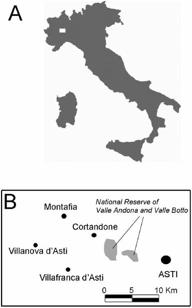
Type locality: Cortandone (geographical coordinates of the town: 44°58′N, 18°27′E), a town located around 20 km WNW from Asti and around 5 km NE from Villafranca d’Asti in Piedmont (north-west Italy; Fig. 1 View Figure 1 ) GoogleMaps .
Condylobasal length 930 Maximum width of skull (at anterior tip 490 of zygomatic processes of squamosals)
Maximum transverse diameter of supraorbital 160 process of frontal
Supraoccipital length
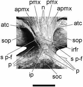
Transverse diameter of foramen magnum 55 Dorsoventral diameter of foramen magnum 65 Posterior width of vomer 120 Length of right basicapsular fissure 120 Width of right basicapsular fissure 65 Length of left basicapsular fissure 105 Width of left basicapsular fissure 50 Length of right palatine 270 Width of right palatine 78 Length of left palatine 260 Width of left palatine 70 premaxilla is located under the posteromedial corner of the maxilla, which is prolonged posteriorly to cover part of the interorbital region of the frontal. The premaxilla is medially concave anterior to the nasal bones on the side of the narial fossa; anterior to the narial fossa, the medial sides of the premaxillae converge toward the longitudinal axis of the skull and project anteriorly. There are no premaxillary foramina.
strained between late Zanclean and early Piacenzian but it is impossible to get a more precise chronostratigraphic assessment.
Diagnosis: As for the genus.
Etymology: The patronymic gastaldii was given by Strobel to the holotype specimen in recognition of the importance of the palaeontological work of Bartolomeo Gastaldi (Torino, 1818–1879).
DESCRIPTION
Skull
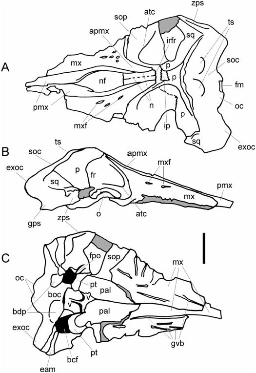
Premaxilla: Most of the anterior portion of the rostrum is lost ( Figs 2 View Figure 2 , 3 View Figure 3 ). The posterior end of the Maxilla: Maxilla horizontal and flat ( Figs 2 View Figure 2 , 3 View Figure 3 ). Lateral borders of the maxillae not preserved and this prevents an assessment of the whole width of the rostrum. Four maxillary foramina are present in the posterior and medial portion of the left maxilla lateral to the narial fossa. Posteromedial corner of the maxilla projecting posteriorly and superimposing onto the posterior end of the premaxilla and the interorbital region of the frontal, forming a long and narrow ascending process ( Fig. 4 View Figure 4 ). Posterior end of the ascending process rounded; its medial and lateral borders are parallel for their main development; more anterior portion of the ascending process of the maxilla wide, differing from balaenopterids in which this portion is narrower. In fact, this condition is closer to that of Cetotherium -like mysticetes. In ventral view, the surface of the maxilla bears only a few grooves for the vasculature of the baleen; among them, two grooves run parallel to the longitudinal axis of the skull very close to the mid-line; a groove is evident in the left maxillary which starts from a hole in the ventral surface of the bone and runs anteriorly. The medial portion of the maxilla gently protrudes ventrally, forming a longitudinal keel which is transversely wide and round. Infraorbital plate with a sharp posterior border directed anterolaterally; this condition is common among the mysticetes, being shared with Eschrichtius robustus and other mysticetes such as Cophocetus oregonensis Packard & Kellogg, 1934 , Aglaocetus moreni Kellogg, 1934 , Pelocetus calvertensis Kellogg, 1965 and Diorocetus hiatus Kellogg, 1968 . Posterior border of the maxilla profoundly indented by the interposition of the palatine.
Nasal: Nasal long; dorsal surface flat; anterior border straight ( Figs 2 View Figure 2 , 3 View Figure 3 ). Anterior end of the nasal located on a transverse line crossing the anterior border of the supraorbital process of frontal; in Parietobalaena palmeri Kellogg, 1924 , Diorocetus hiatus , Pelocetus calvertensis , Aglaocetus moreni , Cophocetus oregonensis and Mixocetus elysius the anterior end of the nasals is anterior to the anterior border of the supraorbital process of the frontal; in Balaenoptera acutorostrata Lacépède, 1804 , B. edeni Anderson, 1878 and B. borealis Lesson, 1828 the anterior border of the nasals is slightly anterior to the anterior border of the supraorbital process of the frontal; in Balaenoptera physalus (Linnaeus, 1758) and B. musculus (Linnaeus, 1758) the anterior border is posterior to the anterior border of the supraorbital process of the frontal. Nasofrontal suture located on a transverse line crossing the middle of the orbit; suture slightly indented. The position of the nasofrontal suture is similar in Cophocetus oregonensis , Aglaocetus moreni , Pelocetus calvertensis and in living balaenopterids. In Parietobalaena palmeri , Diorocetus hiatus and Aglaocetus patulus the nasofrontal suture is located more anteriorly, near the anterior border of the interorbital region of the frontal. Nasal and premaxilla contacting anteriorly; nasal and ascending process of maxilla contacting along the posterior half of nasal, the premaxilla being located beneath the maxilla in that region. This condition can be observed in specimens of Megaptera novaeangliae (Borowski, 1781) as illustrated by True (1904).
Frontal: Supraorbital process of the frontal abruptly depressed from the interorbital region as in eschrichtiids and balaenopterids ( Figs 2 View Figure 2 , 3 View Figure 3 ). Supraorbital process dorsally flat and crossed by an ascending temporal crest whose medial portion projects medially, and whose lateral portion abruptly projects posterolaterally towards the postorbital corner of the process in the proximity of the orbit. Interorbital region of the frontal almost completely hidden by the posteromedial elements of the rostrum: only a subtle sheet of bone is observed, which is located lateral and posterior to the ascending process of the maxilla, resembling the condition found in Cetotherium -like mysticetes. In ventral view, the channel for the optic nerve is highly concave and is surrounded by anterior and posterior crests, which are located under the central body of the supraorbital process of the frontal.
Parietal: In dorsal view, a strong postorbital constriction is observed posterior to the skull vertex ( Figs 2 View Figure 2 , 3 View Figure 3 ). The parietals appear dorsally anterior to the anterior border of the supraoccipital where their dorsolateral borders form short lateral concavities corresponding to the attachment sites for the temporalis muscle. In lateral view, anterior border of the parietal located more anteriorly than the posteriormost portion of the interorbital region of the frontal. Parietal not interdigitating with the posteromedial corner of the maxilla as in balaenopterids.
Squamosal: Only the right squamosal is sufficiently complete to allow a description of almost every feature of the bone ( Figs 2 View Figure 2 , 3 View Figure 3 ). Zygomatic process of the squamosal short and triangular in lateral view; it descends from the posterior apex of the lambdoidal crest with a steep inclination and terminates anterior to the anterior border of the supraoccipital. Posterior portion of the zygomatic process projecting anteriorly and laterally; anterior portion projecting only anteriorly, paralleling the longitudinal axis of the skull. Medial to the zygomatic process there is a broad, shallow, sharp squamosal fossa running along the dorsoventral axis; I observed such a fossa in all the specimens of Eschrichtius robustus that I have examined (Appendix 1) and in some specimens of Balaenoptera borealis . However, although all the examined eschrichtiids have this fossa, only a few specimens of B. borealis display the character, suggesting that this formation is highly variable in this balaenopterid species. The squamosal forms the posteromedial wall of the temporal fossa together with the parietal; both bones bulge into the fossa forming a globular expansion that must have included the brain hemispheres. The dorsal edge of the squamosal forms the posterior portion of the lambdoidal crest, which is rounded and whose posterior apex is triangular in dorsal view and is located on a transverse line anterior to the occipital condyles. In other mysticetes this formation may be shaped differently: in living balaenopterids, for instance, the posterior apex of the lambdoidal crest is triangular and anterior to the occipital condyles; in cetotheriids such as Cophocetus oregonensis , Aglaocetus moreni , Mixocetus elysius and Pelocetus calvertensis the posterior apex of the lambdoidal crest is nearly triangular and is located largely posterior to the occipital condyles; in other cetotheriids (such as Parietobalaena palmeri , Diorocetus hiatus , Aglaocetus patulus and Piscobalaena nana Pilleri & Siber, 1989 ) the posterior apex of the lambdoidal crest is wider and is located barely posterior or at the level of the occipital condyles.
Supraoccipital and exoccipital: The supraoccipital is short and wide; its main width is reached immediately dorsal to the foramen magnum; its anterior border is wide and round. In lateral view, the anterior portion of the supraoccipital is high and forms a sort of dome similar to that observed in the genus Eubalaena Gray, 1864 ( Bisconti, 2002) and in Eschrichtius robustus ( True, 1904) ( Figs 2 View Figure 2 , 3 View Figure 3 ). The dorsal surface of the supraoccipital bears a pair of tubercles located near the anterior border and in a parasagittal position; these tubercles are separated by a low sagittal ridge originating a few centimetres dorsal to the foramen magnum. The exoccipital projects posteriorly and laterally and its posterolateral corner is located at a considerable distance from the postglenoid process of the squamosal. This condition is shared with Eschrichtius robustus , Piscobalaena nana , Metopocetus durinasus , Mixocetus elysius , Aglaocetus moreni and Titanocetus sammarinensis Bisconti, 2006 . In Cophocetus oregonensis the exoccipital protrudes posteriorly very markedly but the posterolateral corner is located more medially. In living balaenopterids the exoccipital does not protrude posteriorly and its posterolateral corner is located much more medially than the postglenoid process. The occipital condyles are nearly flat. The dorsal border of each condyle is transversely wider than the ventral border. The foramen magnum is small and has linear dorsal and ventral borders.
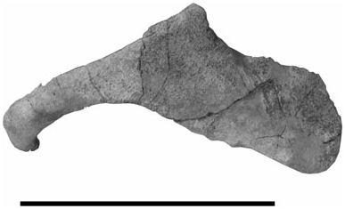
Jugal: The anterior portion of the right jugal is small and projects anteriorly and ventrally ( Fig. 5 View Figure 5 ); at the anteriormost end it protrudes medially. The posterior portion is dorsoventrally broader and flat. At mid length, a triangular projection protrudes distinctly dorsally.
Basicranium
Posterior to the maxilla, the palatine is broadly rectangular and has a posterior portion projecting posteriorly and laterally ( Fig. 6 View Figure 6 ). However, the posterior border of the palatine is not well preserved and it is not possible to assess the true morphology. The palatines are divided along the longitudinal axis by the interposition of the vomer. The posterolateral corner of the palatine is partially superimposed on the ventral surface of the pterygoid whose hamular process is transversely orientated and forms a ventral lamina corresponding to that described by Fraser & Purves (1960) in Balaenidae but developed to a lesser extent. As far as I am aware, a ventral lamina is present only in Balaenidae and Neobalaenidae . The pterygoid projects markedly ventrally and posteriorly. The vomer is interposed between the two pterygoids and is superimposed on the suture between the basioccipital and basisphenoid. The basioccipital is transversely short and bears two large and stocky descending processes forming the posteromedial angle of the basicapsular fissure. The latter is not well preserved and is still partially filled with the matrix. The pars cochlearis of the right periotic is embedded in the matrix and its preservation is so poor that it cannot be fully described. The posterior process of the periotic appears as a strong crest between the postglenoid process of the squamosal and the posteroventral portion of the exoccipital. The exoccipital projects posteriorly and laterally as described above; it appears as a strong structure similar to that observed in Cetotherium -like mysticetes and Eschrichtius robustus .
I was unable to find the tympanic bulla in the MRSN collections. Portis (1885: pl. 2) showed a tympanic bulla associated with the skeleton of E. gastaldii which was transversely broad and anteroposteriorly short, resembling very closely the tympanic bulla of living eschrichtiids (see, for example, specimens USNM 364973, 504305 and 571931). The tympanic bulla of Eschrichtius robustus and Eschrichtioides gastaldii differs from that of living balaenopterids in that it is dorsoventrally higher and anteroposteriorly shorter; the bulla of these eschrichtiids is more compressed in both the anteroposterior and the dorsoventral axes.
Dentary
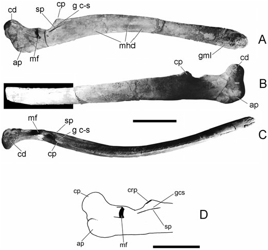
Measurements of the dentary are presented in Table 2. The dentary displays a continuous outward curvature that is interrupted at the level of the coronoid region; the neck of the bone is straight; the Length (straight) 1120 Length (curve) 1140 Height at anterior end 170 Height at condyle 90 Heigth at coronoid process 140 Minimum height between condyle and 140 coronoid process
Height 100 mm behind anterior end 90 Height 200 mm behind anterior end 85 Height 300 mm behind anterior end 85 Height 400 mm behind anterior end 90 Height 500 mm behind anterior end 90 Height 600 mm behind anterior end 95 Height 700 mm behind anterior end 110 Height 800 mm behind anterior end 130 Height 900 mm behind anterior end 95 Height 1000 mm behind anterior end 160 Distance between condyle and coronoid process 200 Distance between coronoid process and 900 anterior end mandibular body exhibits a dorsoventral arc ( Fig. 6 View Figure 6 ). Anteriorly, the dentary does not show any sign of torsion and the groove for the mental ligament is only slightly developed; the dentary terminates anteriorly with a rounded border. Along the ventromedial surface of the ramus, a broad, shallow groove is observed which is located at a position consistent with the attachment of the mylohyoideus muscle; this groove is therefore considered homologous to the mylohyoidal sulcus of Balaenidae . Such a groove is absent in cetotheres and in balaenopterids; in the neobalaenid Caperea marginata Gray, 1873 the mylohyoidaeus muscle is attached to a medial depression rather than a groove as in Balaenidae .
The angular process of the dentary is robust, squared, and high and does not bear any groove for the attachment of pterygoideus muscles. The mandibular condyle is placed at the extremity of a posterodorsal projection, which brings it to a higher level than the top of the coronoid process; its articular surface is mainly orientated dorsally.
The coronoid region is quite complex. In lateral view, the coronoid process is a small and round emergence that is slightly higher than the dorsal border of the ramus; posteriorly, it continues into a postcoronoid crest whose lateral surface forms a wide, long postcoronoid fossa; immediately posterior to the postcoronoid crest, a round, shallow, long concavity is observed in the dorsal border of the dentary. In medial view, posterior to that concavity, there is the opening of a circular, small mandibular foramen which is prolonged into a broad groove projecting posteriorly and dorsally. The coronoid process is paralleled by a medial crest-like emergence which is separated from the coronoid process by a shallow, wide groove. This emergence is homologous to the satellite process described by Bisconti & Varola (2000, 2006) and it is found also in the living Eschrichtius robustus . Bisconti & Varola (2006) suggest that it represents a synapomorphy of Eschrichtiidae .
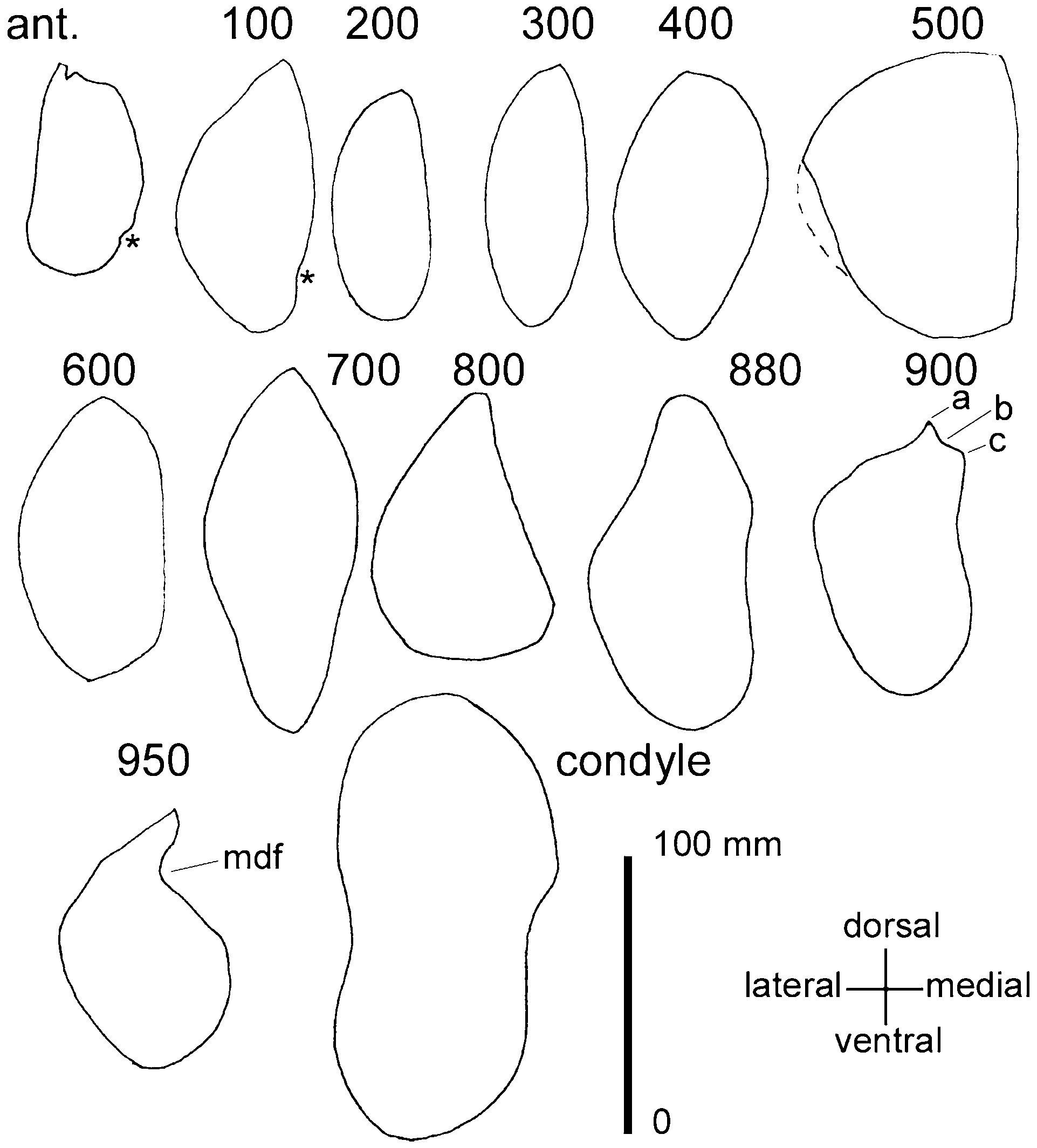
In cross-section, the anterior portion of the dentary has a convex surface both laterally and medially; the medial surface becomes nearly flat around 800 mm behind the anterior apex and shows a medial concavity about 880 mm behind the anterior apex ( Fig. 7 View Figure 7 ). The coronoid process and the parallel satellite process are evident in the section traced 900 mm behind the apex. Approaching the posterior end, the two sides become strongly convex.
Postcrania
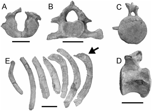
Atlas: The atlas is largely broken. The articular surfaces with the occipital condyles are mainly concave ( Fig. 8A View Figure 8 ); they are elongate dorsoventrally and narrow transversely. The neural channel is wider dorsally than ventrally. The articular surface for the axis is broadly convex. In lateral view, the atlas bears a foramen at the base of the broken dorsal transverse process lateral to the low neural arch. However, the height of the neural process cannot be estimated because it is broken at its base (measurements of postcranial bones are listed in Table 2).
Axis: The axis is almost complete ( Fig. 8B View Figure 8 ). The articular surface with the atlas is concave laterally and convex medially; the border of the articular surface forms a heart-like figure. Ventrally, two massive processes develop which form the ventral border of the foramen transversarium; the distal end of the right process is wide and flat (this process is broken in the left process). The dorsal transverse process is more delicate and its distal end is broken on both sides. Judging from the preservation of the right ventral process, it seems that the foramen transversarium was not completely bordered by bone and that it was laterally incomplete. The neural arch is nearly triangular and is dorsally bordered by a strong, acute neural process which is developed just for a few centimetres due to a breakage of its apical end. The lateral surfaces of the neural arch descend gently laterally and ventrally.
Fourth cervical vertebra: This is largely incomplete, lacking neural arch and ventral transverse processes. The vertebra is tiny, delicate and short. The border of the articular surface is nearly quadrangular.
measurements in mm
No known copyright restrictions apply. See Agosti, D., Egloff, W., 2009. Taxonomic information exchange and copyright: the Plazi approach. BMC Research Notes 2009, 2:53 for further explanation.
|
Kingdom |
|
|
Phylum |
|
|
Class |
|
|
Order |
|
|
Family |
|
|
Genus |
Eschrichtioides gastaldii
| Bisconti, Michelangelo 2008 |
Balaenoptera
| Demere TA & Berta A & McGowen MR 2005: 105 |
Balaenoptera acutorostrata:
| Caretto PG 1970: 57 |