
Sectonema tropicum, Nguyen & Abolafia & Bonkowski & Peña-Santiago & Álvarez-Ortega, 2016
|
publication ID |
https://doi.org/ 10.5852/ejt.2016.171 |
|
publication LSID |
lsid:zoobank.org:pub:2AE15A24-33C0-42F9-BC47-5A44DF36AE9F |
|
DOI |
https://doi.org/10.5281/zenodo.3850207 |
|
persistent identifier |
https://treatment.plazi.org/id/014CA9CB-57EE-43BB-BBFF-0C1A7EBD522D |
|
taxon LSID |
lsid:zoobank.org:act:014CA9CB-57EE-43BB-BBFF-0C1A7EBD522D |
|
treatment provided by |
Valdenar |
|
scientific name |
Sectonema tropicum |
| status |
sp. nov. |
Sectonema tropicum sp. nov.
urn:lsid:zoobank.org:act:014CA9CB-57EE-43BB-BBFF-0C1A7EBD522D
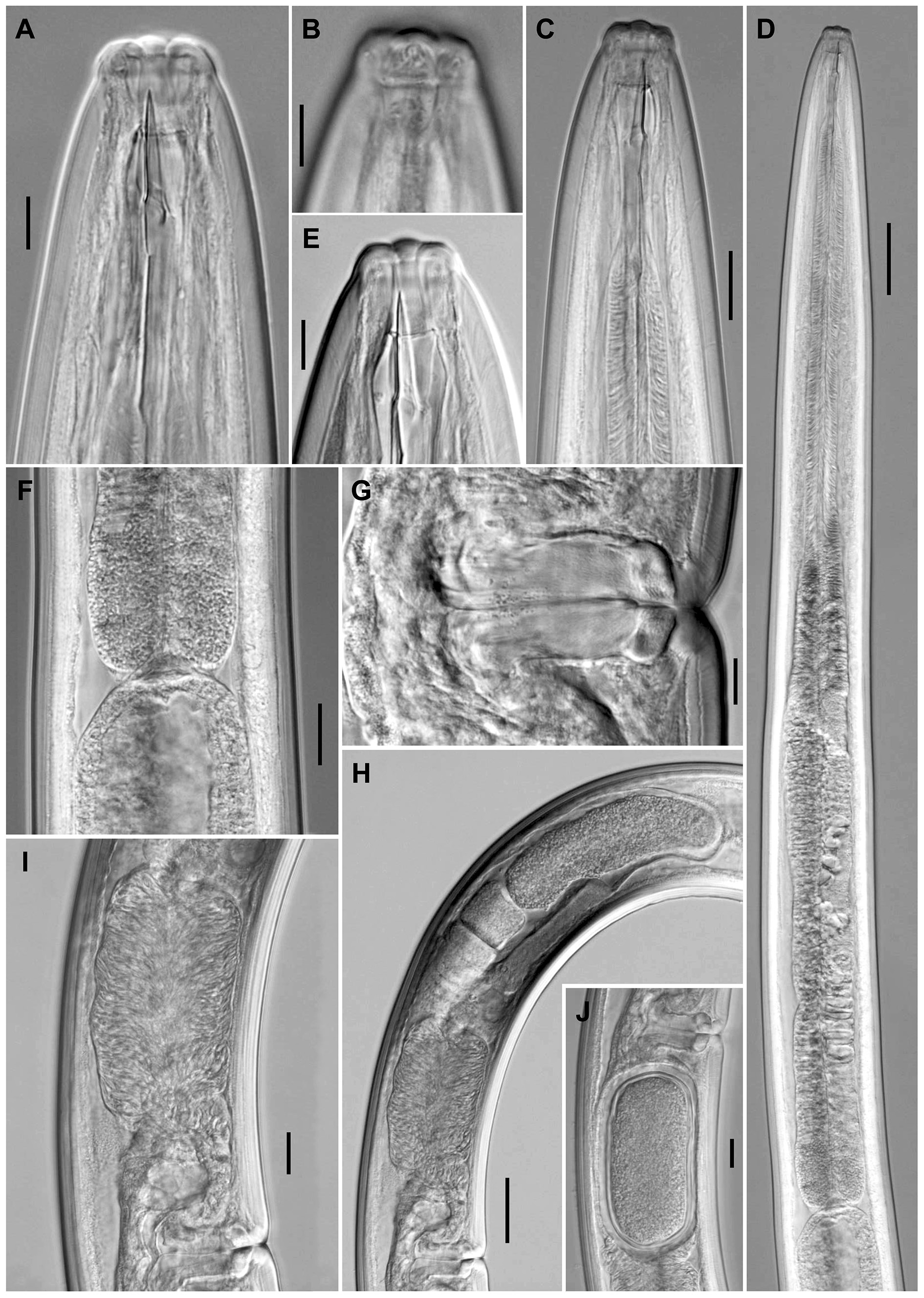
Figs 1–4 View Fig View Fig View Fig View Fig ; Table 1 View Table 1
Diagnosis
The new species is characterized by a 2.56–3.24 mm long body, lip region hardly offset by very weak depression and 19–21 μm broad, odontostyle 20–21 μm long at its ventral side and 6.6–7.1 times as long as wide, 730–834 μm long neck, pharyngeal expansion 403–470 μm long or occupying 52–59% of total neck length, uterus a simple tube-like structure 150–242 μm long or 1.2–2.5 times the body diameter, pars refringens vaginae present, V = 48–52, short (32–39 μm, c = 70–88, c’ = 0.5–0.6) and rounded female tail, male tail similar to that of female (31–40 μm, c = 73–91, c’ = 0.5–0.6), 91–97 μm long spicules, and only one weakly developed ventromedian supplement.
Etymology
The specific epithet refers to the tropical area where the new species was collected.
Type material examined
Holotype GoogleMaps
VIETNAM: ♀, in acceptable state of preservation, Northern Vietnam, Cao Bang Province, Pia Oac Natural Reserve, 22º 36’28’’ N, 105º 52’15’’ E, tropical evergreen forest soil associated with Machilus sp. and Dimocarpus sp., deposited in the nematode collection of the University of Jaén GoogleMaps , Spain GoogleMaps .
Paratypes
VIETNAM: 4 ♀♀, 3 ♁♁, in acceptable state of preservation, same data as holotype; 1 ♀, 1 ♁, in acceptable state of preservation, same locality, deposited in the nematode collection of the Institute of Ecology and Biological Resources, Hanoi, Vietnam; 1 ♁, same locality, used for SEM.
Description
Adult
Moderately slender to slender nematodes of medium to big size, 2.56–3.24 mm long. Body cylindrical, distinctly tapering towards the anterior end, less so towards the posterior end as the caudal region is short and rounded. Habitus curved ventrad after fixation, especially in posterior body region, C-, G- or spiral-shaped. Cuticle 3.0–4.5 μm thick at anterior region, 5–6 μm in mid-body and 8–10 μm on tail; consisting of three layers, especially distinguishable at caudal region: thinner outer layer bearing very fine transverse striation across the entire body, thicker intermediate layer with radial striation, and thin inner layer. Lateral chords 13–21 μm wide at mid-body, occupying about one-sixth (13–18%) of midbody diameter. Two ventral and two dorsal body pores often present at level of odontostyle-odontophore. Lip region hardly offset from the adjacent body by weak, but perceptible depression, 2.8–3.0 times wider than high and up to one-fourth (17–25%) of body diameter at neck base; lips (under SEM) mostly amalgamated, but their perioral part distinctly separated by the existence of six radial, interlabial, deep incisures delimiting six perceptible liplets; button-like labial papillae, the inner ones located at the margin of the oral field and surrounded by two or three concentric annuli, whereas the outer papillae, located a little behind the inner papillae, are surrounded by only one annulus; cephalic papillae pore- rather than button-like, also surrounded by only one ring-like annulus; oral aperture a dorso-ventral, slightly hexagonal orifice, the lip region hence showing a bi-radial symmetry. Amphid fovea cup-shaped, its opening occupying 9–10 μm or less than one-half (43–49%) of lip region diameter. Cheilostom nearly cylindrical, without any differentiation. Stomatal protruding structure apparently a reduced odontostyle 6.6–7.1 times longer than wide (see: Remarks), its ventral side 1.0–1.1 times longer than lip region diameter and 0.65–0.83% of body length. Guiding ring simple, plicate, at 0.8–0.9 lip region diameters from the anterior end. Odontophore linear, rod-like, 1.7–1.9 times the odontostyle length, somewhat irregular at its base and with (in lateral view) the ventral side slightly longer than the dorsal one ( Fig. 2A View Fig ). Anterior region of pharynx enlarging very gradually; basal expansion 7.1–10.7 times longer than wide, 3.6–5.6 times as long as body diameter, and occupying 52–59% of total neck length; gland nuclei often obscure, located as follows: DN = 60–62 (n = 4); S 1 N 1 = 75 (n = 2); S 1 N 2 = obscure; S 2 N = obscure. Nerve ring located at 182–216 μm from anterior end or 25–26% of total neck length. Cardia rounded conoid, 13–17 × 13–17 μm; a weak ring-like structure is present surrounding its junction to pharyngeal base.
Female
Genital system didelphic-amphidelphic, with almost equally and well developed branches, the anterior 320–442 μm or 12–15% of body length and the posterior 348, 440 μm (n = 2) or 14, 16% of body length (447, 509 μm or 15, 17% bearing one uterine egg inside). Reflexed ovaries well developed, often surpassing the sphincter level, the anterior 182–319 μm, the posterior 207–380 μm long; oocytes arranged first in two or more rows, then in a single row. Oviduct 124–174 μm long or 1.1–1.8 times the corresponding body diameter, and consisting of a slender part made of prismatic cells and a moderately developed pars dilatata, with visible lumen and often containing sperm cells inside. Oviduct-uterus junction marked by a sphincter. Uterus a simple, tube-like structure 150–190 μm long or 1.2–1.7 times the corresponding body diameter [223, 242 μm (n = 2) long or 2.2, 2.5 times the corresponding body diameter, with one uterine egg inside; and 231, 240 μm (n = 1) long, with two uterine eggs], always containing abundant sperm cells inside. Vagina extending inwards 52–57 μm or one-half to foursevenths (48–57%) of body diameter: pars proximalis 33–43 × 23–28 μm, with somewhat sigmoid walls surrounded by weak musculature; pars refringens with two trapezoidal pieces measuring (in optical section) 13–16 × 7–9 μm and with a combined width of 24–28 μm; pars distalis 5.5–7.0 μm long. Vulva a nearly equatorial transverse slit. Prerectum 2.0–2.4, rectum 0.7–1.0 anal body diameters long. Tail short and rounded; two pairs of caudal pores, sublateral and close together.
Male
Genital system diorchic, with opposite testes. In addition to the ad-cloacal pair, situated at 18–21 μm from cloacal aperture, one weakly developed ventromedian supplement, lying out the range of retracted spicules and located at 80–131 μm from the ad-cloacal pair. Spicules distinctly robust and massive, especially in its posterior half, 3.6–4.2 times its maximum width, 1.4–1.6 times the body diameter at level of the cloacal aperture: dorsal contour regularly convex, ventral contour slightly concave, with distinct hump and hollow; curvature 121–124°; head occupying 11–16% of spicule total length, its dorsal side visibly curved at its anterior end and longer than the ventral one, which is shorter and almost straight; median piece 5.4–6.2 times longer than wide, occupying 51–59% of spicule maximum width, reaching the posterior tip; posterior end 7–8 μm wide. Lateral guiding pieces 23–29 μm long, 5.3– 7.1 times longer than wide. Prerectum 3.2–4.6, cloaca 0.9–1.3 times the corresponding body width long. Cloacal aperture, as seen under SEM, a curved anteriad, transverse slit. Tail similar to that of female; caudal pores two pairs, one lateral, another subdorsal.
Relationships
In having a long stomatal protruding structure (20–21 μm at its ventral side) and short (c’ up to 1.0) and rounded tail, S. tropicum sp. nov. is morphologically very similar to S. demani Altherr, 1965 , S. heynsi Altherr, 1968 , S. rotundicauda Goodey, 1951 , S. septentrionale Peña-Santiago & Álvarez-Ortega, 2015 and S. sica Clark, 1964 , but it can be distinguished from all of them in its smaller general size (body length 2.56–3.24 mm, neck 730–834 μm long vs more than 5 mm, more than 1000 μm long, respectively), nearly continuous lip region (vs offset by a more or less deep constriction) and stomatal protruding structure of different nature (a reduced odontostyle vs a mural tooth; see Remarks). Besides, the new species differs from S. demani (see recent re-description by Peña-Santiago & Álvarez-Ortega 2014b) in its less slender (a = 21–36 vs a = 48–56) body, narrower (19–21 vs 27–28 μm broad) lip region, shorter pharyngeal expansion (occupying 52–59 vs 68–69% of total neck length), shorter female tail (32–39 vs 51–55 μm, c’ = 0.5–0.6 vs c’ = 0.7–0.8), and male present (vs absent). It differs from S. heynsi (see recent re-description by Peña-Santiago & Álvarez-Ortega 2014b) in its narrower (19–21 vs 28 μm broad) lip region, circumoral area lacking (vs bearing) cilia- or setae-like structures, shorter pharyngeal expansion (occupying 52–59 vs 68% of total neck length), shorter (32–39 vs 54 μm, c’ = 0.5–0.6 vs c’ = 0.7) female tail, and male present (vs absent). It differs from S. rotundicauda in its less slender (a = 21–36 vs a = 80) body, narrower (19–21 vs about 28 μm broad) lip region, shorter (31–40 vs 64 μm) male tail, fewer (one vs four) ventromedian supplements, and shorter (91–97 vs about 155 μm) spicules. It differs from S. septentrionale in its less slender (a = 21–36 vs a = 47–64) body, narrower (19–21 vs 25–29 μm broad) lip region, shorter (occupying 52–59 vs 60–73% of total neck length) pharyngeal expansion, uterus simple (vs tripartite) and shorter (150–242 μm or 1.2–2.5 times the body diameter vs 370–493 μm or 2.9–4.3 times the corresponding body diameter), shorter (31–40 vs 40–62 μm, c’ = 0.5–0.6 vs c’ = 0.6–0.8) tail, fewer (one vs 5–11) ventromedian supplements, and shorter (91–97 vs 100–145 μm) spicules. Finally, it differs from S. sica in its narrower (19–21 vs about 24 μm broad) lip region, shorter (occupying 52–59% vs two-thirds of total neck length) pharyngeal expansion, more posterior (V = 48–52 vs V = 40) vulva, shorter (32–39 vs 54 μm, c’ = 0.5–0.6 vs c’ = 0.7) female tail, and male present (vs absent).
Moreover, in having a lip region nearly continuous with the adjacent body, the new species resembles S. mucrodens Siddiqi, 1984 and S. truxum Siddiqi, 1984 , but it can be easily distinguished from these by the more anterior position of the stomatal protruding structure (its anterior tip in front of the level of guiding ring, just behind the base of lip region vs its anterior tip distinctly behind the level of guiding ring, far from the lip region base) and pars refringens vaginae well (vs faintly) developed. Moreover, it differs from S. mucrodens by its shorter (730–834 vs 1086–1157 μm) neck, shorter (occupying 52–59 vs 69% of total neck length) pharyngeal expansion, shorter (32–39 μm, c’ = 0.5–0.6 vs 44–50 μm, c’ = 0.7) female tail, and male present (vs absent). It differs from S. truxum in its narrower (19–21 vs 24 μm broad) lip region, shorter (730–834 vs 837–1070 μm) neck, more anterior (V = 48–52 vs V = 54–56) vulva, and male present (vs absent).
Molecular characterisation
Two sequences of the D2–D 3 28S rRNA gene were obtained from one female and one male. Both sequences are very similar (99%) and differ by two nucleotides only. The evolutionary relationships of the new species with representatives of the order Dorylaimida are presented in Figs 5–6 View Fig View Fig . The two S. tropicum sp. nov. sequences are clustered together with other available sequences of Sectonema representatives and form a highly supported clade together with members of the genus Metaporcelaimus , thus confirming previous results ( Álvarez-Ortega et al. 2013a, b).
Remarks
The nature of the stomatal protruding structure in the new species is difficult to interpret and deserves further explanation. The specimens examined bear a reduced odontostyle (comparable to that described in the type species S. ventrale Thone, 1930 ; see recent description by Peña-Santiago & Álvarez-Ortega 2014a) rather than a mural tooth as the base of its dorsal side is visibly sclerotized and seems to join the dorsal stomatal wall (vs dorsal side not scletorized at the base, somewhat sigmoid and not joining the dorsal stomatal wall). Nonetheless, some doubt persists in this matter.


No known copyright restrictions apply. See Agosti, D., Egloff, W., 2009. Taxonomic information exchange and copyright: the Plazi approach. BMC Research Notes 2009, 2:53 for further explanation.
|
Kingdom |
|
|
Phylum |
|
|
Class |
|
|
Order |
|
|
Family |
|
|
Genus |