
Plagiosterna aenea ( Linnaeus, 1758 )
|
publication ID |
https://doi.org/ 10.11646/zootaxa.4236.2.12 |
|
publication LSID |
lsid:zoobank.org:pub:43E1D901-D818-4006-B0BD-8FF1B3774C69 |
|
DOI |
https://doi.org/10.5281/zenodo.6019035 |
|
persistent identifier |
https://treatment.plazi.org/id/039C87CE-FFFA-A14B-FF32-FC9D90F7FC5D |
|
treatment provided by |
Plazi |
|
scientific name |
Plagiosterna aenea ( Linnaeus, 1758 ) |
| status |
|
Description of the larva of Plagiosterna aenea ( Linnaeus, 1758)
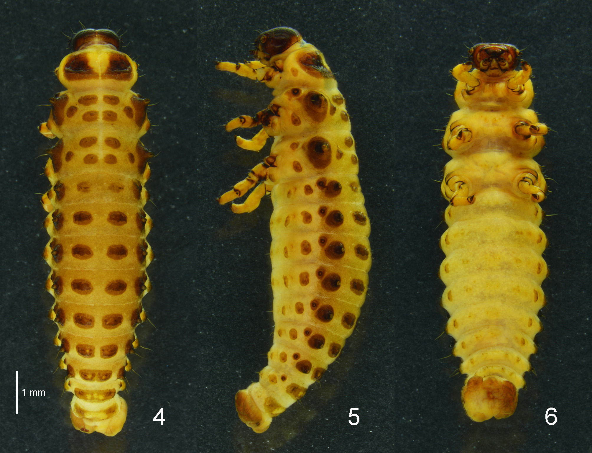
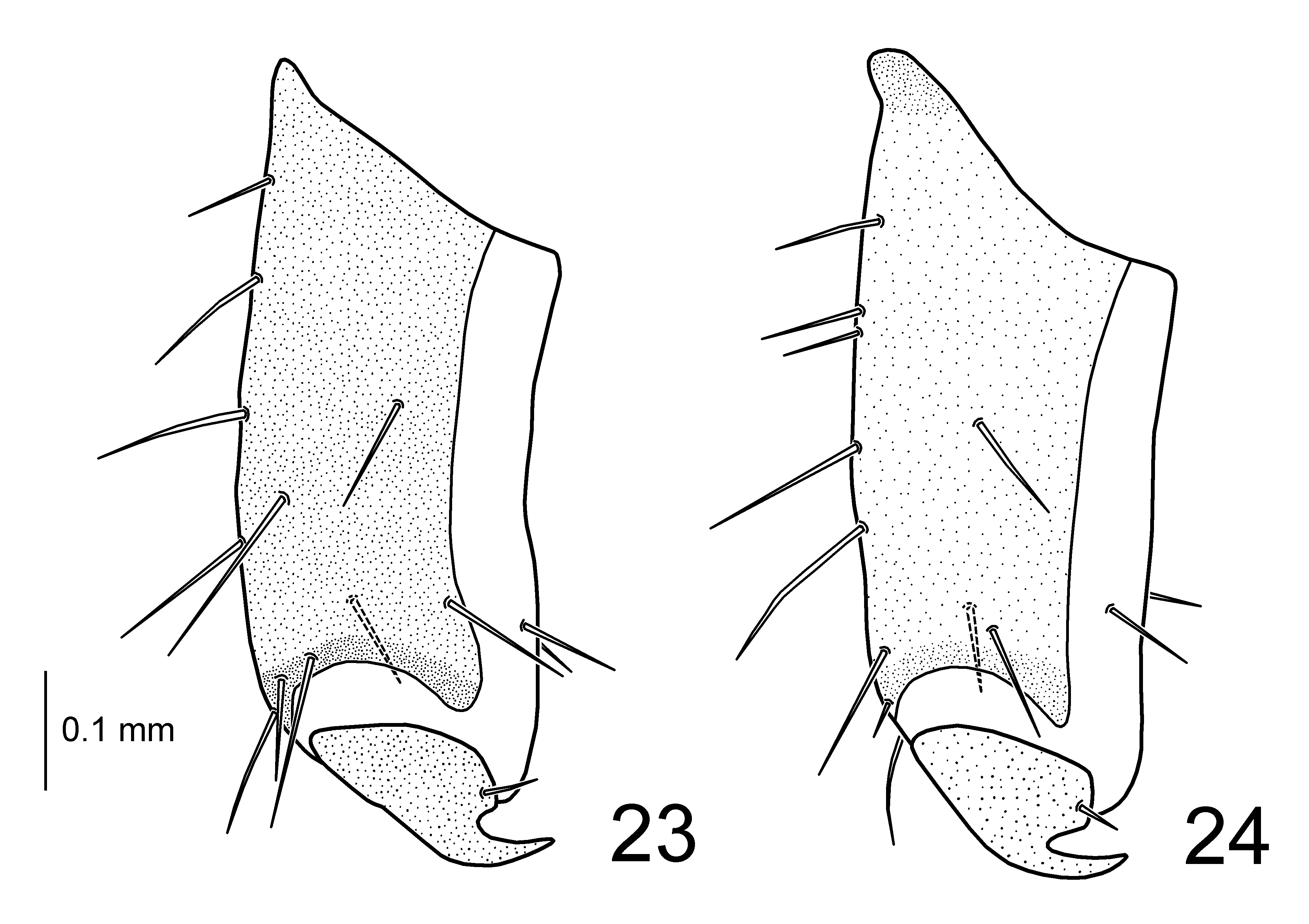
( Figs 4–6 View FIGURES 4 – 6 , 15–22 View FIGURES 15 – 22 , 24 View FIGURES 23 – 24 , 27–29 View FIGURES 25 – 29 )
First instar larva. Body length 2.40–2.60 mm, width 0.70–0.80 mm, head width 0.58 mm (n = 2). Body yellowish-white with head dark brown, tubercles and legs brown. Sclerotized platelets on dorsum rather dense, on venter sparse. Defensive glands present on meso- and metathorax and abdominal segments I–VII. Small egg bursters present on meso- and metathorax. Head and mouthparts similar in the shape and chaetotaxy to those of the last instar larva.
Thorax. Prothorax with D-DL-EPa (8–11L 7–9S 3–4M); EPp (1S 2M); P (3S); ES-SS (1S 2M) ( Fig. 27 View FIGURES 25 – 29 ). Meso- and metathorax with Da (2–4S 0–2M); Dp (1L 0–1S) smaller than Da; DLi (1L 2S) with a small egg burster situated anterior to a long seta; DLe (1L 5–6S 0–1M) conical with a defensive gland; EPa (4–5S); EPp (1L 3–4S); P (2S); SS (1M); ES (1S 1M).
Abdomen. Segments I–VI with D (1L 4–6S); DL (1L 2S 1–2M) with a defensive gland; EP (1L 4–5S); P (1L 2–3S); PS-SS (2–3S 1M) generally divided into 2 tubercles; ES (2S) on both sides fused; as1 (1M). Segment VII with D on both sides fused; DL with a defensive gland. Segment VIII with dorsal and dorsolateral tubercles fused. Segment IX with D-DL-EP (5S 4–5M) fused. Segment X with pygopod well developed.
Third (last) instar larva. Body length 8.6–10.1 mm, width 2.3–3.3 mm, head width 1.27–1.37 mm (n = 8). Body elongate, widest at abdominal segments II–III and moderately convex dorsally ( Figs 4–5 View FIGURES 4 – 6 ). Head brown to dark brown with mouthparts partially yellowish-white; dorsal tubercles brown to pale brown, whereas ventral ones unpigmented ( Fig. 6 View FIGURES 4 – 6 ); median region of D-DL-EPa on prothorax unpigmented; sometimes median region of D on abdominal segments VI–IX unpigmented; very rarely all tubercles pigmented ( Fig. 29 View FIGURES 25 – 29 ); dorsal integument yellowish-brown except median line of thorax on account of dense and moderately sclerotized platelets, whereas ventral one pale yellow on account of sparse and weakly sclerotized platelets; legs pale yellow with apex of each segment dark brown. Defensive glands present on meso- and metathorax and abdominal segments I–VII.
Head. Hypognathous, rounded, strongly sclerotized ( Fig. 15 View FIGURES 15 – 22 ). Vertex with 4 pairs of minute setae; epicranium with 7–9 pairs of setae; temporal side of head with 4 pairs of setae. Epicranial suture distinct; frontal suture short, not reaching antennal socket; endocarina well developed. Frons with 3 pairs of setae. Clypeus trapezoidal with 3 pairs of setae. Labrum deeply emarginate with 2 pairs of short and 1 pair of minute setae and 2 pairs of campaniform sensilla placed medially ( Fig. 20 View FIGURES 15 – 22 ); epipharynx with 3 pairs of stout setae at anterior margin ( Fig. 21 View FIGURES 15 – 22 ). Mandible palmate, 5-toothed, with 2 setae and 2 campaniform sensilla ( Figs 16–17 View FIGURES 15 – 22 ). Maxillary palp 3-segmented; palpomere I transverse with 1 seta and 1 campaniform sensillum; II rectangular with 2 setae and 1 campaniform sensillum; III subconical with 1 seta, 1 digitiform sensillum and 1 campaniform sensillum on sides and a group of peg-like sensilla at the apex; palpifer distinct with 2 setae ( Fig. 22 View FIGURES 15 – 22 ). Mala rounded with 10 setae and 1 campaniform sensillum; stipes longer than wide with 3 setae; cardo without setae. Labial palp 2-segmented; palpomere I rectangular with 1 minute seta and 1 campaniform sensillum; II subconical with 1 campaniform sensillum on outer side and a group of peg-like sensilla at the apex. Hypopharyngeal area with 3 pairs of setae (2 of them placed internally, not visible on Fig. 22 View FIGURES 15 – 22 ) and 1 pair of campaniform sensilla. Prementum with 2 pairs of setae; postmentum with 3 pairs of setae. Stemmata 6 on each side, 4 of them located above antenna and 2 behind antenna. Antenna short, 3-segmented; antenomere I transverse with 3 campaniform sensilla; II stout with a conical sensorium and 5 setae; III subconical with 5 setae and 1 campaniform sensillum ( Figs 18–19 View FIGURES 15 – 22 ).
Thorax. Prothorax with D-DL-EPa (1L 17–19S) largest; EPp (3–4S) small and rounded; P (2S); ES-SS (2–4S) not sclerotized ( Fig. 28 View FIGURES 25 – 29 ). Meso- and metathorax with Da (3–4S 2M) rarely divided into large Dai and small Dae; Dp (2–3S) smaller than Da; DLi (2–3S) smaller than Dp; DLe (1L 7–9S 1M) large and conical with a defensive gland; EPa (3–6S); EPp (1L 3–5S); P (1–3S); SS (1S) and ES (2S) not sclerotized. Mesothoracic spiracles annuliform; peritreme fused with EPa. Legs rather stout; tibia with 13 setae; tarsungulus strongly curved, basal tooth well developed, with 1 minute seta ( Fig. 24 View FIGURES 23 – 24 ).
Abdomen. Segments I–VI with D (0–1L 6–8S) wider than long; DL (1L 2–3S) conical with a defensive gland; EP (1L 5–6S); P (1L 1–3S) smaller than EP, rarely equal in size; PS-SS (2–4S) sometimes divided into 2–3 tubercles, slightly sclerotized; ES (2S) on both sides fused or separated, slightly sclerotized; as1 (1S) very small. Segment VII with D on both sides fused; DL with a defensive gland. Segments VIII–IX each with dorsal and dorsolateral tubercles fused (2L 6–7S 2M and 9S 2–4M respectively). Segment X with pygopod well developed. Spiracles present on segments I–VIII.
Diagnosis. The last instar larva of Plagiosterna aenea is similar to that of P. aeneipennis in the pale brown dorsum on account of weakly sclerotized platelets. However, this species can be distinguished from P. aeneipennis by the dorsal tubercles well sclerotized, tubercle D on abdomen well developed and legs pale yellow (dorsal tubercles weakly sclerotized, tubercle D on abdomen hardly recognizable and legs dark brown in P. aeneipennis ).
Material examined. 4 first and 10 last instar larvae, South Korea, Gyeongbuk Prov., Cheongsong, Mt. Bohyeonsan, 17.V.2006, H.W. Cho; 3 last instar larvae, Poland, Złotoryja, Wojcieszów, 3.VII.2013, H.W. Cho.
Distribution. Transpalaearctic species: Albania, Austria, Bosnia Herzegovina, Bulgaria, Belarus, Croatia, Czech Republic, Denmark, Estonia, Finland, France, Germany, United Kingdom, Hungary, Italy, Latvia, Lithuania, Macedonia, Norway, Poland, Romania, Slovakia, Slovenia, Spain, Sweden, Serbia and Montenegro, Turkey, Ukraine, Russia (Central & North European Territories, West & East Siberia, Far East), China (Heilongjiang, Jilin, Liaoning), Korea, Japan ( Kippenberg 2010). This species is excluded from the Taiwanese fauna (see comments under P. formosana ).
Host plant. Betulaceae : Alnus spp.
Remarks. Plagiosterna aenea is divided into three subspecies on the basis of coloration: P. aenea aenea from United Kingdom to Japan (Hokkaido, Honshu, Kyushu, Shikoku), P. aenea insularis Chûjô from Japan (Shikoku, Kyushu) and P. aenea tsutsuii Nakane from Japan (South Honshu). Diagnostic characters of larvae of these subspecies were provided by Takizawa (1976a) and Kimoto & Takizawa (1994). These subspecies generally occupy different regions of Japan (Takizawa 2016, personal communication). Plagiosterna aenea aenea is very common in Hokkaido and middle Honshu, but is gradually diminished and restricted to montane areas in Kyushu and Shikoku. Plagiosterna aenea tsutsuii is scattered in the southern area of Honshu (Sizuoka, Yamanasi, Nagano and Aiti Prefectures). The hybrid zone of P. aenea aenea and P. aenea tsutsuii is known from the Izu Peninsula.
Larval characters of Korean specimens of Plagiosterna aenea are identical to those of Polish specimens. A remarkable variation in pigmentation of tubercle is found in one specimen from Poland ( Fig. 29 View FIGURES 25 – 29 ), but this phenomenon seems to be extremely rare. Recently, Park et al. (2015) wrote “In the Korean specimens, the tubercles on the dorsal surface, except glanduliferous tubercle (DL), disappear in the last instar larval stage. This character is consistent with those noted in European specimens ( Lipp 1935)”. However, the description and illustration of P. aenea in their text clearly shows dorsal tubercles, and Lipp (1935) mentioned that sternal tubercles are disappeared in the last instar larva. The color polymorphism with disappearance of dorsal tubercles has been known only from Japanese population of P. aenea aenea .


No known copyright restrictions apply. See Agosti, D., Egloff, W., 2009. Taxonomic information exchange and copyright: the Plazi approach. BMC Research Notes 2009, 2:53 for further explanation.
|
Kingdom |
|
|
Phylum |
|
|
Class |
|
|
Order |
|
|
Family |
|
|
Genus |
Plagiosterna aenea ( Linnaeus, 1758 )
| Cho, Hee-Wook & Świętojańska, Jolanta 2017 |
Plagiosterna aenea (
| Linnaeus, C. 1758: 384 |