
Prionospio rotunda, Delgado-Blas, Víctor Hugo, 2015
|
publication ID |
https://doi.org/ 10.11646/zootaxa.3905.1.4 |
|
publication LSID |
lsid:zoobank.org:pub:6C454B4B-E32D-4B55-B195-32A575BCC858 |
|
DOI |
https://doi.org/10.5281/zenodo.6113897 |
|
persistent identifier |
https://treatment.plazi.org/id/039D3B4F-812F-125C-FF3A-DEE0F783FBF1 |
|
treatment provided by |
Plazi |
|
scientific name |
Prionospio rotunda |
| status |
sp. nov. |
Prionospio rotunda View in CoL sp. nov.
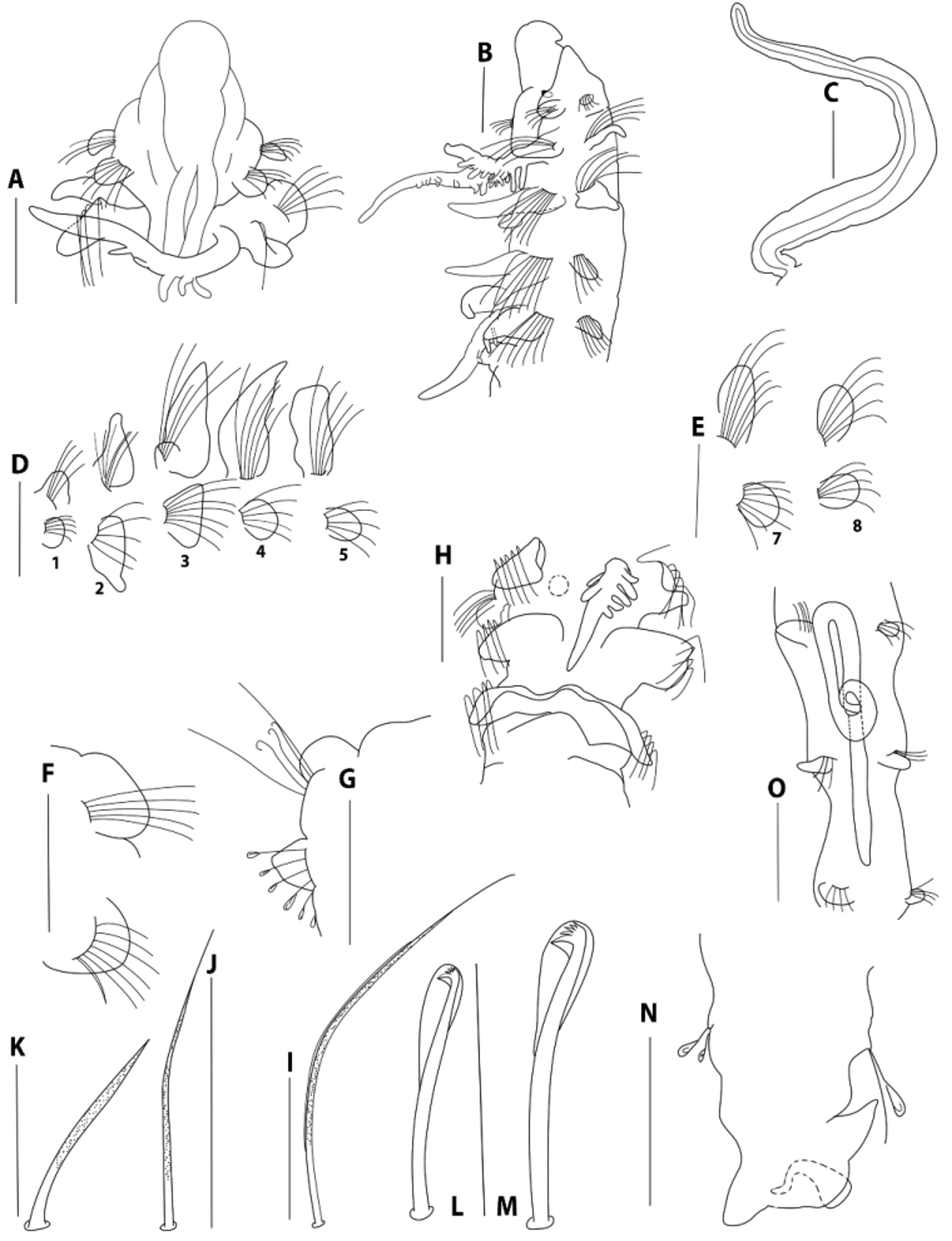
Figure 8 View FIGURE 8 A – O
Material examined. CARIBBEAN SEA. Quintana Roo: Mahahual 24 October 2008, coll. V.H. Delgado-Blas, holotype (LACM-AHF-POLY 6601); E31, on the coast at Yalahau, 22º30’990’’N, 087º05’010’’W, November 2001, 2 paratypes (ECOSUR0170); GULF OF MEXICO. Yucatan: E5, on the coast at Progreso, 22º47’035’’N, 90º20’15’’W, October 2001, 1 paratype (LACM-AHF-POLY 6602).
Description. Holotype complete, 5.0 mm long for 49 chaetigers, 0.2 mm wide. Paratypes complete, 45–47 chaetigers, 5.5 mm long, 0.2 mm wide. Color in alcohol pale white. Prostomium pyriform-shaped, rounded anteriorly ( Fig. 8 View FIGURE 8 A), tapered posteriorly, with long, blunt caruncle extending to the posterior edge of chaetiger 2 with nuchal organs fused on either side ( Fig. 8 View FIGURE 8 A). Specimens with two pairs of red-brown subdermal eyes or eyes absent (1 paratype); anterior pair small, rounded ( Fig. 8 View FIGURE 8 B); posterior pair cup-shaped. Palps lost (Holotype), except one specimen with a pair of long palps, grooved, lacking basal sheaths ( Fig. 8 View FIGURE 8 C) extending up to chaetiger 12. Peristomium moderate in size, collar-like, surrounding the prostomium, fused dorsally with large, rounded notopodial lamellae of chaetiger 1 ( Fig. 8 View FIGURE 8 A, B, D). Neuropodial postchaetal lamellae of chaetiger 1 large, rounded ( Fig. 8 View FIGURE 8 B, D), much smaller than half the size of the notopodial lamellae.
Four pairs of long branchiae present on chaetigers 2–5 ( Fig. 8 View FIGURE 8 B); first pair longer than fourth pair. Pairs 1 and 4 with a few long, digitiform pinnules on posterior face, branchiae with very long naked, smooth distal tips ( Fig. 8 View FIGURE 8 A–B) and naked basal region; pinnules thick, blunt, long on middle region of the branchiae ( Fig. 8 View FIGURE 8 A); the central stem of branchial pairs 1 and 4 pinnate, elongate. Pairs 2 and 3 apinnate, cirriform, long with a dense lateral ciliation ( Fig. 8 View FIGURE 8 B); subequal in length, shorter than pinnate pairs, but longer than notopodial lamellae.
Notopodial postchaetal lamellae triangular and thinner on chaetigers 2–5 ( Fig. 8 View FIGURE 8 D); lamellae larger on chaetiger 4, largest on chaetigers 3–4 and with triangular tips ( Fig. 8 View FIGURE 8 D); becoming more oval on chaetigers 7–14 ( Fig. 8 View FIGURE 8 E); progressively decreasing in size on middle chaetigers ( Fig. 8 View FIGURE 8 F) and becoming more rounded on posterior chaetigers ( Fig. 8 View FIGURE 8 G). Notopodial lamellae united across dorsum of chaetiger 7 only, forming a high dorsal crest ( Fig. 8 View FIGURE 8 H); chaetiger 8 and subsequent chaetigers without dorsal fold. Ventral and dorsal edges of notopodial and neuropodial lamellae not touching on anterior chaetiger ( Fig. 8 View FIGURE 8 B, D). Notopodial prechaetal lamellae very short in branchial region ( Fig. 8 View FIGURE 8 B, D), inconspicuous thereafter.
Anterior neuropodial postchaetal lamellae rounded throughout, except the neuropodia on chaetigers 2–3; neuropodium on chaetiger 2 large and subtriangular, ventrally pointed ( Fig. 8 View FIGURE 8 D), neuropodium on chaetiger 3 trapezoid, slightly dorsally pointed ( Fig. 8 View FIGURE 8 D); second and third pairs larger than the other neuropodial lamellae; subsequent neuropodial lamellae small and rounded on middle chaetigers ( Fig. 8 View FIGURE 8 D – F) and subtriangular on posterior chaetigers ( Fig. 8 View FIGURE 8 G). Neuropodial prechaetal lamellae very short ( Fig. 8 View FIGURE 8 D), rudimentary throughout. Interparapodial pouches lacking.
All capillaries on the anterior chaetigers granulated ( Fig. 8 View FIGURE 8 I, J); notopodial and neuropodial capillaries of chaetiger 1 arranged in one row, with short, slender chaetae; notopodial chaetae longer. Notopodial capillaries of chaetigers 2–12 arranged in two rows, with very long and very acute unilimbated chaetae ( Fig. 8 View FIGURE 8 I); upper chaetae much longer than lower ones, chaetae curving outwards and upwards, in posterior segments becoming shorter. Neuropodial capillaries of chaetigers 2–9 arranged in two rows, with short and very acute alimbated chaetae ( Fig. 8 View FIGURE 8 J), anterior row shorter than posterior row, later becoming one row; longer, slender and non-granulated capillaries on median and posterior chaetigers. Sabre chaetae in neuropodia from chaetiger 10, up to two per fascicle, stout, curved, moderately granulated, without sheaths ( Fig. 8 View FIGURE 8 K). Neuropodial hooded hooks ( Fig. 8 View FIGURE 8 L) from chaetigers 11–12, up to six per fascicle, accompanied by capillaries. Notopodial hooded hooks ( Fig. 8 View FIGURE 8 M) from chaetigers 28–32, up to four per fascicle. Accompanied by up to two thin capillaries. All hooks with four pairs of teeth above the main tooth, with large secondary hoods ( Fig. 8 View FIGURE 8 L, M).
Pygidium with one long median cirrus and two longer lateral lobes ( Fig. 8 View FIGURE 8 N).
One specimen had up to 12 gregarine parasites within the gut, between chaetigers 7 to 24 ( Fig. 8 View FIGURE 8 O).
Remarks. Examination of the 35 previously known species of the P. steenstrupi group ( Sigvaldadóttir & Mackie, 1993: Table 2; Blake, 1996; Sigvaldadóttir 1997; Zhou & Li 2009) reveals that Prionospio rotunda sp. nov. is very similar to P. fallax Södertröm, 1920 and P. membranacea Imajima 1990 in having a dorsal crest on chaetiger 7 and the same shaped neuropodial lamellae on chaetiger 2. However, Prionospio rotunda differs from the redescription of P. fallax by Sigvaldadóttir & Mackie (1993) and the original description of P. membranacea in that the former has an anteriorly rounded prostomium; a long, blunt caruncle extending to the posterior edge of chaetiger 2 with nuchal organs fused over either side, cirriform second and third branchial pairs; trapezoid, ventrally pointed neuropodial lamellae on chaetiger 3, subtriangular neuropodial lamellae on the posterior chaetigers, and all hooks with four pairs of teeth above the main tooth. In addition, P. ro t u nd a differs from P. fallax in that the former has branchiae with pinnules arranged along the posterior margins of the stems, and large, rounded neuropodial lamellae on chaetiger 1. Finally, P. rotunda differs from P. membranacea in that the former has neuropodial hooded hooks from chaetigers 11–12 and notopodial hooded hooks from chaetigers 28–32. The differences between P. rotunda and the other species examined are provided in the key and Table 1.
Gregarine parasites were found in the digestive tract of one specimen of P. ro t u nd a sp. nov. Douglas & Jones (1991) reported on gregarine associated with eight spionid species ( Boccardia proboscidea Hartman, 1940 , Boccardiella hamata (Webster, 1879) (cited as Boccardia hamata ), Polydora cornuta Bosc, 1802 (cited as Polydora ligni Webster, 1879 ), Polydora nuchalis Woodwick, 1953 , Dipolydora socialis (Schmarda, 1861) (cited as Polydora socialis ), Streblospio benedicti Webster, 1879 , Spio maculata (Hartman, 1961) (cited as Scolelepis maculata ), and Pygospio elegans Claparède, 1863 ) where the gregarine were found in the gut lumen.
Etymology. The species name is from the Latin rotundus meaning rounded and refers to the anteriorly rounded prostomium.
Type locality. Gulf of Mexico: Yucatan, Caribbean Sea: Mahahual.
No known copyright restrictions apply. See Agosti, D., Egloff, W., 2009. Taxonomic information exchange and copyright: the Plazi approach. BMC Research Notes 2009, 2:53 for further explanation.