
Leptonetela latapicalis, He & Liu & Xu & Yin & Peng, 2019
|
publication ID |
https://doi.org/ 10.11646/zootaxa.4554.2.10 |
|
publication LSID |
lsid:zoobank.org:pub:90C205A6-B1CC-4377-990E-E511A4F1989A |
|
DOI |
https://doi.org/10.5281/zenodo.5928047 |
|
persistent identifier |
https://treatment.plazi.org/id/039D87A4-AC02-201E-FF33-7A19DF528ACF |
|
treatment provided by |
Plazi |
|
scientific name |
Leptonetela latapicalis |
| status |
sp. nov. |
Leptonetela latapicalis View in CoL sp. nov.
Figures 5–8 View FIGURES 5 View FIGURES 6 View FIGURES 7 View FIGURES 8 , 12 View FIGURE 12
http://zoobank.org/NomenclaturalActs/ 9D53BD4D-BCDF-4DA2-9369-6AADA3C89151
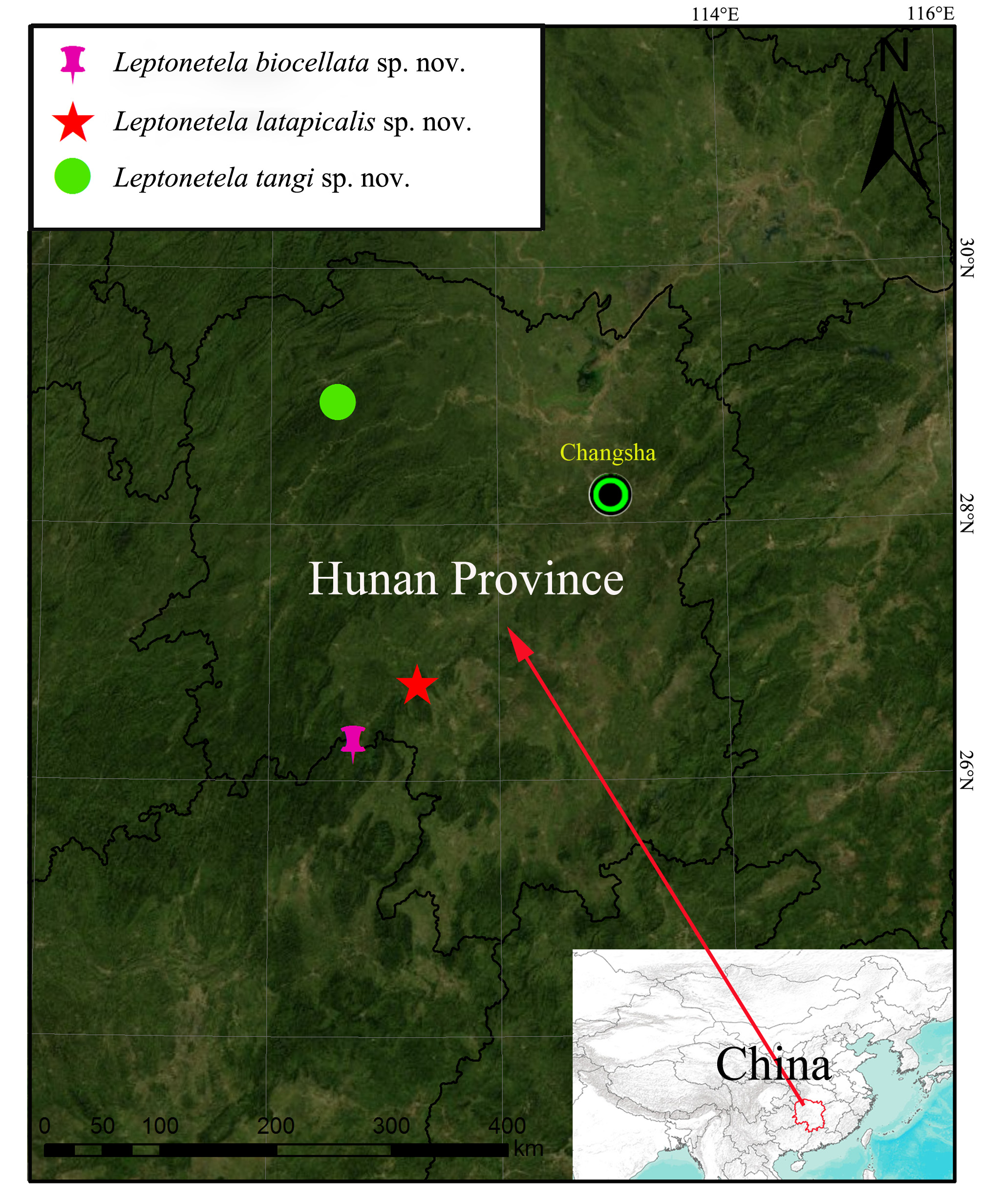
Type material. Holotype: GoogleMaps male (HNU) China, Hunan Province, Shaoyang County, Hebo Town, Chengbei Village GoogleMaps , Jigong Cave GoogleMaps , 111°17.460'E, 26°45.438'N, 576 m, 23 November, 2011, Xiang Xu, Jinlong Wan, Yi Zhao, Shihong Peng leg. Paratypes: 14 females, 15 males, same data as holotype GoogleMaps .
Etymology. The specific name is an adjective in apposition and derived from the Latin words “lata” (broad) and “apicalis” (apical), in reference to the presence of broad and peak-shaped median apophysis.
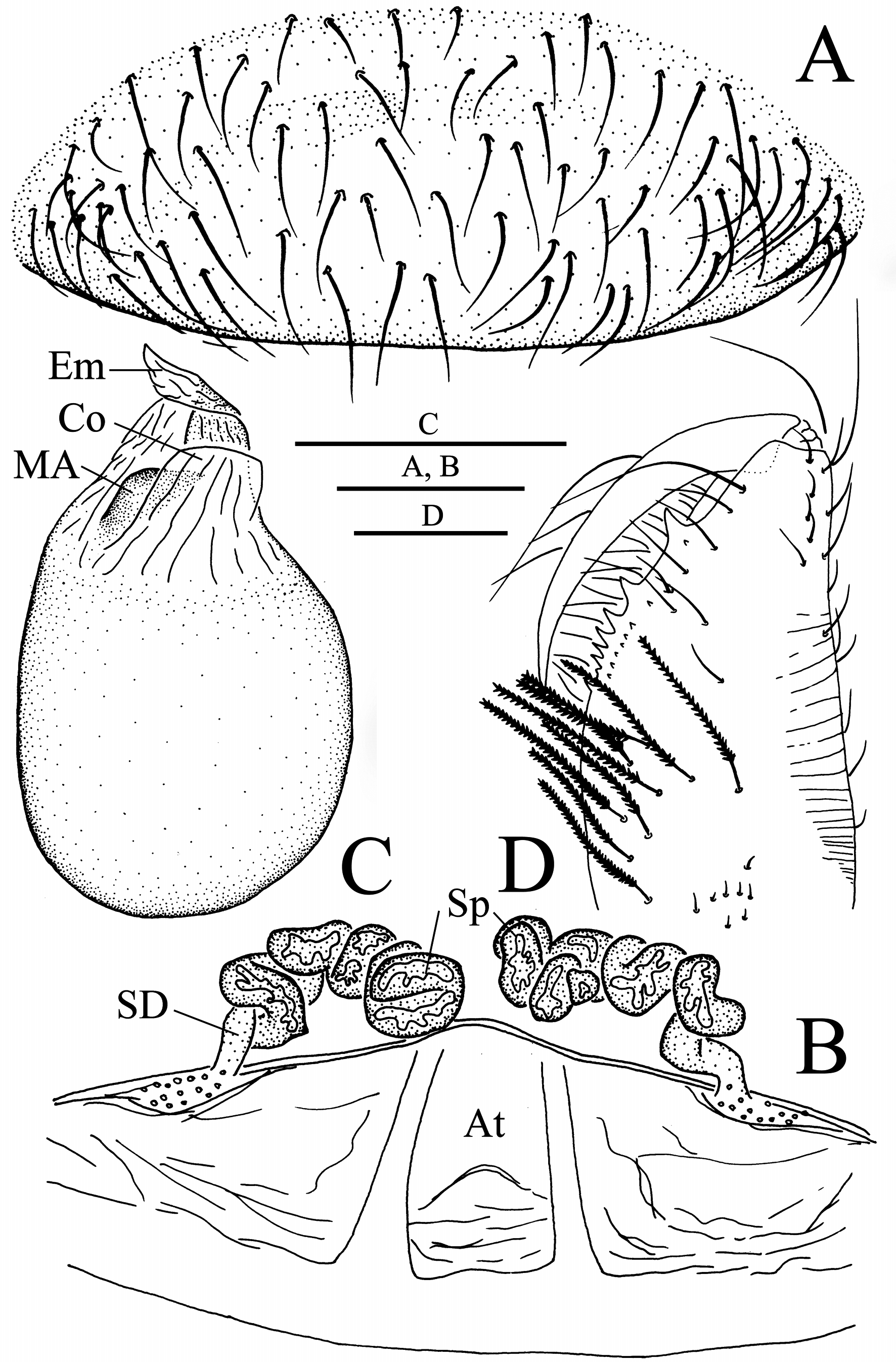
Diagnosis. Males resemble those of Leptonetela hexacantha Lin & Li, 2010 , L. jinsha Lin & Li, 2010 , L. reticulopecta Lin & Li, 2010 , L. kanellisi (Deeleman-Reinhold, 1971) , and L. tianxingensis Wang & Li, 2011 by having depressed and strongly contracted structure in the middle of the pedipalpal tarsus ( Figs 5C, D View FIGURES 5 , 7A, B View FIGURES 7 in the present paper; figs 24A, B, 26A, B, 45A, B in Lin & Li 2010; figs 18A, B, 63A, B in Wang & Li 2011); short distance between apical tarsus and pedipalpal bulb ( Figs 5C, D View FIGURES 5 , 7A, B View FIGURES 7 in the present paper; figs 24A, B, 26A, B, 45A, B in Lin & Li 2010; figs 18A, B, 63A, B in Wang & Li 2011), the new species is distinguished from these species by presence of three prolateral, nine retrolateral tibial spines on male pedipalpus; broad and peak-shaped median apophysis; short, wide, translucent conductor on male bulb ( Figs 5C, D View FIGURES 5 , 7A, B View FIGURES 7 ). The new species resembles L. quinquespinata (Chen & Zhu, 2008) in being eyeless, having membranous embolus, slightly twisted prolaterally ( Figs 5A, B View FIGURES 5 , 8B View FIGURES 8 in this present paper; figs 44A, B, 47D in Wang & Li 2011); it can be distinguished by nine strong spines located in retrolateral side of pedipalpal tibia ( Figs 5D View FIGURES 5 , 7A View FIGURES 7 in this present paper; figs 44B, 46C in Wang & Li 2011); nine small retromarginal teeth on the chelicerae in the new species ( Fig. 8D View FIGURES 8 in this present paper; fig. 47C in Wang & Li 2011), while six strong spines retrolaterally-directed on the pedipalpal tibia; five small retromarginal teeth on the chelicerae in L. quinquespinata ( Figs 5D View FIGURES 5 , 7A View FIGURES 7 in this present paper; figs 44D, 46B in Wang & Li 2011). Females resemble those of L. quinquespinata in being eyeless; having highly twisted sperm, but can be distinguished by the distal ends of sperm ducts closer placed to each other in the new species ( Figs 6C View FIGURES 6 , 8B View FIGURES 8 ), slightly further away from each other in L. quinquespinata (figs 45C, 47B in Wang & Li 2011).
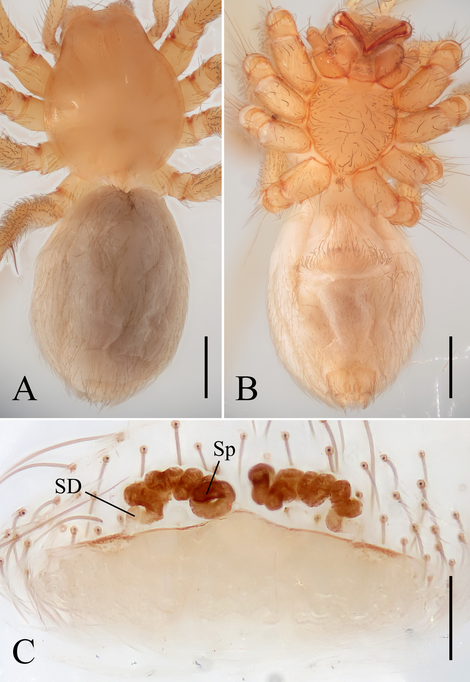
Description. Male. Total length 2.69 ( Fig. 5A View FIGURES 5 ). Carapace 1.26 long, 1.12 wide. Opisthosoma 1.37 long, 1.03 wide. Prosoma reddish brown, with several setae near the anterior margin of carapace. Ocular area with a pair of setae, eyes absolutely absent. Median groove short, cervical grooves and radial furrows light brown. Clypeus 0.24 high. Chelicerae brown, with nine promarginal and eight small retromarginal teeth, promarginal row of teeth gradually becoming smaller and denser from the base to distal end of fang furrow ( Fig. 8D View FIGURES 8 ). Endites brown. Labium brown and plump, fused with sternum. Sternum and legs yellowish. Leg measurements: I 12.72 (3.53, 0.45, 3.98, 2.97, 1.79); II 10.03 (2.91, 0.41, 3.09, 2.12, 1.50); III 8.66 (2.52, 0.42, 2.41, 2.10, 1.21); IV 10.34 (3.12, 0.36, 2.98, 2.53, 1.35). Leg formula: I–IV–II–III. Opisthosoma pale brown, ovoid, lacking distinctive patterns. Male pedipalpus ( Figs5 View FIGURES 5 B–D, 7A, B, 8C): femur covered with long and thin hairs; tibia with three trichobothria dorsally; three slender spines prolaterally and nine strong spines retrolaterally (six spines along the tibia form a longitudinal row and the other three spines along distal margin of the tibia form a transversal row) exist on the pedipalpal tibia ( Figs 7A, B View FIGURES 7 ). Tarsus rugose and contracted mesially, attaching to an earlobe-shaped process retrolaterally, with long spines distally ( Figs 5C, D View FIGURES 5 , 7A, B View FIGURES 7 ). Pedipalpal bulb nearly round; embolus membranous, slightly twisted towards the prolateral side; conductor translucent, broad, flat; median apophysis broad, peakshaped ( Figs 5B View FIGURES 5 , 8C View FIGURES 8 ). Prolateral lobe cuspate ( Fig. 7B View FIGURES 7 ).
Female. Similar to male in coloration of opisthosoma and general features, but larger body size, shorter legs, and prosoma yellowish. Total length 3.06 ( Figs 6A, B View FIGURES 6 ). Carapace 1.23 long, 1.15 wide. Opisthosoma 1.70 long, 1.24 wide. Clypeus 0.24 high. Leg measurements: I 10.94 (3.06, 0.40, 3.33, 2.42, 1.73); II 9.46 (2.62, 0.41, 2.88, 2.13, 1.42); III 7.93 (2.38, 0.33, 2.21, 1.86, 1.15); IV 10.26 (2.95, 0.36, 2.95, 2.48, 1.52). Leg formula: I–IV–II–III. Genital area densely covered with long hairs ( Figs 6B View FIGURES 6 , 8A View FIGURES 8 ). Internal genitalia with a pair of spermathecae and sperm ducts: spermathecae dark brown, sclerotized and highly twisted, with the distal ends very close to each other; sperm ducts pale brown, less sclerotized ( Figs 6C View FIGURES 6 , 8B View FIGURES 8 ). The atrium broad, nearly triangular, slightly procurved at anterior median margin ( Fig. 8B View FIGURES 8 ).
Distribution. Known only from the type locality ( Fig. 12 View FIGURE 12 ).


No known copyright restrictions apply. See Agosti, D., Egloff, W., 2009. Taxonomic information exchange and copyright: the Plazi approach. BMC Research Notes 2009, 2:53 for further explanation.
|
Kingdom |
|
|
Phylum |
|
|
Class |
|
|
Order |
|
|
Family |
|
|
Genus |