
Leptonetela tangi, He & Liu & Xu & Yin & Peng, 2019
|
publication ID |
https://doi.org/10.11646/zootaxa.4554.2.10 |
|
publication LSID |
lsid:zoobank.org:pub:90C205A6-B1CC-4377-990E-E511A4F1989A |
|
DOI |
https://doi.org/10.5281/zenodo.5928051 |
|
persistent identifier |
https://treatment.plazi.org/id/039D87A4-AC0F-2005-FF33-7DB5DF5289B4 |
|
treatment provided by |
Plazi |
|
scientific name |
Leptonetela tangi |
| status |
sp. nov. |
Leptonetela tangi View in CoL sp. nov.
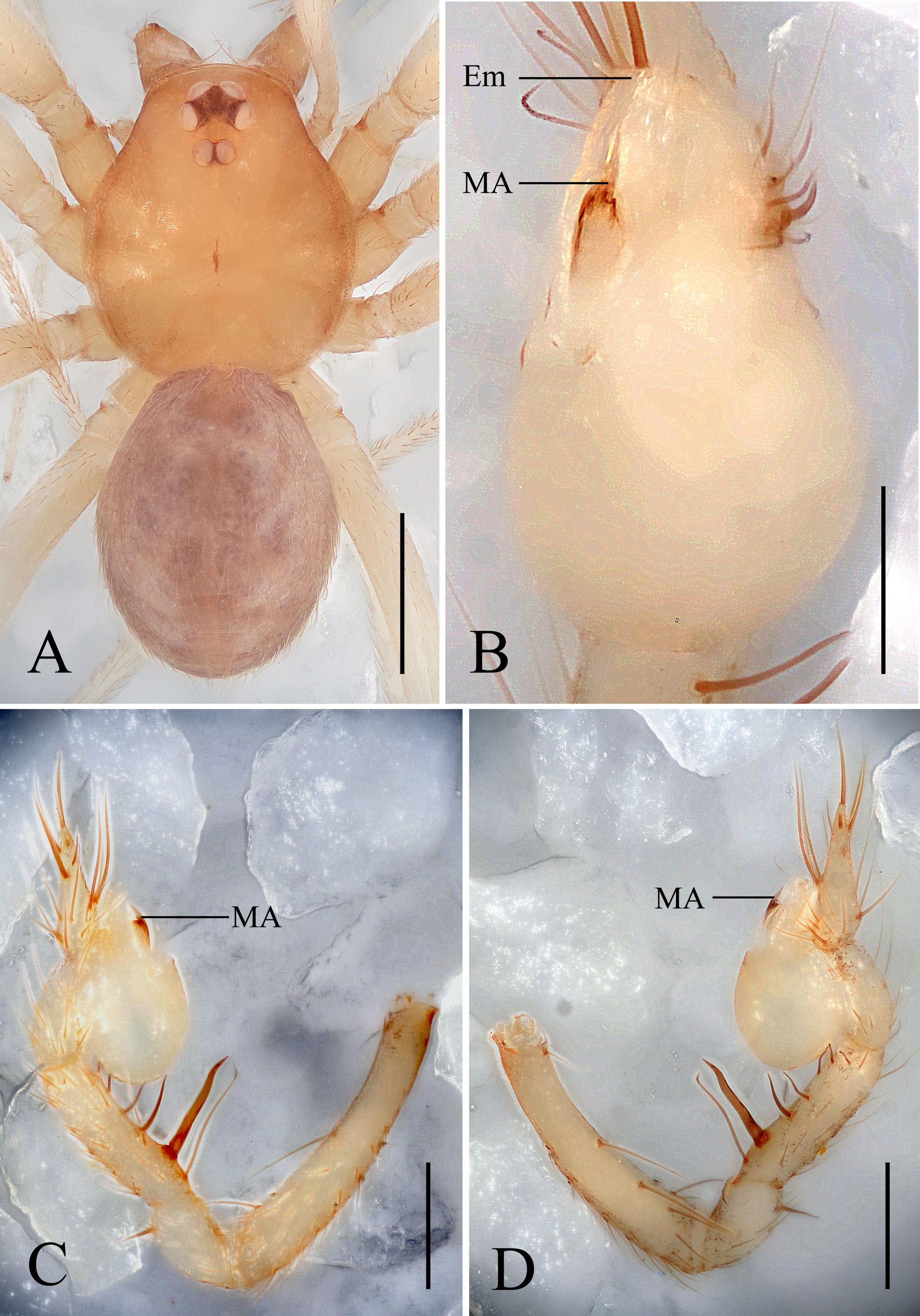
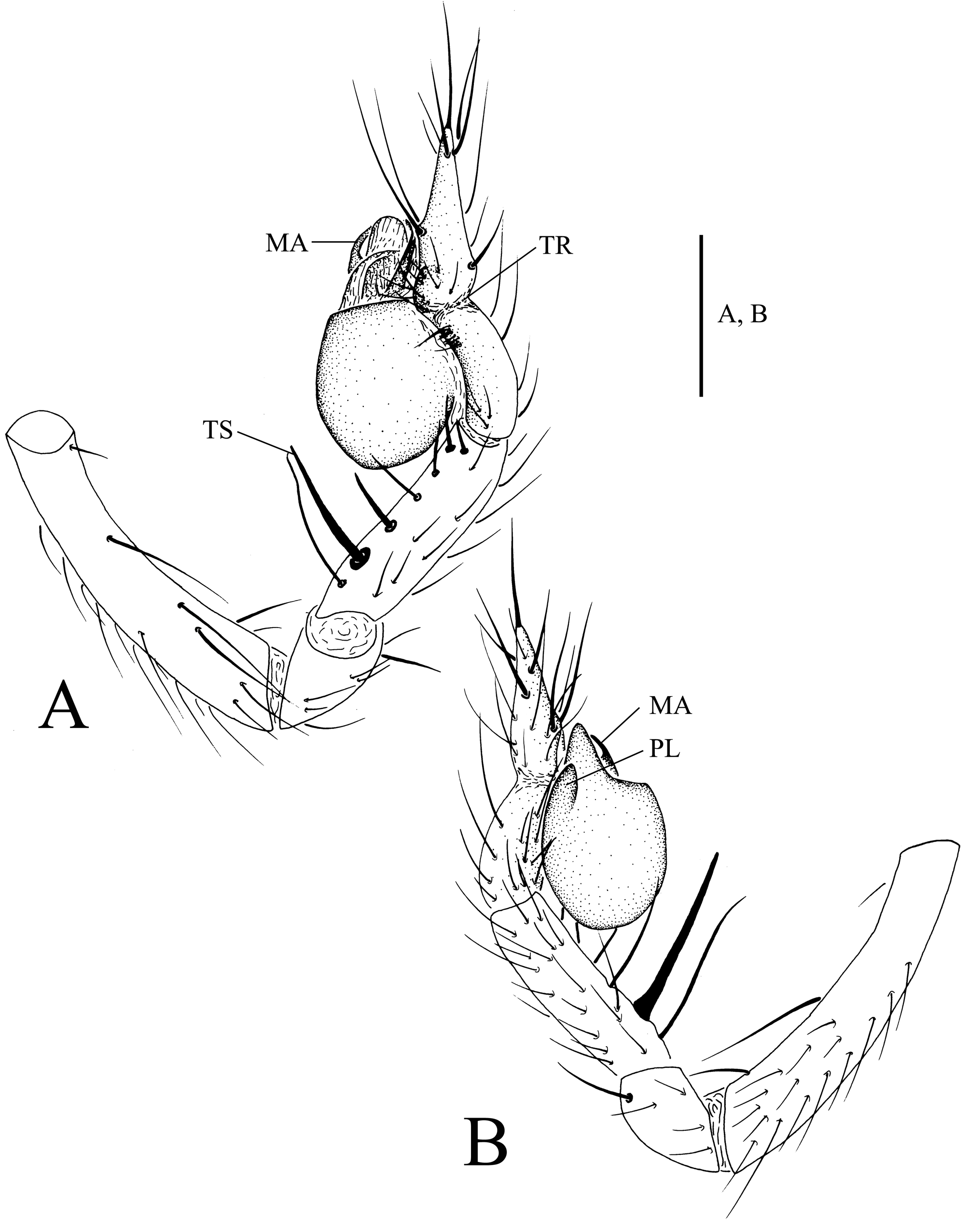
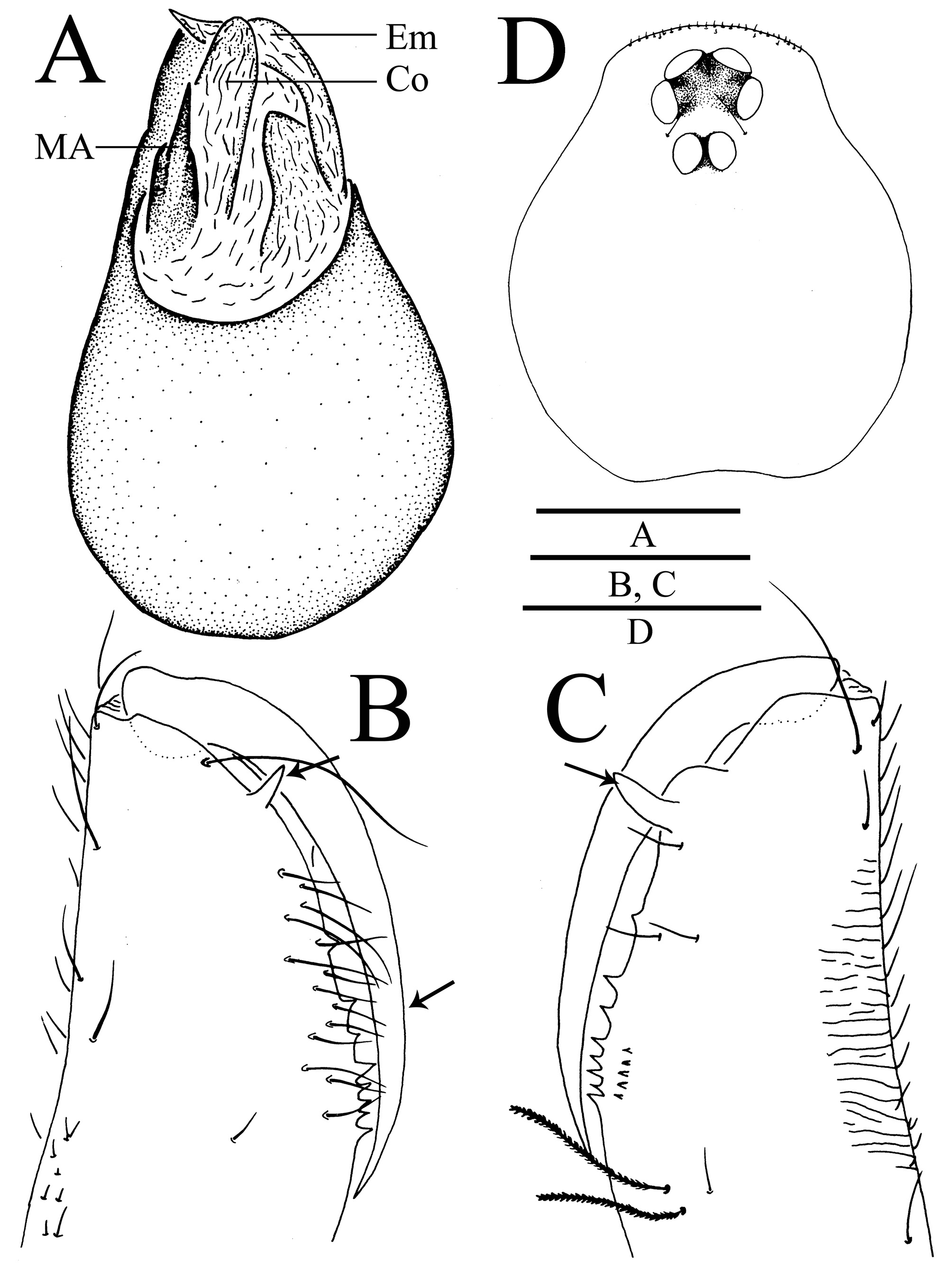
Figures 9–12 View FIGURES 9 View FIGURES 10 View FIGURES 11 View FIGURE 12
http://zoobank.org/NomenclaturalActs/ 59BAAE5B-7E35-44E8-9576-5F8F9CEBED3D
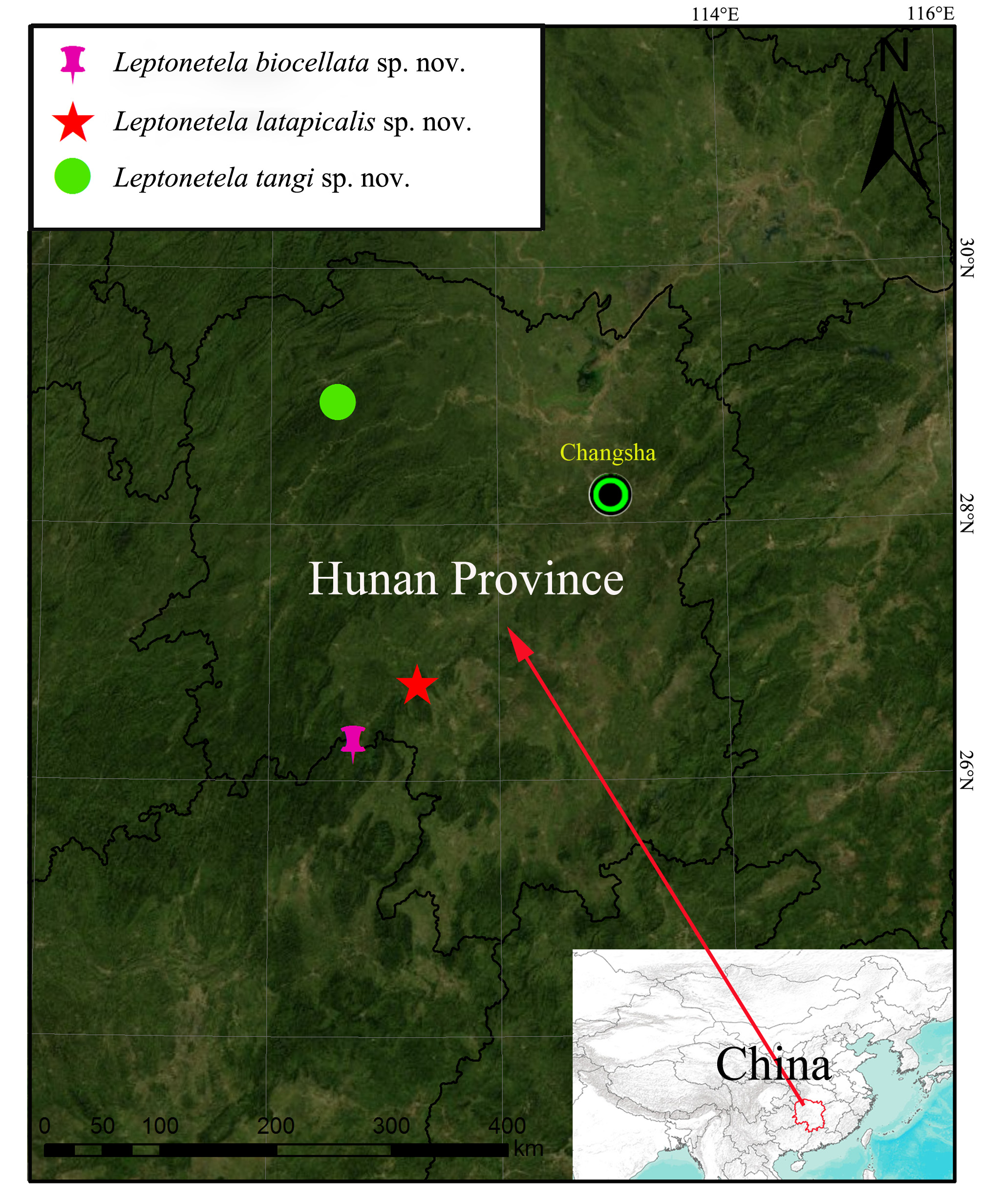
Type material. Holotype: GoogleMaps male (HNU) China, Hunan Province, Yuanling County, Xiazhai Village, Wuyuan cave GoogleMaps , 110°35.518’E, 28°57.537’N, 315 m, 1 November, 2006, Xiang Xu and Guo Tang leg.
Etymology. The species is named after one of the collectors, Mr. Guo Tang, a deceased colleague at Hunan Normal University, China; name is in genitive case.
Diagnosis. Leptonetela tangi sp. nov. resembles L. gigachela ( Lin & Li, 2010) by strong chelicerae; male pedipalp tibia subequal to tarsus in length ( Figs 9C, D View FIGURES 9 , 10A, B View FIGURES 10 , 11B, C View FIGURES 11 in the present paper; figs 1A, B, 2D, E in Lin & Li, 2010); it can be distinguished by presence of median apophysis in new species, absent in L. gigachela ; mid pedipalpal tarsus ventral apophysis absent in L. tangi sp. nov.; present in L. gigachela ( Figs 9B View FIGURES 9 , 10A, B View FIGURES 10 , 11A View FIGURES 11 ); Leptonetela tangi sp. nov. also resembles L. chakou Wang & Li, 2017 , L. kangsa Wang & Li, 2017 , L. langdong Wang & Li, 2017 , L.qiangdao Wang & Li, 2017 , and L. shanji Wang & Li, 2017 by having a strong second spine close to proximal male palp tip ( Figs 9C, D View FIGURES 9 , 10A, B View FIGURES 10 in the present paper; figs 4C, D, 20C, D, 22C, D, 54C, D, 58C, D in Wang, Xu & Li, 2017); it can be distinguished by median apophysis with thick base and sharp tip in new species ( Fig. 9B View FIGURES 9 in the present paper; figs 4B, 20B, 22B, 54B, 54B in Wang, Xu & Li, 2017).
Description. Male. Total length 1.91 ( Fig. 9A View FIGURES 9 ). Carapace 0.97 long, 0.88 wide. Opisthosoma 0.93 long, 0.77 wide. Prosoma yellowish brown and pear-shaped, with two setae at anterolateral area of PME and many short setae in the anterior margin of clypeus. Thoracic median groove dark-brown, needle-shaped. Cervical groove and radial furrows light brown. Clypeus 0.17 high, slightly sloped anteriorly. Six eyes, ALE and PLE connected to each other by the black bases, PME separated from ALE and PLE ( Figs9A View FIGURES 9 , 11D View FIGURES 11 ). Eye sizes: ALE 0.10, PLE 0.09, PME 0.06. ALE-ALE 0.01, ALE-PME 0.14, PLE-PLE 0.13, PLE-PME 0.05, AER 0.19, PER 0.24. Chelicerae yellowish, long and strong, fang furrow with nine promarginal (the first tooth sturdy) and six retromarginal teeth (the first tooth especially sturdy, even stronger than the first promarginal tooth) ( Figs 11B, C View FIGURES 11 ). Endites pale yellow. Labium yellowish and plump, fused to sternum. Sternum yellowish, peltate. Legs yellowish. Leg measurements: I 7.25 (2.01, 0.35, 2.12, 1.68, 1.09); II 5.22 (1.53, 0.30, 1.28, 1.20, 0.91); III 4.41 (1.27, 0.29, 1.16, 0.93, 0.76); IV 6.25(1.82, 0.29, 1.74, 1.43, 0.97). Leg formula: I–IV–II–III. Opisthosoma deep brown, ovoid, covered with short hairs. Male pedipalpus ( Figs 9 View FIGURES 9 B–D, 10A, B, 11A): tibia and tarsus subequal in length; femur slightly longer than tibia and tarsus, with four short ventral spines; patella with one dorsal spine distally; tibia with seven long spines ventrally (the second one near the basal end especially strong), but without any trichobothria; tarsus with a long seta ventrally, a strong apical seta, three prolateral, and three retrolateral setae at the distal half, and attaching to an earlobe-shaped process medially ( Figs 9C, D View FIGURES 9 , 10A, B View FIGURES 10 ); bulb ovoid, with a smooth surface; embolus and conductor thin and translucent; median apophysis distinct, strongly cuticularized, with a thick base and sharp end ( Figs 9B View FIGURES 9 , 11A View FIGURES 11 ).
Female. Unknown.
Distribution. Known only from the type locality ( Fig. 12 View FIGURE 12 ).
No known copyright restrictions apply. See Agosti, D., Egloff, W., 2009. Taxonomic information exchange and copyright: the Plazi approach. BMC Research Notes 2009, 2:53 for further explanation.
|
Kingdom |
|
|
Phylum |
|
|
Class |
|
|
Order |
|
|
Family |
|
|
Genus |