
Scaphiophryne madagascariensis (Boulenger, 1882)
|
publication ID |
https://doi.org/ 10.1111/j.1096-3642.2007.00329.x |
|
DOI |
https://doi.org/10.5281/zenodo.10544915 |
|
persistent identifier |
https://treatment.plazi.org/id/039D87F8-FFB7-FFFC-771C-FA717EC6C9A9 |
|
treatment provided by |
Felipe |
|
scientific name |
Scaphiophryne madagascariensis |
| status |
|
SCAPHIOPHRYNE MADAGASCARIENSIS View in CoL
(BOULENGER, 1882)
Specimens were collected in a shallow, ephemeral pond in the grasslands of the Andohariana plateau, Andringitra National Park. The bottom of the pond was covered with grass and the maximum depth was about 30–40 cm. The water was clear and cold. The description of external morphology was based on a specimen at a young stage 35, ZSM 595/2004 (TL and BL are 23.0 mm and 10.0 mm, respectively). Buccopharyngeal features are described based on a tadpole at stage 38, ZSM 599/2004.
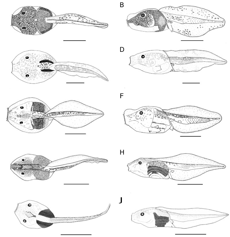
External morphology: In dorsal view ( Fig. 1A View Figure 1 ), body ovoid, widest at the level of gills, snout truncate. In profile ( Fig. 1B View Figure 1 ), body depressed, almost flat below, BW 124% of BH, snout small and round. Eyes moderately small, ED 14% of BL, slightly bulging (caused by the presence of a space between the outer integument and the organs), not visible in ventral view, positioned more dorsally than dorsolaterally and directed laterally. Nares not open, positioned dorsally, closer to pupils than to snout, RN 127% of NP, very close to each other, NN 26% of PP. Spiracle sinistral but very low, very slightly conical, moderately large, entirely attached to body wall, inner wall absent, orientated almost posteriorly, closer to end of body than to tip of snout, SS 76% of BL; spiracular opening crescent-shaped, closer to the level of the opening of ventral tube than to the insertion of hindlimb. Tail musculature moderately weak, TMH 34% of BH and 33% of MTH, its maximum height reached before the proximal third of caudal muscle, then gradually tapering, not reaching tail tip. Tail fins of moderate height, UF 35% of MTH, LF 37% of MTH, convex, upper fin not extending onto body, SU 93% of BL; point of maximum height of tail located before the middle of tail length, MTH 102% of BH, tail tip round. Ventral tube moderately large, medial, conical but its posterior part folded against ventral fin, directed posteroventrally, its posterior part linked to ventral tail fin, opening medial. Neither lateral line organs nor glands visible.
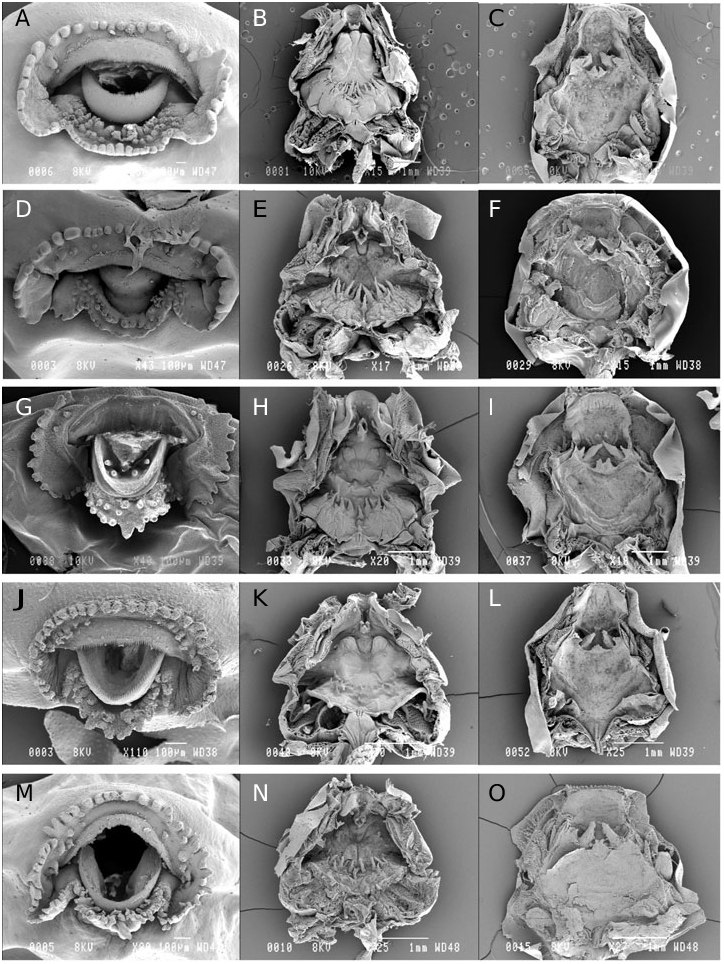
Oral disc ( Fig. 2A View Figure 2 ) in position and orientation subterminal, emargination very low, of moderate size, ODW 27% of BL and 39% of BW. An uninterrupted row of marginal papillae; a few submarginal papillae on a row laterally on the upper labium, a double row on the lower labium, the external one being interrupted shortly medially, a small group of smaller submarginal papillae at point of emargination on each side; marginal and submarginal papillae round, large, stocky, some of them blunt, those on the top of upper labium very small. No denticulate papillae. No keratodonts. Jaw sheaths of moderate breadth, very finely serrated; upper jaw sheath a large arch, flat on the most part with a weak median convexity, white; lower jaw sheath V-shaped, its distal third white, its proximal two-thirds light brown.
Coloration in preservative: Tadpole transparent, all underlying organs visible. External integument of upper side transparent except the snout, which is speckled with light brown; underlying tissues densely speckled with spots of the same colour. Flanks speckled in the same way as upper side, but with a dorsoventral gradation. Ventral side immaculate, except the part anterior to gills slightly speckled and the digestive tract brown coloured. Oral disc brown. Caudal
A
C
E
G
I
muscle coloured with the same tint. Fins bearing some scarce small spots, more numerous on the upper fin than on the lower. Upper side of hind limbs coloured with the same tint.
Variation: The ratios taken on three tadpoles at stages 34–36 (ZSM 696/2004-ZSM 698/2004) vary in the following proportions: BW 116–144% of BH; RN 129–133% of NP; NN 24–29% of PP; SS 78–79% of BL;
TMH 30–44% of BH; TMH 29–34% of MTH; UF 33– 38% of MTH; SU 86–94% BL; MTH 104–128% of BH; ODW 24–27% of BL; ODW 35–39% of BW.
Buccal floor ( Fig. 2B View Figure 2 ): Prelingual arena small; a pair of small prelingual papillae on the lateral wall of the arena, directed medially; a single medial curved gutter-shaped papilla originating from the base of the lower beak, bearing a vertical medial ridge on its distal part resulting in a short medial stub in posterior view. Tongue anlage prominent, without lingual papillae. Buccal floor arena round delimited anteriorly by few small papillae and posteriorly to the buccal pockets by a dense transversal row of large pustulate papillae, the largest medial; interior of arena smooth. Buccal pockets long, narrow, almost straight and obliquely orientated; four or five prepocket papillae of different size. Ventral velum with spicular support, bearing a pair of projections on each half above the 2nd and 3rd filter plate; medial notch present allowing the glottis to be fully exposed; glottis behind the velum, a papilla in front of the glottis and posterior to the row of the buccal floor arena papillae; secretory pits limited to the projections. Branchial baskets oblique, wider than long; with three filter cavities, filter plates obliquely arranged, filter mesh dense with tertiary folds.
Buccal roof ( Fig. 2C View Figure 2 ): Prenarial arena rectangular, bearing two very small papillae in a transverse row in the centre of the arena. Choanae large, drop-shaped; anterior wall slightly elevated, smooth, without papilla; narial valve greatly enlarged posteromedially into a triangular structure curved dorsally and orientated anteromedially, extending above the choana, its lateral edge slightly jagged. Postnarial arena small bearing a pustule in central position. Median ridge triangular with an irregular median cleft. Two pairs of lateral ridge papillae, the larger one posterior to the narial valve, triangular with a jagged edge; the second pair (missing from Fig. 2C View Figure 2 , visible on the buccal floor picture, Fig. 2B View Figure 2 ) lateral to the first, smaller, smooth and elongate. Buccal roof arena oval elongate; buccal roof arena papillae absent. A few small pustulations and papillae scattered across the buccal roof posteriorly to medial ridge; two small papillae and three pustules posteriorly in the arena. Posterolateral ridge present, lying relatively far anteriorly. Glandular zone present laterally, anterior to the dorsal velum. Dorsal velum interrupted medially, lateral edges curved anteriorly; secretory pits present on its posterior side. Two pressure cushions on each side.
No known copyright restrictions apply. See Agosti, D., Egloff, W., 2009. Taxonomic information exchange and copyright: the Plazi approach. BMC Research Notes 2009, 2:53 for further explanation.
|
Kingdom |
|
|
Phylum |
|
|
Class |
|
|
Order |
|
|
Family |
|
|
Genus |