
Scaphiophryne menabensis, Glos, Glaw & Vences, 2005
|
publication ID |
https://doi.org/ 10.1111/j.1096-3642.2007.00329.x |
|
DOI |
https://doi.org/10.5281/zenodo.5484761 |
|
persistent identifier |
https://treatment.plazi.org/id/039D87F8-FFBC-FFFD-7708-FA727FEBC8E4 |
|
treatment provided by |
Felipe |
|
scientific name |
Scaphiophryne menabensis |
| status |
|
SCAPHIOPHRYNE MENABENSIS View in CoL
GLOS, GLAW AND VENCES, 2005
The tadpoles were collected from an ephemeral breeding pool in closed forest. This pool was medium sized (150 m 2), shallow (<10 cm) over 75% of its area, with clear to slightly muddy water and a sparse coverage of aquatic vegetation. The external morphological description is based on a specimen at stage 35, ZSM 413/2004 (TL and BL are 24.5 and 11.9 mm, respectively). Buccopharyngeal features are described on the basis of a tadpole at stage 36 included in the batch ZSM 413/2004.
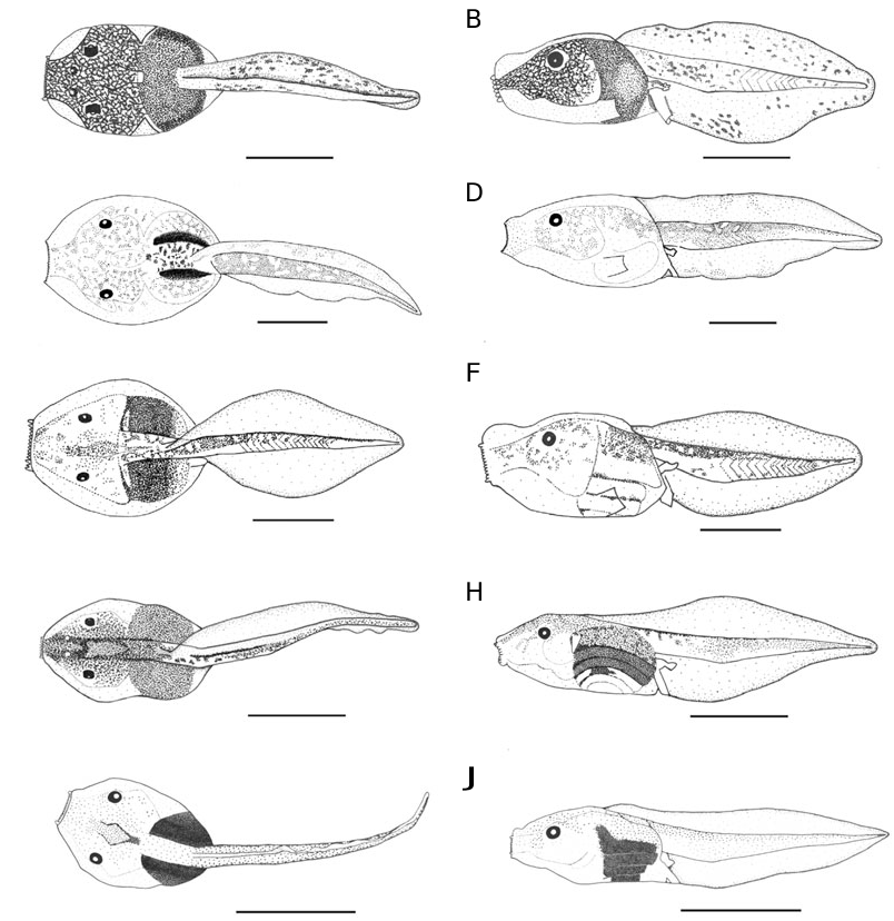
External morphology: In dorsal view ( Fig. 1C View Figure 1 ), body roughly ovoid. In profile ( Fig. 1D View Figure 1 ), BW 131% of BH, snout small, vertical and directed slightly upward. Eyes small, ED 8% of BL, not bulging, directed almost laterally. RN 184% of NP, NN 23% of PP. Spiracle formed by a large square of skin, orientated posteriorly; spiracular opening clinging to the body wall, on a plane situated just above the opening of ventral tube. TMH 31% of BH and 32% of MTH, maximum height of tail musculature reached at the proximal quarter then slightly tapering to end, abruptly very finely. UF 35% of MTH, LF 35% of MTH, upper fin increasing slowly in height before decreasing abruptly in the distal third, extending slightly onto body, SU 79% of BL, lower fin roughly convex; point of maximum height of tail located at the proximal third of the tail length, MTH 97% of BH. Ventral tube short and relatively large, directed almost ventrally, its anterior part linked to body wall.
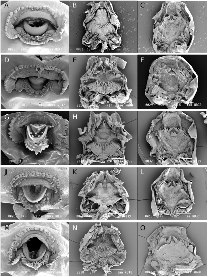
Oral disc ( Fig. 2D View Figure 2 ) moderately small, in position and orientation terminal, ODW 20% of BL and 29% of BW. A row of four submarginal papillae separated from the row of marginal papillae on each side on the upper labium leaving a large medial gap anteriorly, a row of submarginal papillae separated from the row of marginal papillae on the lower labium; papillae small, marginal papillae round, submarginal papillae pointed. Jaw sheaths white; upper jaw sheath a large arch, flat on the most part; lower jaw sheath Ushaped.
Coloration in preservative: External tegument of the upper side and underlying tissues heavily pigmented by dark brown spots which form dots. Upper part of flanks less densely pigmented as the upper side of body except the snout which is well pigmented. Lower part of flanks and ventral side immaculate. Caudal muscle heavily pigmented with brown spots leaving some small parts immaculate which form white dots. The external half of fins slightly coloured with brown, the internal half immaculate. Upper part of hindlimbs pigmented with the same tint.
Variation: The ratios taken on 12 tadpoles at stages 25–39 vary in the following proportions: BW 115– 164% of BH; SS 79–92% of BL; TMH 24–38% of BH; TMH 25–33% of MTH; UF 29–41% of MTH; LF 33– 40% of MTH; MTH 84–136% of BH; ODW 24–35% of BL; ODW 28–40% of BW.
Buccal floor ( Fig. 2E View Figure 2 ): Prelingual arena very narrow, gutter-shaped, two pustules anteriorly on the internal wall of the beak and two others laterally; a single medial curved gutter-shaped papilla originating just anterior to tongue anlage, directed posterodorsally, its distal part diamond-shaped with three projections corresponding to three angles, a small projection inside the gutter medially, edges jagged. Buccal floor arena diamond-shaped, without ornamentation anteriorly, delimited posteriorly by a transversal row of about 15 large papillae, the largest medial. Buccal pockets wide, deep, transversely orientated, unperforated; two small prepocket papillae. Ventral velum bearing a pair of projections on each half medially (the most lateral above the second filter plate); medial notch present, a vertical papillae in front of the glottis; secretory pits not obvious. Branchial baskets wide, the fourth filter plate vertical, filter mesh very dense with tertiary folds.
Buccal roof ( Fig. 2F View Figure 2 ): Prenarial arena small and round, bearing a small median papilla adjoining a small knob on each side laterally. Anterior wall of choanae pustulate; narial valve greatly enlarged posteromedially into a large and elongate structure, slightly jagged distally on its lateral side, dorsally and anteromedially directed, covering partially the choana. Postnarial arena small and concave, without ornamentation. Median ridge triangular, its extremity bifid. The larger pair of lateral ridge papillae posterolateral to the narial valves, triangular, stocky and smooth. Buccal roof arena non-existent, a few pustulations scattered within; one small papillae on each side anterolaterally, directed medially. Posterolateral ridges elevated, present through the buccal roof (if not a preservational artefact). Glandular zone present laterally between posterolateral ridges and dorsal velum, formed of only a few secretory pits wide. Dorsal velum straight; secretory pits not obvious on its posterior side.
No known copyright restrictions apply. See Agosti, D., Egloff, W., 2009. Taxonomic information exchange and copyright: the Plazi approach. BMC Research Notes 2009, 2:53 for further explanation.
|
Kingdom |
|
|
Phylum |
|
|
Class |
|
|
Order |
|
|
Family |
|
|
Genus |