
Satonius stysi Hájek & Fikáček, 2008
|
publication ID |
https://doi.org/ 10.5281/zenodo.5341906 |
|
DOI |
https://doi.org/10.5281/zenodo.5444486 |
|
persistent identifier |
https://treatment.plazi.org/id/039D87F9-FF98-FFD6-FE68-3A9BAB6EF9B6 |
|
treatment provided by |
Felipe |
|
scientific name |
Satonius stysi Hájek & Fikáček, 2008 |
| status |
|
Satonius stysi Hájek & Fikáček, 2008
( Figs. 2 View Figs , 11-17 View Figs , 20-26, 28, 30 View Figs View Figs ) Material examined. 13 larvae of penultimate instar (NMPC): CHINA: YUNNAN: Shanzhi env., Jizu Shan Mt., Jade Dragon waterfall, 27°57.8′N 100°23.2′E, 2250 m a.s.l., 23.-24.vi.2007, J. Hájek & J. Růžička leg. Larvae collected together with the adults designated above as type specimens.
Description. Body oniscoid, strongly flattened and ovoid in shape ( Fig. 2 View Figs ), dorsal surface distinctly sclerotized. Head partly covered by pronotum. Legs not visible in dorsal view.
Measurements. Head width 0.52 mm; body length 1.50-1.80 mm; maximum body width 1.20-1.32 mm.
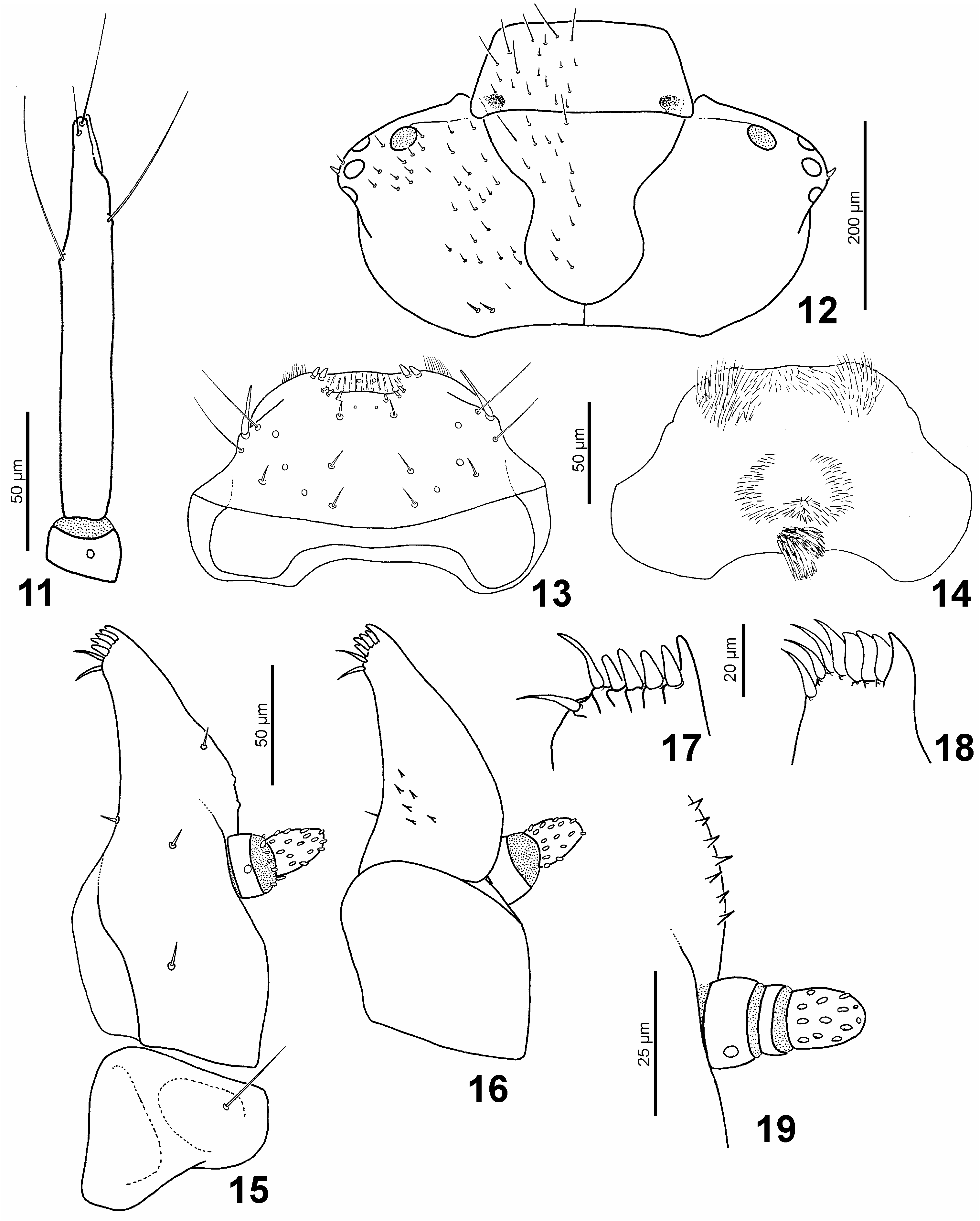
Head capsule ( Fig. 12 View Figs ) subprognathous, transverse, ca. 2× as wide as long. Dorsal surface brown, ventral parts paler, weakly sclerotized. Clypeus separated from frons by narrow ‘suture’; trapezoid in shape, with distinct darker attachment of anterior tentorial arms. Frontal sutures lyriform, joining clypeofrontal membrane at clypeal sides anteriorly; sulci distinctly shifted mesally by attachments of dorsal tentorial arms. Coronal suture short but distinct. Four stemmata situated on lateral prominence. Antenna inserted on anterolateral part of parietale, insertion area not connected with frontal suture or clypeofrontal membrane. Maxillary groove large, anteriorly limited by narrow projection of parietale with ventral mandibular articulation. Gula broad, trapezoid, not separated from submentum; laterally with posterior tentorial pits. Chaetotaxy of head capsule consisting of numerous short setae dorsally, with longer setae situated only on anterior parts of clypeus and frons; each lateral prominence with one short stout seta among stemmata; posterior part of parietale with two stout setae dorsally; ventral surface with few long hair-like setae anterolaterally.
Labrum ( Figs. 13-14 View Figs ) transverse, separated from clypeus by membranous fold. Chaetotaxy of disc consisting of eight pores and eight short setae; lateral margin with two long hair-like setae and one long and stout seta situated on angle of lateral labral fold. Anterior part with transverse ridge; three pairs of small stout setae situated posteriorly of this ridge. Surface between the ridge and anterior margin of labrum finely longitudinally ridged with two small submedian pores. Anterior margin with two pairs of small stout setae. Frayed setae absent. Ventral surface (epipharynx) membranous, with anterior area covered with microtrichia; posterior part with semicircular microtrichial area and dense mesal cluster of microtrichia situated posteriorly.
Antenna ( Fig. 11 View Figs ) directed anterolaterally, composed of two antennomeres. Antennomere I short, ring-like, bearing a pore dorsally. Antennomere II 10.5× as long as antennomere I; two long trichoid sensilla situated in apical third, one long and one shorter trichoid sensilla situated at antennal apex. Sensorium well developed, ca. 0.15× as long as antennomere II, inserted in shallow groove on inner surface of antennomere.
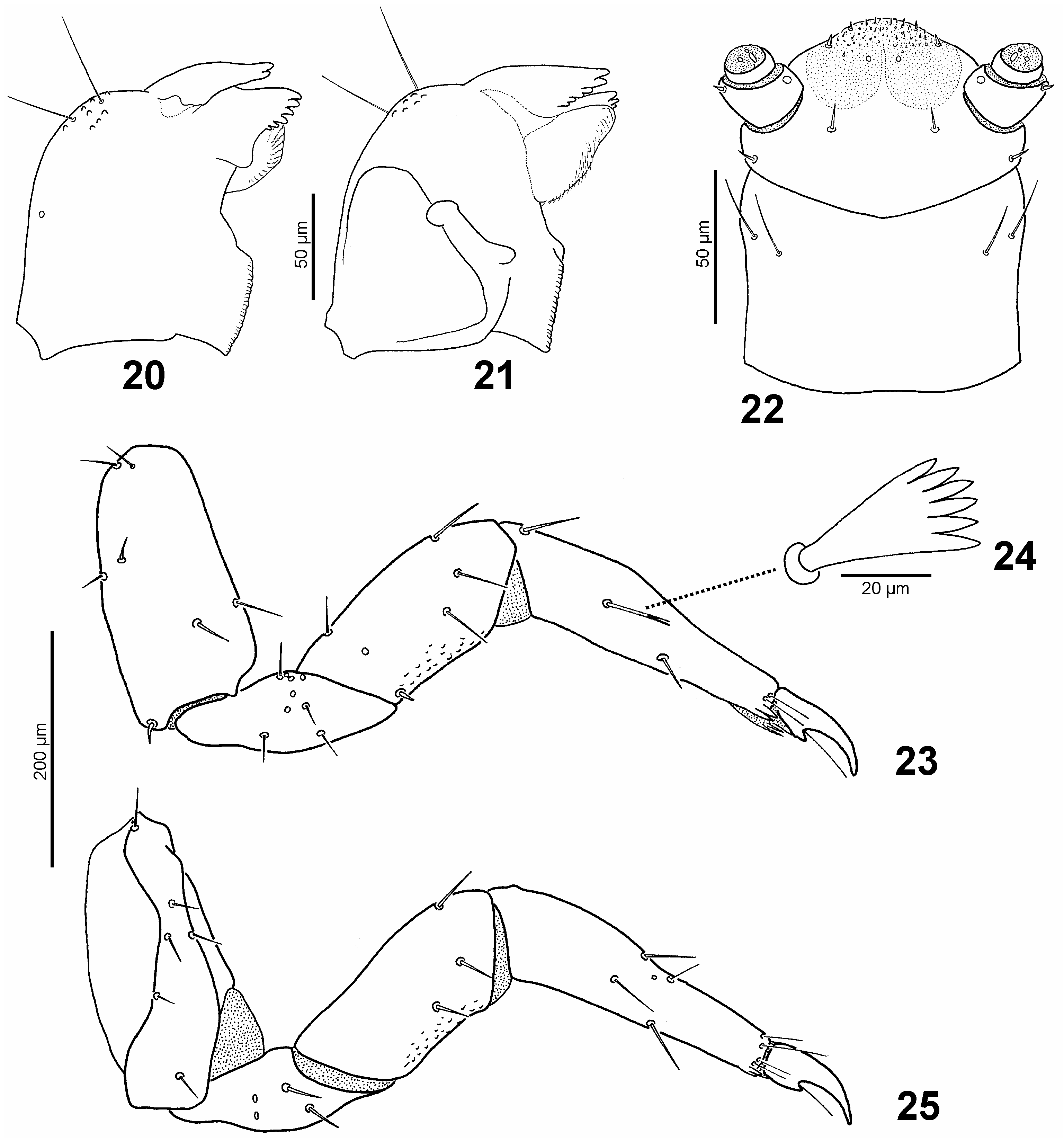
Mandibles ( Figs. 20-21 View Figs ) symmetrical, rather wide and short. Each mandible with one pore and two long hair-like setae dorsally on outer margin. Area bearing setae with numerous cuticular tubercles. Apex divided into two lobes, each with six denticles mesally; basal lobe slightly projecting mesad, largely covering ventral membranous fold (pseudomola sensu BEUTEL (1998)) with numerous microtrichia arranged in transverse series. Basal part of mandible with large, strongly sclerotized mola. Mandibular acetabulum (mandibular part of secondary joint) situated laterally; ventral condyle (mandibular part of primary joint) shifted anteromesally.
Maxilla ( Figs. 15-16 View Figs ). Cardo as wide as basal portion of stipes, ca. 0.4× as long as remaining part of maxilla, slightly wider than long, bearing one hair-like seta. Stipes not separated from mala, largely opened dorsally to interior of head capsule; ventral surface bearing two short setae in basal half, one short mesally directed seta on inner surface and one short seta on apical third of outer face; dorsal surface with few cuticular spines near seta on inner face. Apical part of mala with five teeth and two long setae; lateral tooth fixed, remaining teeth represented by stout setae. Palpus situated laterally, small, consisting of two palpomeres; palpomere I short, ring-like, bearing one pore ventrally; palpomere II ca. 2.7× as long as palpomere I, slightly membranous apically, bearing numerous oval sensilla.
Labium ( Fig. 22 View Figs ). Submentum fused with gula. Mentum short, slightly wider than long, lateral margins nearly parallel-sided; anterior part with two pairs of long, hair-like setae. Prementum short and wide, with one pair of short setae situated in posterolateral corners and another pair between insertions of labial palpi. Apical part (ligular area) membranous, with numerous minute sensilla. Labial palpus with two palpomeres; palpomere I ca. 2.5× as long as palpomere II, bearing one pore mesally and one short seta laterally; palpomere II very short, ring-like, with apical membranous field bearing three sensilla.
Thorax as long as abdomen. Dorsal surface dark, strongly sclerotized. Pronotum 3.1× as wide as long, narrowing anteriad, anterolateral corners slightly projecting anteriad. Mesonotum shorter but slightly broader than pronotum, 4.8× as wide as long. Metanotum widest, ca. as long as mesonotum, 5.0× as wide as long. Lateral margins of all thoracic segments with fringe of alternating long and shorter hair-like setae. Chaetotaxy of pronotum consists of sparsely distributed, minute hair-like setae; chaetotaxy of meso- and metanotum with only a few minute hair-like setae. Ventral parts membranous, insertions of right and left coxae well separated from each other.
Legs ( Figs. 23-25 View Figs ) pentamerous, moderately long, prothoracic legs shorter than meso- and metathoracic legs. Coxa elongate, bearing 13 short, hair-like setae. Trochanter elongate-triangular, with a row of pores on both anterior and posterior surface; in addition with four setae and one pore on anterior surface and two setae on posterior surface. Femur 1.2× as long as trochanter (1.1× so in prothoracic leg), bearing five setae and one pore on anterior surface and three setae on posterior surface; ventral part with numerous cuticular tubercles. Tibiotarsus 1.3× as long as femur (as long as femur in prothoracic leg), bearing three setae on anterior and four setae and one pore on posterior surface; apical part with six fine and long setae and two pairs of cuticular spines. Seta situated on basal third of anterior surface of tibiotarsus different in morphology among leg pairs: scale-like and palmate on prothoracic leg, simple on mesothoracic leg, and bifid or rarely simple on metathoracic leg. Claw ca. 0.3× as long as tibiotarsus, with distinct sharp basal tooth and one long hair-like seta.
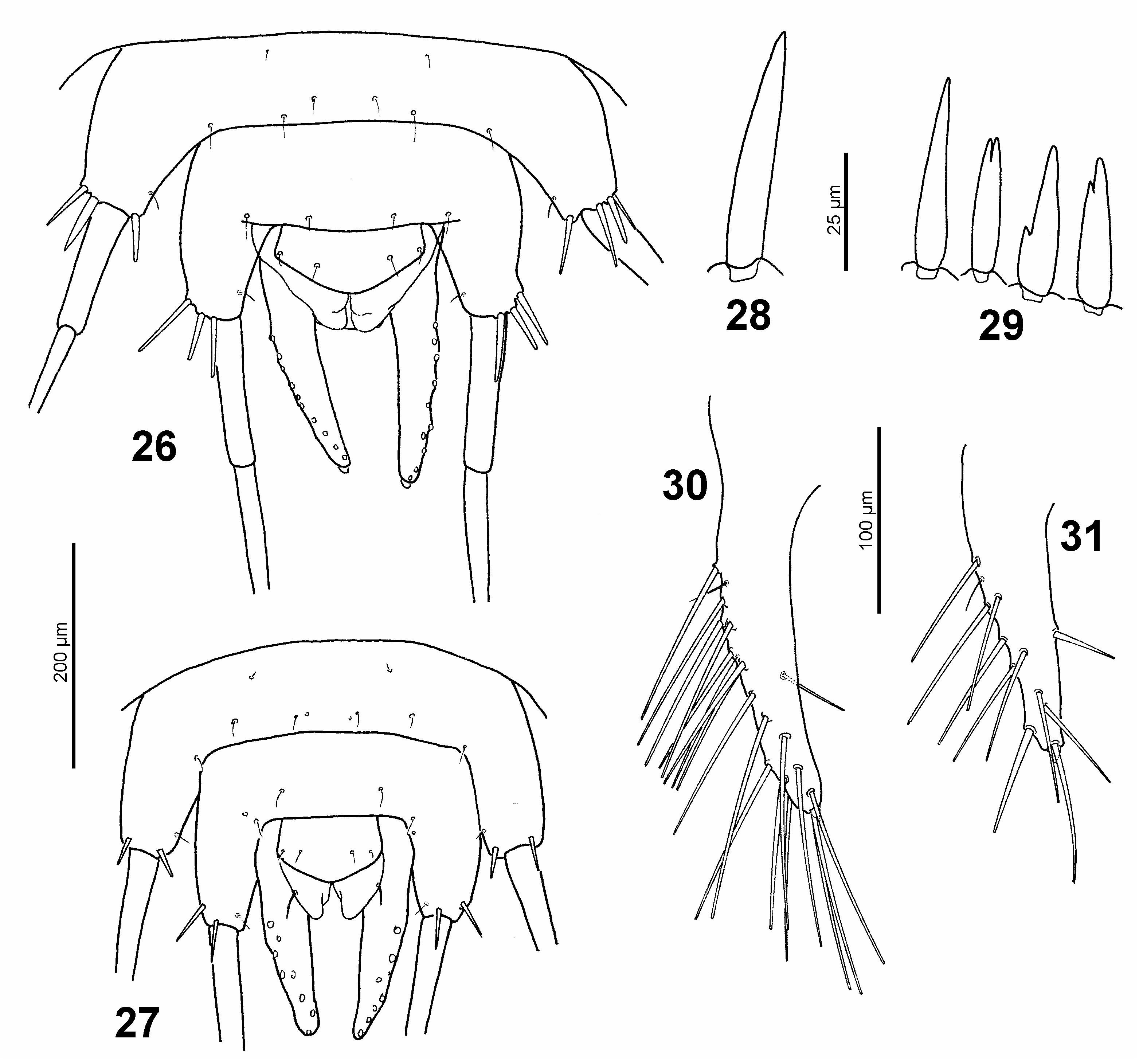
Abdomen ( Figs. 26 View Figs ) with 10 segments. Segment I slightly narrower than metanotum, each following segment slightly narrower than the previous one. Segments I-VIII short and transverse, projecting into lateral processes; tergum and sternum sclerotized and equally wide; dorsal chaetotaxy consisting of two minute hair-like setae situated along anterior margin, and 8 similar setae situated along posterior margin. Lateral processes bearing long, two segmented tracheal gills; first gill segment very short on abdominal segments I-V, slightly longer on abdominal segment VI and moderately long on segment VII-VIII; second gill segment often with secondary subapical constrictions; trachea opened at base of second gill segment into shallow groove. Lateral processes with few spine-like setae at base of tracheal gills ( Table 1), all setae simple ( Fig. 28 View Figs ). Abdominal segment IX bearing pair of fixed urogomphi; each urogomphus with numerous long and thin hair-like seta, one fine short subbasal seta and one short but very wide apical seta. Ventral part of abdominal segment IX developed as subpentagonal plate bearing two pairs of fine hair-like setae, the plate ca. 2× as wide as long. Abdominal segment X developed as pair of membranous ventral flaps posteriorly to sternite IX.
Note. All larvae collected together with the adults represent the same larval instar based on their body measurements. In order to identify the instar, we have compared the ratio of length of larvae and adults (LAR) in S. stysi sp. nov. (LAR = 0.85) and S. kurosawai (ultimate instar LAR = 0.95, penultimate instar LAR = 0.80). These measurements therefore suggest that the examined larvae of S. stysi sp. nov. probably represent the penultimate instar. This is corroborated by the simple shape of the setae of lateral abdominal lobes (i.e. lacking bifid apices), as only simple setae are found in the penultimate instar of S. kurosawai (part of these setae is bifid in the ultimate instar, see below, Fig. 29 View Figs ). Number or larval instars is not known for most torridincolids except for Ytu zeus and Delevea namibiensis , which have four instars ( REICHARDT (1973) and ENDRÖDY- YOUNGA (1997), respectively).

No known copyright restrictions apply. See Agosti, D., Egloff, W., 2009. Taxonomic information exchange and copyright: the Plazi approach. BMC Research Notes 2009, 2:53 for further explanation.
|
Kingdom |
|
|
Phylum |
|
|
Class |
|
|
Order |
|
|
Family |
|
|
Genus |