
Autogneta aokii, Behan-Pelletier, Valerie M., 2015
|
publication ID |
https://doi.org/10.11646/zootaxa.3946.1.2 |
|
publication LSID |
lsid:zoobank.org:pub:25788BA8-0C84-4B71-A28C-D6A922BC924C |
|
DOI |
https://doi.org/10.5281/zenodo.5684790 |
|
persistent identifier |
https://treatment.plazi.org/id/039DE80E-FF8F-AF51-FF11-FBFFFED569A5 |
|
treatment provided by |
Plazi |
|
scientific name |
Autogneta aokii |
| status |
sp. nov. |
Autogneta aokii View in CoL sp. nov.
Figs 10 View FIGURES 10, 11 –22
Material Examined. Holotype: adult male, USA, California, Sonoma Co., Salt Point State Park, 38.570°N 123.319°W, Pygmy Forest Trail, 4.iii.2009 (V. Behan-Pelletier), deposited in the CNC, type number 24201. Paratypes: 9 males, 10 females with same collecting data as holotype; Marin Co., Taylor State Park, Trail nr. S. Bridge, 38.023°N, 122.725°W, 11.xii.1986 (R.A. Norton and J.B. Kethley) 1 male, 1 female from pulpy outer wood and bark of redwood stump. Paratypes deposited in the CNC, USNM, and RNC.
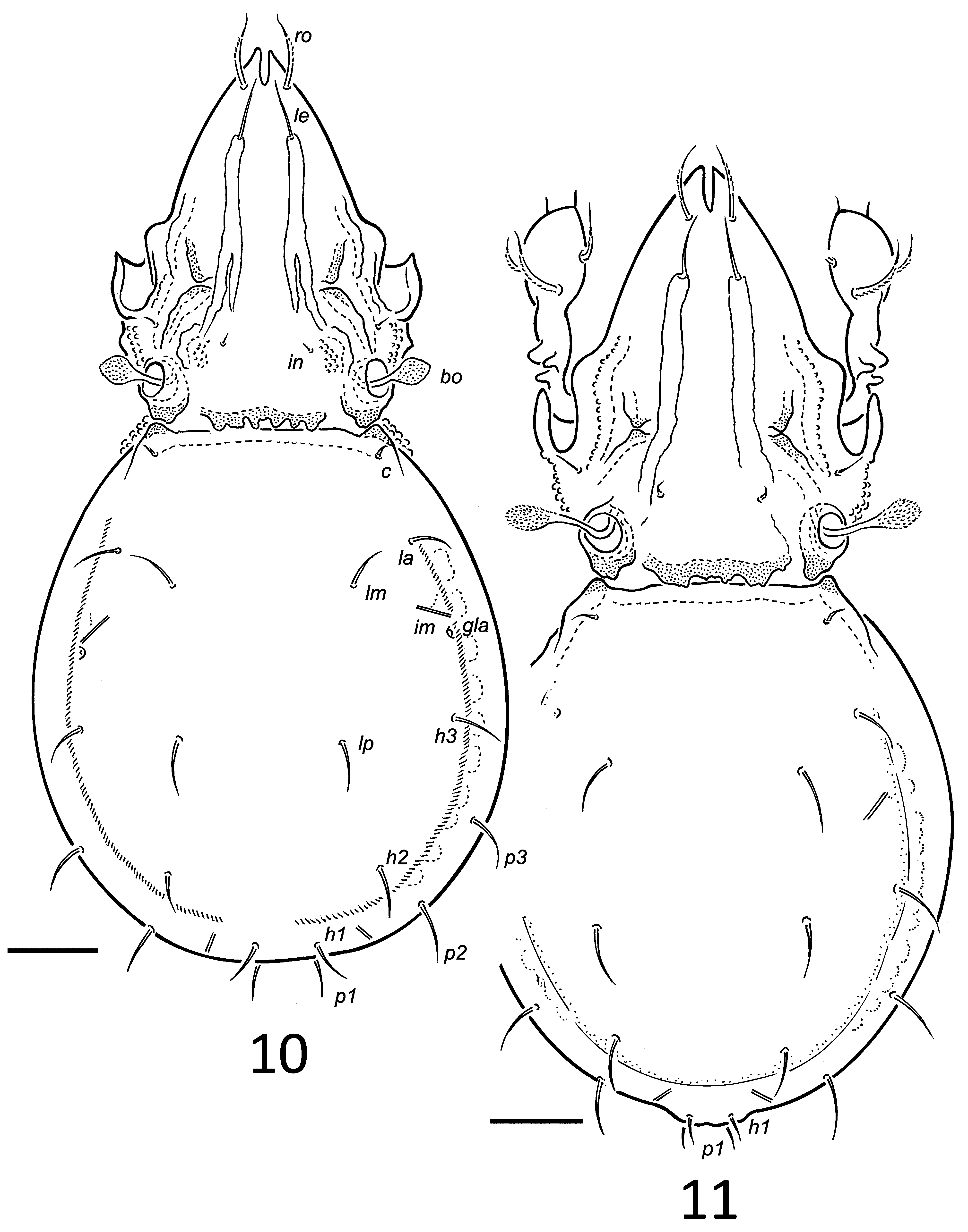
Diagnosis. Adult. Apophyses posteriorly on prodorsum expressed as 3–5 pairs of discrete or fused tubercles overhanging dorsosejugal scissure. Bothridial seta about 47, clavate, head about 17, with minute spicules. Genital setae 5 pairs. Femur I with 3 tubercles positioned antiaxially and dorsally in proximal third of leg. Genua I and II with seta v’ present. Sexual dimorphism expressed as small, convex porose area posteromedially in male, positioned between lyrifissures ips, bearing setae h1 and p1. Mutual distance (male) h1–h1 about 13, p1–p1 about 10; (female) h1–h1 about 25, p1–p1 about 23.
Description. Adults. Dimensions: Total length: females (n = 10) 312 (298–336); males (n = 10) 295 (range 278–307). Notogastral length: females (n = 9) 184 (168–192); males (n = 10) 173 (range 163–187).
Integument: Generally microtuberculate. Pleural region strongly tuberculate from base of pedotectum I to acetabulum IV, circular patch of tubercles present posterior of acetabulum IV. Microtubercles on trochanters III, IV and femora I–IV more evident than on other leg segments. Margin of epimere IV with concave depressions medially.
Prodorsum: Rostral incision about 17. Costulae about 82–90, medial and lateral edges with microtubercles (Fig. 16); bearing setae le anteriorly, about 19 long, acuminate, weakly barbed. Longitudinal ridge weakly developed lateral to costula; small ridges associated with enantiophysis E (Fig. 17). Enantiophysis E well developed lateral to proximal third of costula (Figs 16, 17). Apophyses posteriorly on prodorsum expressed as 3–5 pairs of discrete or fused tubercles overhanging dorsosejugal scissure ( Figs 10, 11 View FIGURES 10, 11 , 16). Rostral, lamellar, interlamellar and exobothridial setae thin, smooth, acuminate; ro about 81, le about 19, in about 11, ex 6–10. Mutual distance ro–ro about 13, le–le about 19, in–in about 34. Humeral enantiophysis well-developed (Fig. 17, arrow). Bothridial seta about 47, clavate, head about 17, head with spicules mostly minute but 6–10 longer spicules distally (Fig. 17).
FIGURES 16–22. Autogneta aoki sp. nov. Differential interference contrast microscope images of adult male: 16, prodorsum and anterior of notogaster; 17, posterolateral of prodorsum and anterolateral of notogaster showing humeral enantiophysis (arrow), bothridial seta displaced; 18, femur I showing proximal tubercles (arrow) (8 layers combined); 19, posterior of notogaster showing extent of projection; 20, posterior of notogaster (arrow to seta h1) (2 layers combined); 21, posterior of notogaster (arrow to porose area) (2 layers combined); 22, genital region showing genital papillae on one side (2 layers combined). Scale bar: 16–18 = 20, 19–22 = 10.
Notogaster: Notogaster with U-shaped furrow weakly developed, outlined by microtubercles. Notogastral setae thin, smooth acuminate, 10–19 long, with c shortest, positioned as in Figures 10, 11 View FIGURES 10, 11 . Sexual dimorphism expressed as small, convex, oval porose area posteromedially positioned between lyrifissures ips in male, bearing setae h1 and p1 (Figs 19–21). These setae (about 10) shorter than in female and closer together: mutual distance (male) h1–h1 about 13, p1–p1 about 10; (female) h1–h1 about 25, p1–p1 about 23.
Ventral Region: Epimeral setae 6–10 long, tapered, slightly roughened. Genital setae 5 pairs, 6–10 long acuminate, smooth. Aggenital and anal setae 10–12 long, smooth, acuminate; ad longest setae 15–20, tapered, with few barbs, ad3 anterolateral to anal plate; iad on level with seta an2.
Gnathosoma: As for genus.
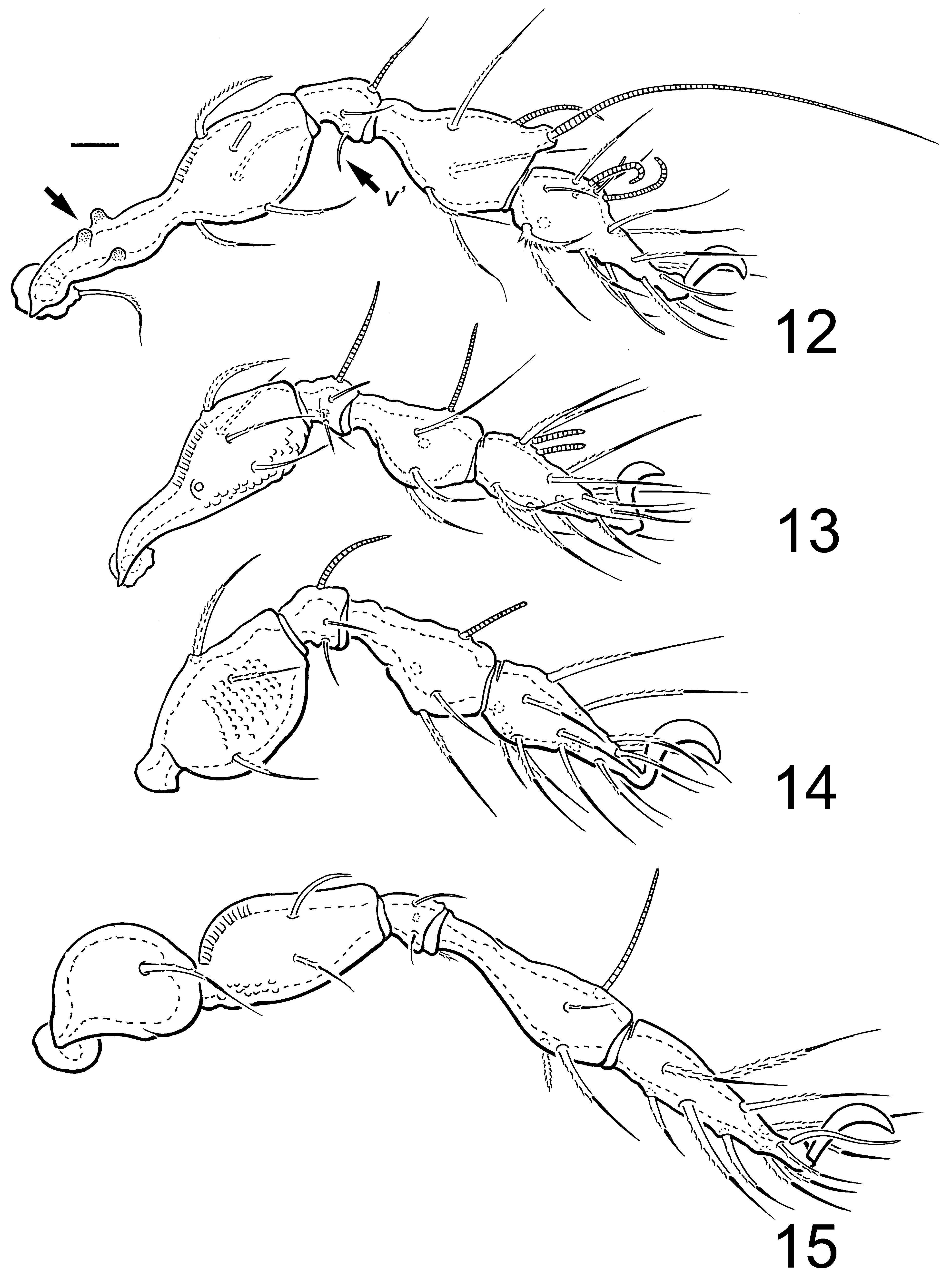
Legs ( Figs 12–15 View FIGURES 12 – 15 ): Leg setation: leg I: 1-5-3(1)-4(2)-18(2); leg II: 1-5-3(1)-4(1)-15(2); leg III: 2-3-1(1)-3(1)- 15; leg IV: 1-2-2-3(1)-12. Femur I with 3 tubercles positioned antiaxially and dorsally in proximal third of leg ( Figs 12 View FIGURES 12 – 15 , 18). Genua I and II with seta v’ present ( Figs 12, 13 View FIGURES 12 – 15 ).
Immatures: Unknown.
Etymology. This species is named in honour of the eminent oribatid expert Professor Dr. Jun-Ichi Aoki .
Remarks. Both males and females of this species are easily recognized by the large tubercles on femur I, unique in the genus. Sexual dimorphism consists of a small convex, oval, weakly porose area positioned medially between lyrifissures ips. Setae h1 and p1 of the male are shorter than those of the female and positioned more closely together.
Autogneta aokii , A. penicillum and A. parva are the only Autogneta species known to have 5 pairs of genital setae.
Gravid females carry up to 2 large eggs. Gut contents are primarily darkly pigmented fungal hyphae and spores (Fig. 19).
No known copyright restrictions apply. See Agosti, D., Egloff, W., 2009. Taxonomic information exchange and copyright: the Plazi approach. BMC Research Notes 2009, 2:53 for further explanation.
|
Kingdom |
|
|
Phylum |
|
|
Class |
|
|
Order |
|
|
SubOrder |
Oribatida |
|
Family |
|
|
Genus |