
Pseudophaeocytostroma bambusicola Monkai & Phookamsak, 2022
|
publication ID |
https://doi.org/ 10.11646/phytotaxa.571.1.3 |
|
DOI |
https://doi.org/10.5281/zenodo.7270450 |
|
persistent identifier |
https://treatment.plazi.org/id/039E87BD-7D5E-8027-FC9B-FCB1FF71FC86 |
|
treatment provided by |
Plazi |
|
scientific name |
Pseudophaeocytostroma bambusicola Monkai & Phookamsak |
| status |
sp. nov. |
Pseudophaeocytostroma bambusicola Monkai & Phookamsak View in CoL , sp. nov. FIGURE 3 View FIGURE 3
Index Fungorum number: IF559821; Facesoffungi number: FoF 12717
Etymology: Refers to the host bamboo from which the holotype was collected.
Holotype: KUN-HKAS 124569 ; living culture, KUMCC 22-12407 .
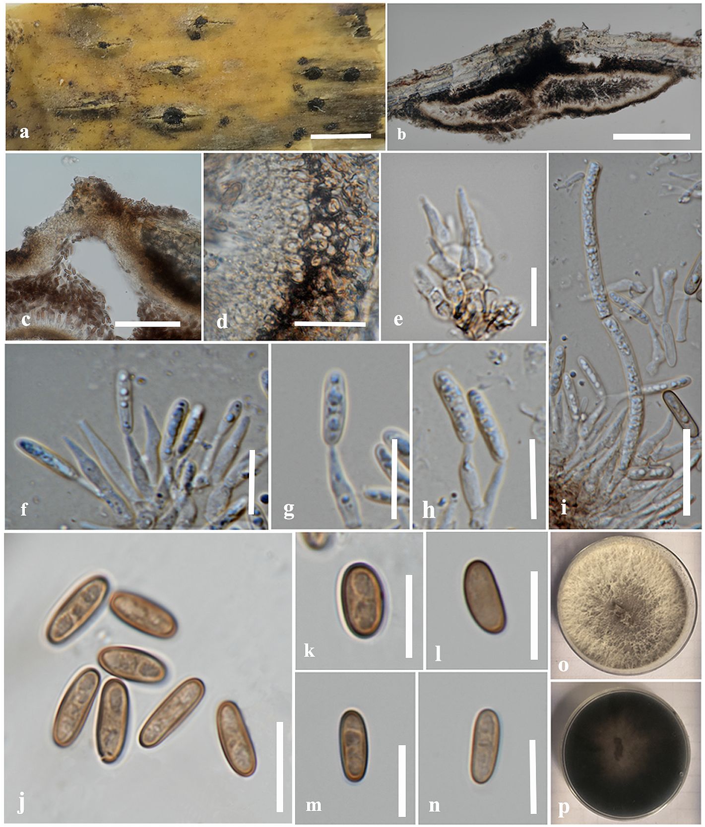
Saprobic on dead bamboo culms. Sexual morph: Undetermined. Asexual morph: Coelomycetous. Conidiomata 89–420 high × 250–704 μm diam., pycnidial, immersed in the clypeus, becoming raised, erumpent, penetrating on host surface, with small black dots of conidial masses, hemispherical to subconical or lenticularis, uni- to bi-loculate, with an ostiole at the center, occasionally produced 2 ostioles in a locule, glabrous. Ostioles up to 80 μm wide, minutely papillate, immersed in host epidermis, circular. Conidiomatal wall up to 60 μm wide, consist of several layers of pseudoparenchymatous cells, arranged in a textura angularis, with dark brown outer layers and hyaline to pale brown towards the inner layers. Paraphyses 59–148 long × 2–3 μm wide (x̅ = 99 × 2 μm, n = 20), intermingled between conidiophores, broadly filiform, septate, hyaline, unbranched, obtuse at the apex, with small granules. Conidiophores 4–10 × 2–5.5 μm (x̅ = 6 × 3 μm, n = 20), tightly aggregated, subcylindrical to ampulliform, or irregular in shape, septate, hyaline to pale brown, branched only at the base. Conidiogenous cells 8–12.5 × 2–3 μm (x̅ = 10 × 2.5 μm, n = 30), enteroblastic, phialidic, determinate, integrated, subcylindrical, tapering towards the apex, hyaline, smoothwalled. Conidia 9–13 × 3–4.5 μm (x̅ = 11 × 4 μm, n = 30), oblong to ellipsoid, obtuse at both ends, aseptate, brown, thick and smooth-walled, guttules.
Known distribution:— Yunnan, China
Culture characteristics:— Conidium germinating on water agar within 24 h at 25 °C. Colonies on PDA reached 5 cm diam. after 7 days at 25 °C, effuse, sparse, entire edge, circular, white in surface, dark gray to black in reverse.
Material examined:— China, Yunnan Province, Honghe Autonomous Prefecture, Honghe County, Honghe Hani Rice Terraces , on dead bamboo culms, 26 January 2021, R . Phookamsak & S. C . Karunarathna , bn17 ( KUN-HKAS 124569 , holotype), ex-type living culture = KUMCC 22-12407 ; ibid., Honghe Autonomous Prefecture, Honghe County, Honghe Hani Rice Terraces , on dead bamboo culms, 26 January 2021, R . Phookamsak & S. C . Karunarathna , bn14 ( KUN-HKAS 124568 ), living culture = KUMCC 22-12410
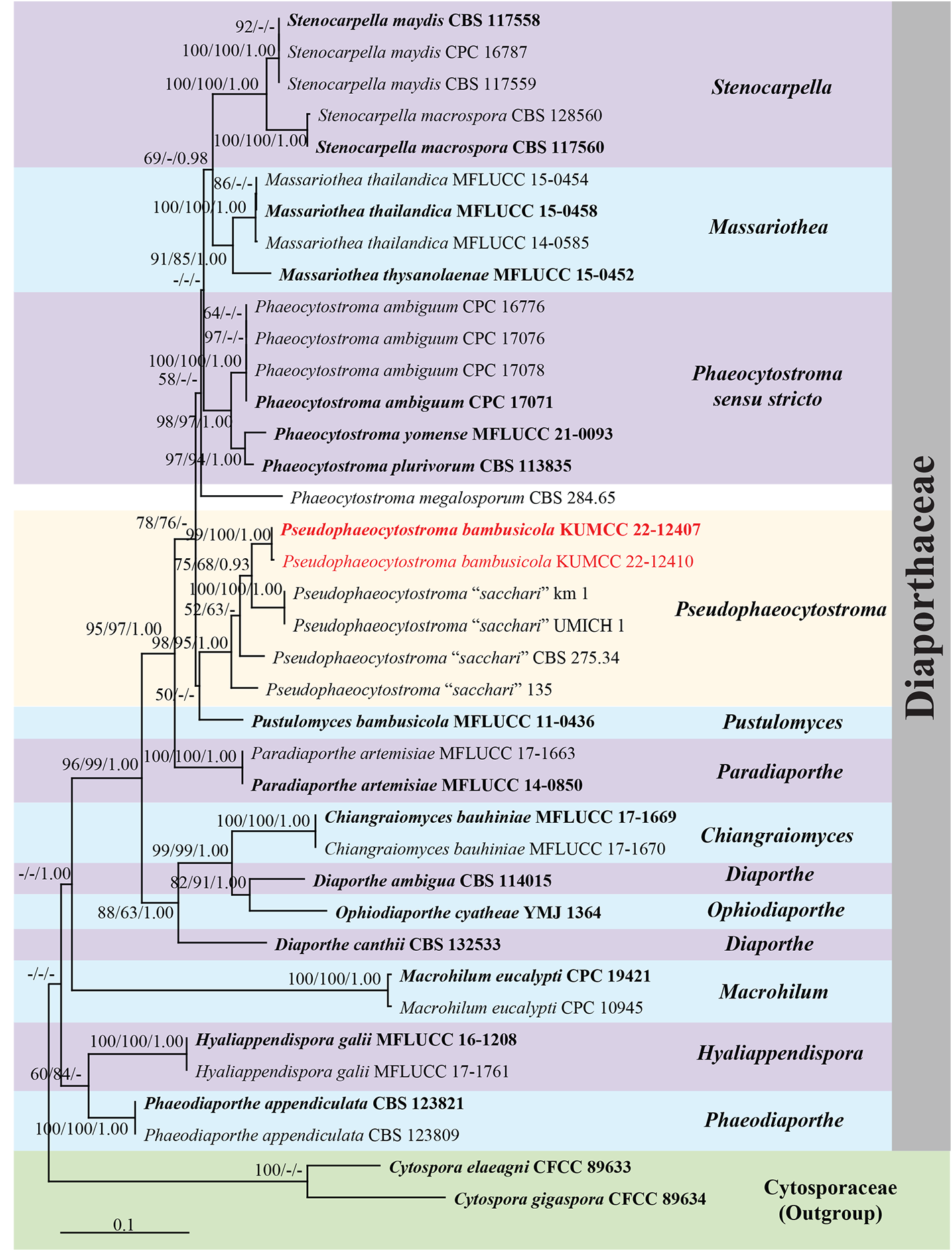
Notes:— The nucleotide BLAST search of ITS sequence indicated that Pseudophaeocytostroma bambusicola (KUMCC 22-12407) has the closest similarity with Phaeocytostroma sacchari strain CBS 275.34 with 96.26% similarity (Identities = 566/588, with 12 gaps), and is similar to Stenocarpella maydis strain Sm.A1-1, CBS 117558 (ex-epitype strain), CPC 16787, CPC 16786, CPC 16782, CPC 16781, CPC 16779, CPC 16778, CPC 16777 with 95.54% similarity (Identities = 552/579, with 5 gaps). The nucleotide BLAST search of LSU sequence indicated that Ps. bambusicola (KUMCC 22-12407) has the closest similarity with Massariothea thysanolaenae strain MFLUCC 15- 0452 (ex-type strain) with 99.64% similarity (Identities = 829/832, with no gap), and is similar to P. ambiguum strain CBS 128561, CPC 17077, CPC 17076, CPC 17074, CPC 16776 with 99.64% similarity (Identities = 829/832, with one gap). The nucleotide BLAST search of TEF1-α sequence indicated that Ps. bambusicola (KUMCC 22-12407) has the closest similarity with P. sacchari strain CBS 275.34 with 88.86% similarity (Identities = 303/341, with 11 gaps), and is similar to P. ambiguum strain CFMS_1294, CPC 17071 (ex-epitype strain), CPC 16776, CPC 17075 CPC 17074, CPC 17072, CPC 16775 with 84.83% similarity (Identities = 179/211, with 12 gaps).
Pseudophaeocytostroma bambusicola is morphologically similar to Phaeocytostroma sacchari in having filiform paraphyses, aseptate, oblong to ellipsoid, brown, conidia with overlapping size (9–13 vs 9–14.5 μm) ( Sutton 1964, 1980, TABLE 2 View TABLE 2 ). However, our new species differs from P. sacchari in having uni- to bilocular conidiomata, minutely papillate ostiole with wider conidiomata (250–704 vs 350 μm) and not abundant, septate, longer paraphyses (59–148 vs 15–35 μm), while P. sacchari has abundant and aseptate paraphyses ( Sutton 1964, 1980, TABLE 2 View TABLE 2 ). The host preference of P. sacchari is Saccharum sp. , Sorghum sp. and Zea mays ( Sutton 1964, Farr & Rossman 2022, TABLE 2 View TABLE 2 ), whereas our new species was found on bamboo.
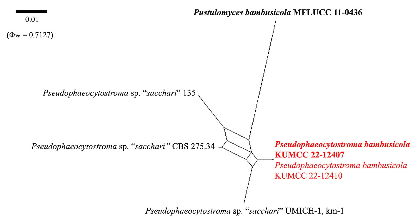
In the phylogenetic analyses, Pseudophaeocytostroma bambusicola (KUMCC 22-12407, KUMCC 22-12410) formed a distinct subclade adjacent to Ps. “ sacchari ” strains CBS 275.34, UMICH-1, km-1 and 135 with high support (98% ML/95% MP/1.00 PP, FIGURE 1 View FIGURE 1 ). The pairwise nucleotide comparison of ITS and TEF1-α sequence data revealed significant differences (more than 1.5%) between Ps. bambusicola (KUMCC 22-12407) and Ps. “ sacchari ” strains CBS 275.34, km-1, UMICH-1 and 135 for each gene region ( TABLE 3 View TABLE 3 ). The results of PHI test also supported the intraspecific variation among these strains and indicated the conspecific between strain UMICH-1 and km-1 ( FIGURE 2 View FIGURE 2 ). Thus, Ps. bambusicola was proposed as a new species based on morphological and phylogenetic evidence.
Phaeocytostroma sacchari was introduced and described by Sutton (1964). Lamprecht et al. (2011) later provided the sequence data for P. sacchari , strain CBS 275.34 which was isolated from Japan without a mention of host substrates and no description provided. Carabez et al. (2014) isolated a fungus strain UMICH-1 from sugarcane displaying stalk rot symptoms in Mexico and identified it as P. sacchari based on morphology and the high similarity of the ITS blast search. Whereas, two strains of P. sacchari (km-1 and 135) were unpublished sequences from the GenBank. Thus, morphological descriptions are not available for these strains. Therefore, we treated these strains as Ps. “ sacchari ” until their correct identification could be confirmed. The examination of ex-type or epitype specimens, more fresh collections and additional sequences are needed for further studies on morphological and genetic variation in this group.
| R |
Departamento de Geologia, Universidad de Chile |
| S |
Department of Botany, Swedish Museum of Natural History |
| C |
University of Copenhagen |
No known copyright restrictions apply. See Agosti, D., Egloff, W., 2009. Taxonomic information exchange and copyright: the Plazi approach. BMC Research Notes 2009, 2:53 for further explanation.