
Altica koreana (Ogloblin)
|
publication ID |
https://doi.org/ 10.11646/zootaxa.3694.5.4 |
|
publication LSID |
lsid:zoobank.org:pub:3E0A832E-77AF-4264-BEBF-CC65EC42CD4E |
|
DOI |
https://doi.org/10.5281/zenodo.6152138 |
|
persistent identifier |
https://treatment.plazi.org/id/039ECE58-FFFE-2E33-ABB6-FB99FAF39F89 |
|
treatment provided by |
Plazi |
|
scientific name |
Altica koreana (Ogloblin) |
| status |
|
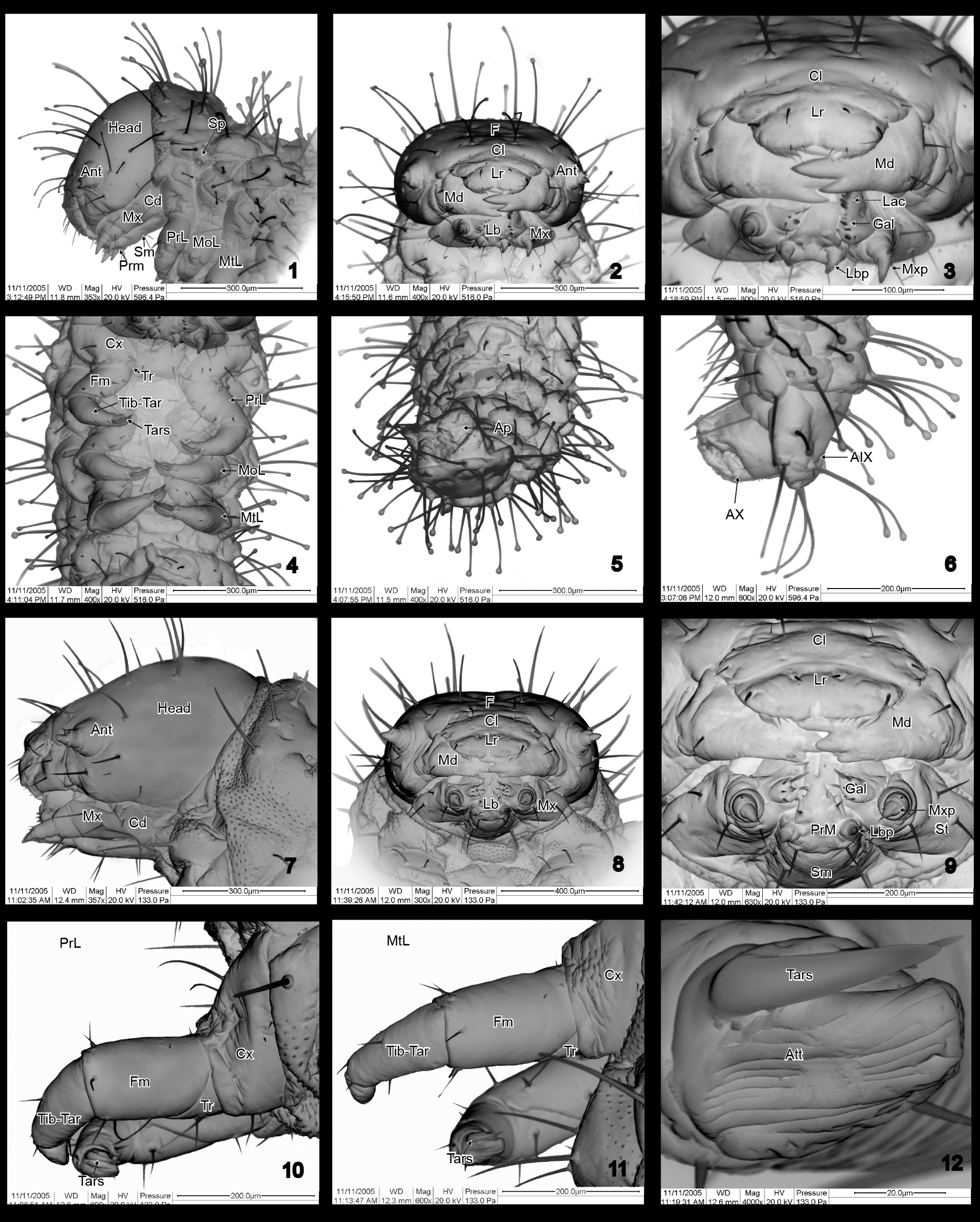
First instar larva ( Figs 1–6 View FIGURES 1 – 12. 1 – 6 , 13, 14 View FIGURES 13 – 16. 13 – 14 ). General body shape: eruciform ( Figs 13, 14 View FIGURES 13 – 16. 13 – 14 ). Capitate setae on dorsal and lateral sides.
Color: dark yellow in life; deep dark yellow after fixation in ethanol.
Body length: 1.5–1.8 mm.
Mature larva ( Figs 7 – 12 View FIGURES 1 – 12. 1 – 6 , 15, 16 View FIGURES 13 – 16. 13 – 14 , 17, 18 View FIGURES 17 – 19. 17 – 18 , 25 View FIGURES 20 – 28. 20 – 24 ). General body shape: eruciform ( Figs 15, 16 View FIGURES 13 – 16. 13 – 14 ); slightly curved when preserved in ethanol.
Color: body mostly dark yellow, with dark brown head; pronotum, legs and postcephalic sclerites black.
Body length: 5.3–5.7 mm.
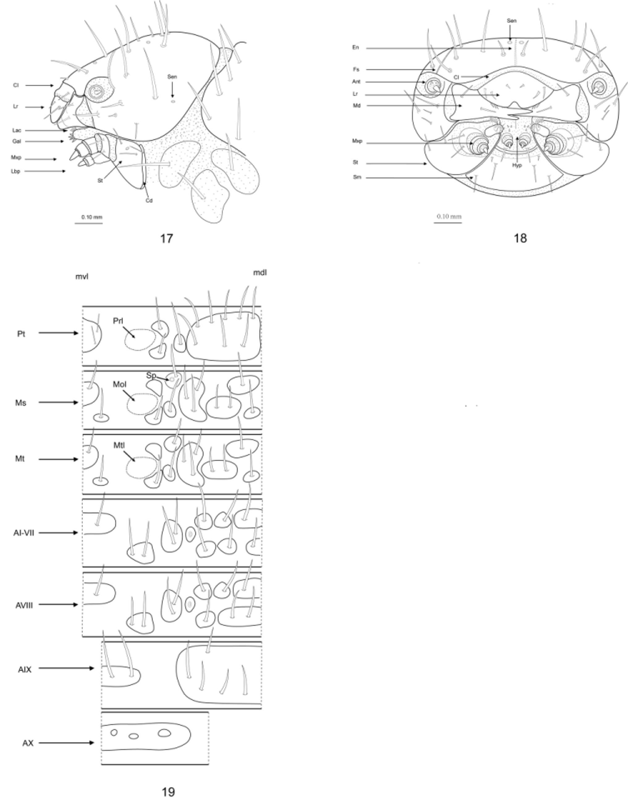
Head: globular, hypognathous, heavily sclerotized. Epicranial suture Y-shaped; coronal suture short; frontal sutures reaching antennal sockets ( Figs 17, 18 View FIGURES 17 – 19. 17 – 18 ); median endocarina robust, extending from base of frontal sutures to clypeus; frons with three pairs of long setae (two pairs of them inserted along endocarina) and pair of frontal campaniform sensilla; vertex strongly convex, with pair of setae on middle of epicranial region; three long setae inserted along frontal suture. Stemmata absent.
Clypeus ( Figs 9 View FIGURES 1 – 12. 1 – 6 , 17, 18 View FIGURES 17 – 19. 17 – 18 ): transverse, with rounded lateral edges, bearing pair of sensilla and three pairs of setae (one long, two short) at its base. Labrum ( Figs 9 View FIGURES 1 – 12. 1 – 6 , 17, 18 View FIGURES 17 – 19. 17 – 18 ): transverse, sclerotized, lateral edges rounded, with deep anteromedial notch; dorsal surface bearing two pairs of long setae and pair of sensory pores near median emargination. Anterior epipharynx with patch of microtrichia grouped in short transverse row close to anteromedian notch; four pairs of epipharyngeal setae arranged in straight longitudinal line laterally at anterior margin. Antenna: articulating area prominent, strongly convex, membranous; located at ends of frontal sutures; two-segmented ( Figs 7, 8 View FIGURES 1 – 12. 1 – 6 ); first antennomere partly membranous, with large conical sensory papilla; second antennomere conical, slightly sclerotized basally. Mandibles ( Figs 8, 9 View FIGURES 1 – 12. 1 – 6 ): symmetrical, palmate, with four teeth; external face bearing two prominent setae; penicillus present, formed by short stout setae; mola absent. Maxilla ( Figs 17, 18 View FIGURES 17 – 19. 17 – 18 ): with large transverse cardo (hardly visible in lateral view) bearing seta; stipes elongate with two sclerotized areas; basal area with lateral and ventrolateral seta and sensory pore anterior to ventrolateral seta and mesad to lateral seta; other sclerotized area smaller, close to palp, bearing seta laterally and seta ventrally; mala ( Figs 17, 18 View FIGURES 17 – 19. 17 – 18 ) distinctly divided into two parts, suggesting presence of galea and lacinia (fused in groundplan of Cucujiformia); apical part (“galea”) with eight setae arranged in circle around stout pedunculate seta (appearing two segmented); proximal part (“lacinia”) with straight longitudinal row of long stout setae; maxillary palpi three segmented; first palpomere with three setae, second palpomere with two setae, third palpomere with one seta. Labium ( Figs 8, 9 View FIGURES 1 – 12. 1 – 6 ): with large trapezoid submentum with light brown coloration, with two pairs of long paramedian setae and short seta at each ventrolateral corner; mentum not well defined; prementum short, sclerotised, dark brown, with large membranous articulatory areas for palps and four setae (one long and three short) and two sensory pores on each side; labial palpi small, two segmented; basal palpomere with two ventromedial sensory pores, and two ventrolateral setae; distal palpomere with ventrolateral sensory pore, two lateral setae (hidden in groove), and lateral elongate placoid sensillum. Hypopharyx: present above prementum, forming subparallel steep structure; slightly narrowing dorsally, with very distinct lateral edges; with two short setae above labial palps.
Thorax ( Figs. 15, 16 View FIGURES 13 – 16. 13 – 14 ): prothorax narrower than other thoracic segments; pronotum well sclerotized, plate-like, divided by median zone of weakness; with five pairs of setae anteriorly, four pairs posteriorly, one unisetose tubercle laterally; two unisetose tubercles present on ventro-lateral pleural region very close to coxal articulation; prosternum with two pairs of paramedian setae.
Meso and metathorax ( Figs 15, 16 View FIGURES 13 – 16. 13 – 14 ): subequal, wider than prothorax; both nota represented by isolated tubercles; pair of unisetose tubercles located anteriorly; pair of unisetose tubercles and pair of large bisetose tubercles arranged in transverse row posteriorly; lateral region with pair of large trisetose tubercles and two pairs of unisetose tubercles (mesothoracic spiracle situated on anterior tubercle); pair of nonsetose tubercles and pair of unisetose tubercles present on ventro-lateral pleural region very close to coxal articulation. Meso- and metasterna with one antero-median bisetose tubercle and pair of postero-median unisetose tubercles. Mesothoracic spiracle annuliform, situated on anterior pleural region close to mesocoxal articulation (epipleural part of Lee 1992).
Legs ( Figs 10, 11, 12 View FIGURES 1 – 12. 1 – 6 , 16 View FIGURES 13 – 16. 13 – 14 ): increasing in size from pro- to metathorax, five-segmented; coxa largely trapezoidal bearing 12 setae (four long and eight short); trochanter largely membranous distally, with four setae; femur strongly sclerotized dorsally, membranous ventrally, with nine long setae and several microtrichia; tibia with four setae; tarsungulus with unpaired membranous attachment structure (pulvillus), with long, setiform sensillum basiconicum at its base.
Abdomen ( Figs 15, 16 View FIGURES 13 – 16. 13 – 14 ): abdominal segments I – VII bearing well defined sclerites arranged dorsally in two transverse rows; posterior row parallel to anterior, with similar chaetotaxy ( Figs 15, 16 View FIGURES 13 – 16. 13 – 14 ); each row with bisetose tubercle in middle, and two pairs of dorso-lateral unisetose tubercles; lateral region with one row of subspiracular trisetose tubercles ( Figs 25, 26 View FIGURES 20 – 28. 20 – 24 ); ventro-lateral region with pair of bisetose tubercles; ventral side with large anteromedian bisetose tubercle and two posterior bisetose tubercles. Segment VIII similar to preceding abdominal segments, except for fusion of posterior bisetose and two unisetose tubercles which form single tubercle with four setae. Segment IX dorsally forming an undivided semicircular pygidium with five pairs of setae. Ventral sclerites fused, forming narrow transverse band with two pairs of setae. Segment X not visible in dorsal view, bearing pygopod. Spiracles present on segments I – VIII, similar to mesothoracic spiracles but smaller.
Material examined: About 19 larvae of various instars, reared from adults collected on buds of Potentilla chinensis in June – July 2004, in Botanical Garden, Beijing, by Huai-Jun Xue, Shu-Yong Wang and Wen-Zhu Li.


No known copyright restrictions apply. See Agosti, D., Egloff, W., 2009. Taxonomic information exchange and copyright: the Plazi approach. BMC Research Notes 2009, 2:53 for further explanation.