
Trichadenotecnum kojimai Yoshizawa & Lienhard, 2014
|
publication ID |
https://doi.org/ 10.11646/zootaxa.3835.4.3 |
|
publication LSID |
lsid:zoobank.org:pub:AD845CF3-CB19-4924-891F-330AAE283D07 |
|
DOI |
https://doi.org/10.5281/zenodo.4921953 |
|
persistent identifier |
https://treatment.plazi.org/id/039F5A5F-FFF6-FFF0-FF20-F8ABFCB339C3 |
|
treatment provided by |
Felipe |
|
scientific name |
Trichadenotecnum kojimai Yoshizawa & Lienhard |
| status |
sp. nov. |
Trichadenotecnum kojimai Yoshizawa & Lienhard View in CoL , sp.n.
( Figs 1G View FIGURE 1.1 , 8 View FIGURE 8 , 9 View FIGURE 9 )
Holotype. Male, Malaysia, Gnung Berembun, Path 3, Cameron Highlands , Pahang, 14.vii.2003, H. Kojima, S. Nomura, N. Takahashi & K. Yoshizawa (canopy fogging, St. 6) ( UKM).
Paratypes. 2 males 1 female, same data as holotype ; 1 male 1 female, type locality, 15.vii.2003, HK.etal (canopy fogging, St. 9) ( SEHU & UKM) .
Description. Male. Head. Yellowish white in ground color; vertical markings blackish brown, each marking touching with neighbors; with pair of triangular blackish brown markings anterior to vertical markings; orbital markings blackish brown; coronal suture black; epicranial suture bordered with blackish brown band dorsally; frons with central pair of brown bands reaching to oceller region dorsally, and with lateral pair of blackish brown broader band not reaching to epicranial suture dorsally, central and lateral bands fused with each other ventrally; eye dark gray, IO/D= 0.6; ocelli white, ocellar field black; gena mostly blackish brown, with pair of oval whitish areas arranged vertically; postclypeus mostly blackish brown ventrally except ventrolateral corner white, dorsal region white with four rows of blackish brown spots; anteclypeus blackish brown. Antenna brown, scape and pedicel blackish brown. Mouthparts brown.
Thorax. Prothorax blackish brown. Mesonotum mostly brown, scutum with yellowish white bands along posterolateral margins of anterior lobe, at middle from posterior end to middle of anterior lobe of scutum, and along anterior margin of lateral lobe. Metanotum brown, median part and anterior region of lateral lobe of scutum yellowish white. Meso- and metapleuron brown except membranous regions.
Legs. Mostly blackish brown, ventral surface of distal half of fore femur and tip of hind femur white, tibiae paler.
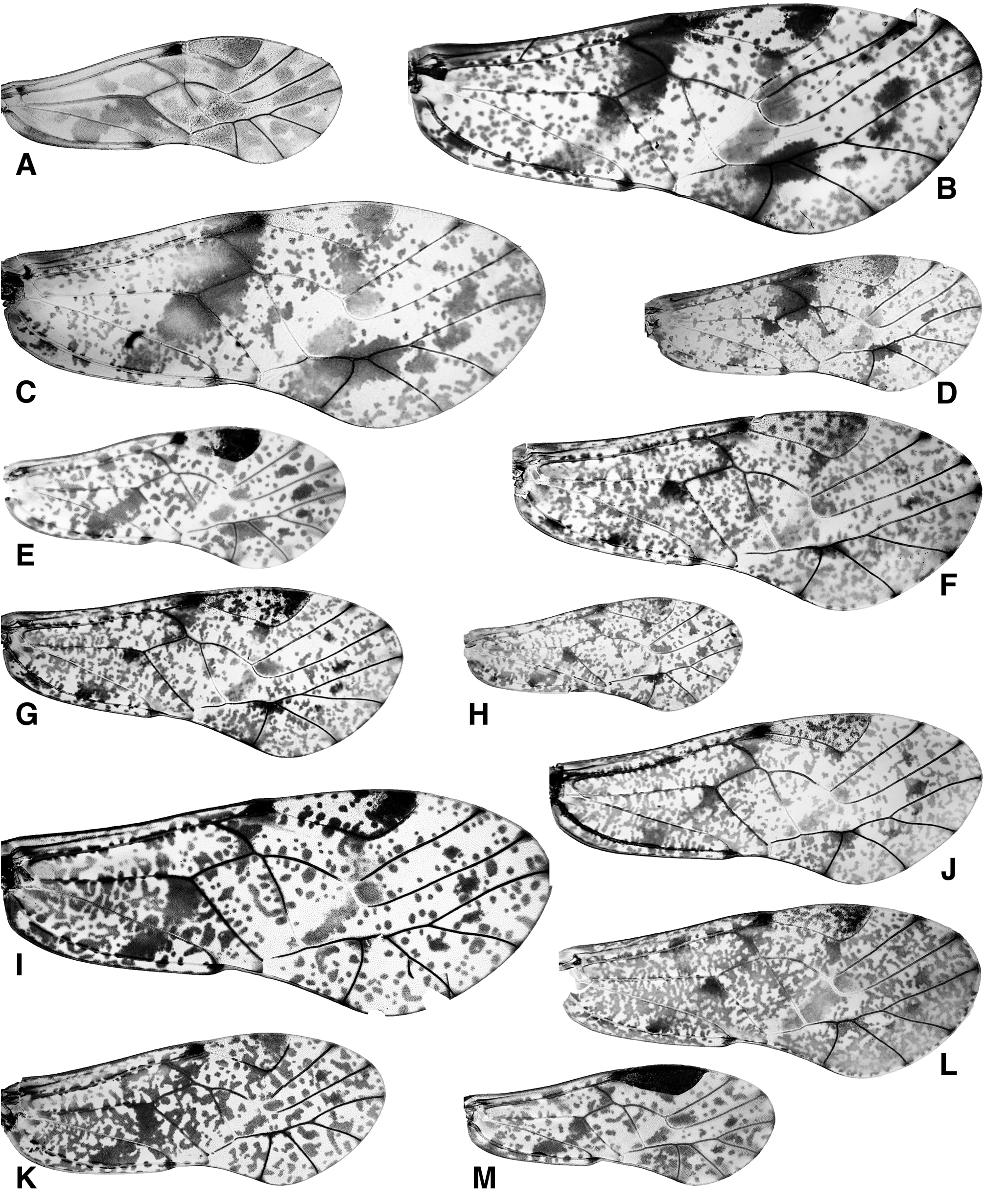
Forewing ( Fig. 1G View FIGURE 1.1 ). Extensively and densely covered with tiny spots, spots rather sparse in cell r3. Distal spot in cell a1 distinct, basal spots distinct. Opposing spots in cell r obscure. Basal band narrow and faint except for dark spots around Rs-M fusion, below M-Cu fork, and in posterior half of cell cup. Distal band faint. Spot on roof of cell m3 narrow but distinct. Submarginal spots distinct in cells r3, r5, and m1, very small but distinct in cell m2, obscure in other cells. Marginal clouds faint. Hindwing hyaline; veins brown.
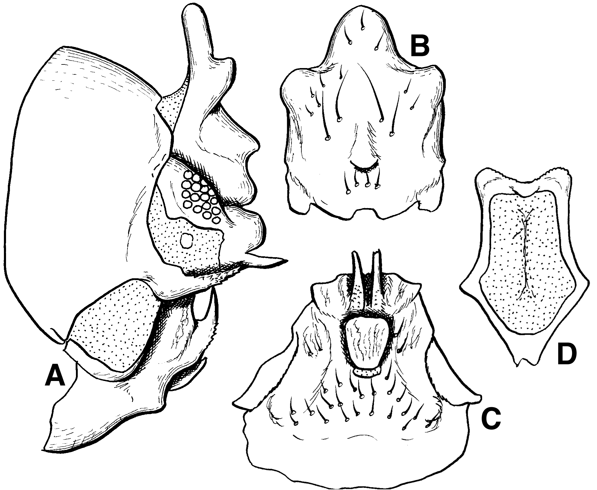
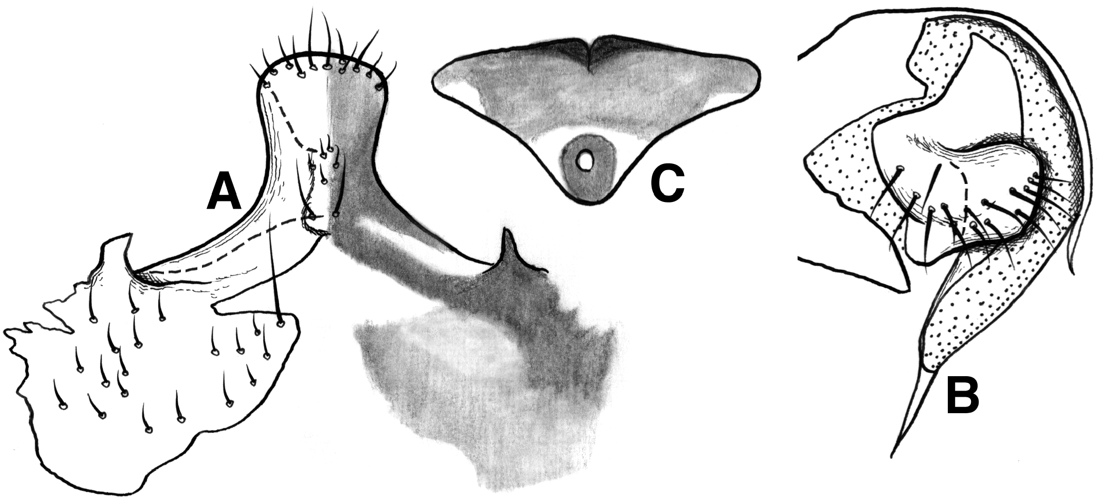
Terminalia. 8th sternum with triangular sclerite completely fused to hypandrium posteriorly ( Fig. 8C View FIGURE 8 ). Clunial arm ( Fig. 8A View FIGURE 8 ) directed posteroventrally in basal half, directed posterodorsally and with some denticles in distal half. Epiproct ( Fig. 8 View FIGURE 8 AB) with strong posterodorsal extension, triangular in lateral view; epiproct lobe strongly expanded dorsally, single-lobed but with weak lateral expansion medially. Paraproct ( Fig. 8A View FIGURE 8 ) with weakly developed triangular basal process; distal lobe broad and rounded; distal process long and fine, almost straight and directed posterodorsally. Hypandrium ( Fig. 8C View FIGURE 8 ) symmetrical, roughly triangular in shape, distal region wrinkled; left and right processes closely associated, narrow, directed posteriorly; median tongue broad, parallel sided, posterior margin shallowly notched medially, ventral surface wrinkled. Phallosome ( Fig. 8D View FIGURE 8 ) with slightly rugous posterior margin, medially with shallow notch, lateral margins slightly broadened toward lateral projection and acutely narrowing toward pointed anterior tip.
Measurements. B 1.9–2.0, Fw 2.3–2.5, Hw 1.9–2.1.
Female. General morphology almost as in male. IO/D = 1.9.
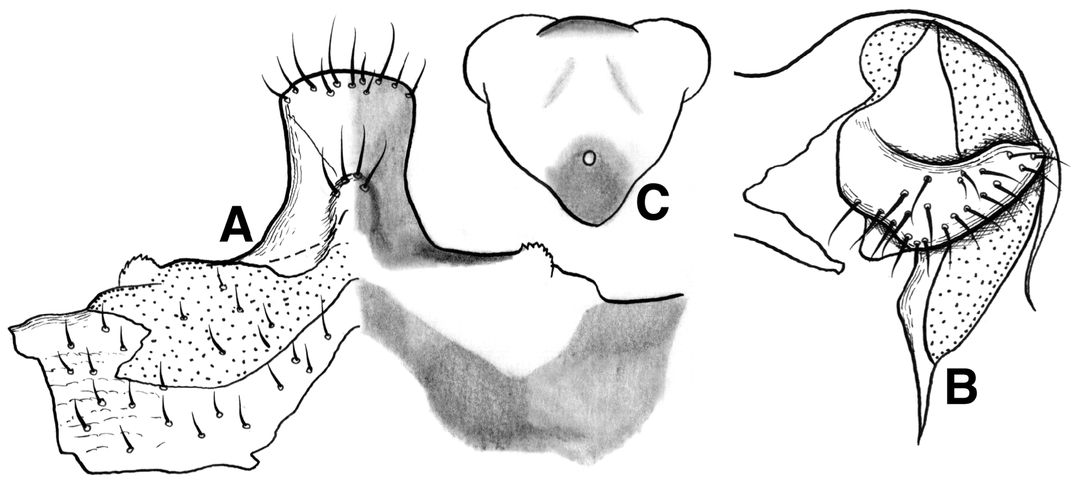
Genitalia. Egg guide ( Fig. 9A View FIGURE 9 ) gradually extended from body of subgenital plate, basally constricted, anterior margin rounded, ventral surface with slight swelling basally; body of subgenital plate widely sclerotized, posteriorly with strongly projecting triangular processes, postero medially with triangular membranous region continuing to anterior trapezoidal membranous region. Gonapophyses ( Fig. 9B View FIGURE 9 ). Ventral valve long. Dorsal valve with long distal process. Posterior lobe of external valve broad but weakly projecting; internal lobe short. Spermapore plate ( Fig. 9C View FIGURE 9 ) broad and short, triangular in shape, anterior margin with small notch, broadly pigmented but darker around spermapore and median region of anterior margin.
Measurements. B 2.0, Fw 2.5–2.6, Hw 1.9–2.0.
Etymology. The species epithet refers to Prof. Hiroaki Kojima who led the Canopy Fogging investigation at Cameron Highland.
Remarks. This species is close to T. yatai , described above, but differs by the shape of male epiproct ( Figs 6A View FIGURE 6 , 8A View FIGURE 8 ) and female subgenital plate ( Figs 7A View FIGURE 7 , 9A View FIGURE 9 ). T. kojimai differs also from T. yatai in the possession of the posterolateral projections on the subgenital plate in the female and the shapes of the lateral clunial arm and epiproct lobe in male. In having a pair of posterolateral projections on the female subgenital plate ( Fig. 9A View FIGURE 9 ), T. kojimai is similar to T. adika and T. paradika Endang & New, 2005 from Indonesia. However, T. kojimai differs clearly from T. adika , by the shape of the lateral clunial arms, epiproct lobe, and distal part of the hypandrium. T. paradika is known from female only, and it differs from T. kojimai by the shapes of the posterolateral projection on the subgenital plate and the external valve of the gonapophyses.
| UKM |
Universiti Kebangsaan Malaysia |
No known copyright restrictions apply. See Agosti, D., Egloff, W., 2009. Taxonomic information exchange and copyright: the Plazi approach. BMC Research Notes 2009, 2:53 for further explanation.
|
Kingdom |
|
|
Phylum |
|
|
Class |
|
|
Order |
|
|
Family |
|
|
Genus |