
Pseudomallada prasinus (Burmeister)
|
publication ID |
https://doi.org/10.11646/zootaxa.4571.4.4 |
|
publication LSID |
lsid:zoobank.org:pub:22B93B2A-8C45-4686-92BD-882183C6B029 |
|
DOI |
https://doi.org/10.5281/zenodo.5928001 |
|
persistent identifier |
https://treatment.plazi.org/id/039F87CE-FFA8-505E-E8B8-069BFE768FB8 |
|
treatment provided by |
Plazi |
|
scientific name |
Pseudomallada prasinus (Burmeister) |
| status |
|
Pseudomallada prasinus (Burmeister) View in CoL
Chrysopa prasina Burmeister, 1839 View in CoL
Chrysopa aspersa Wesmael, 1841 View in CoL
Chrysopa coerulea Brauer, 1850 View in CoL
Chrysopa mariana Navás, 1905 View in CoL
Chrysopa sachalinensis Matsumura, 1911 View in CoL
Chrysopa caucasica Navás, 1914
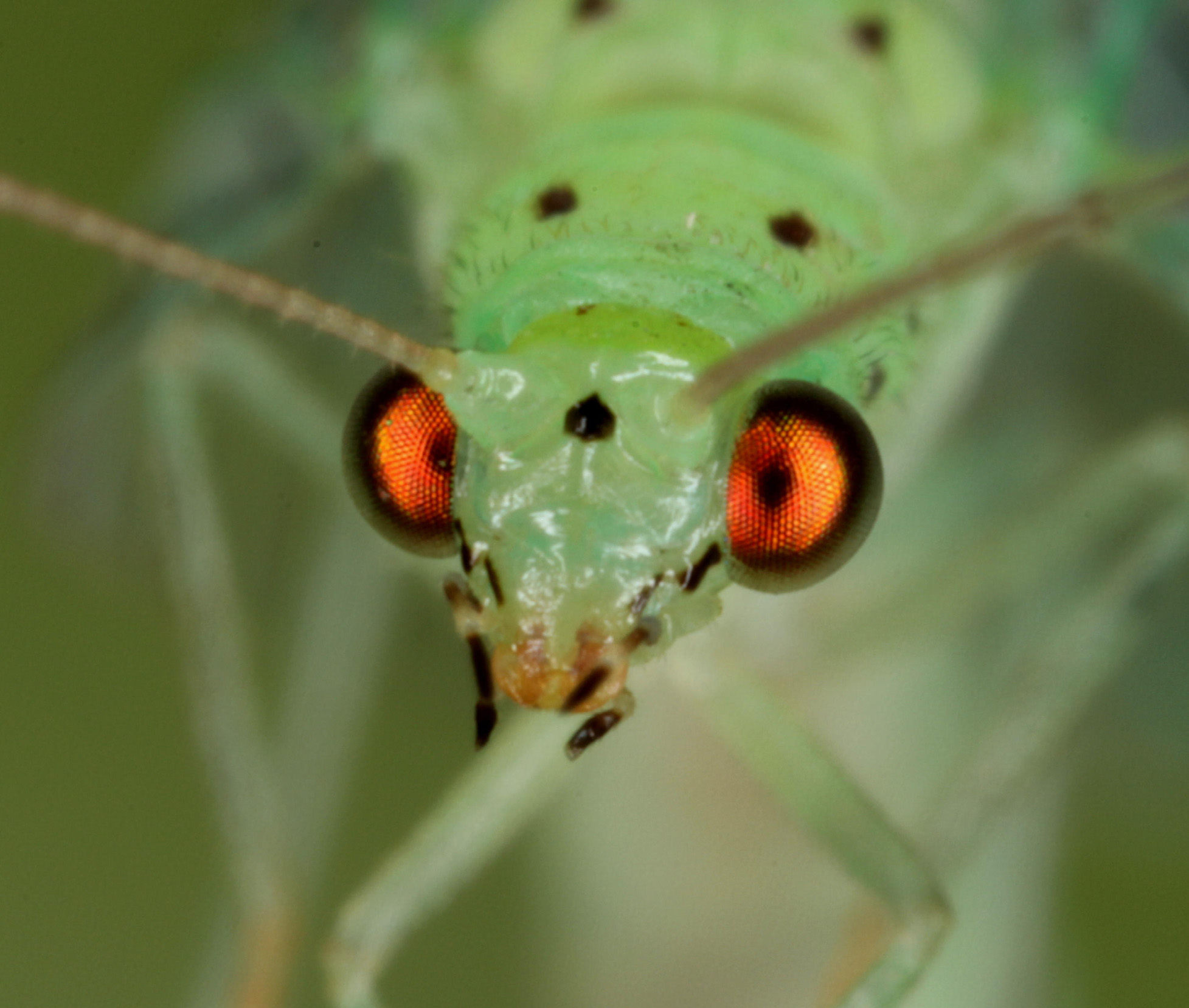
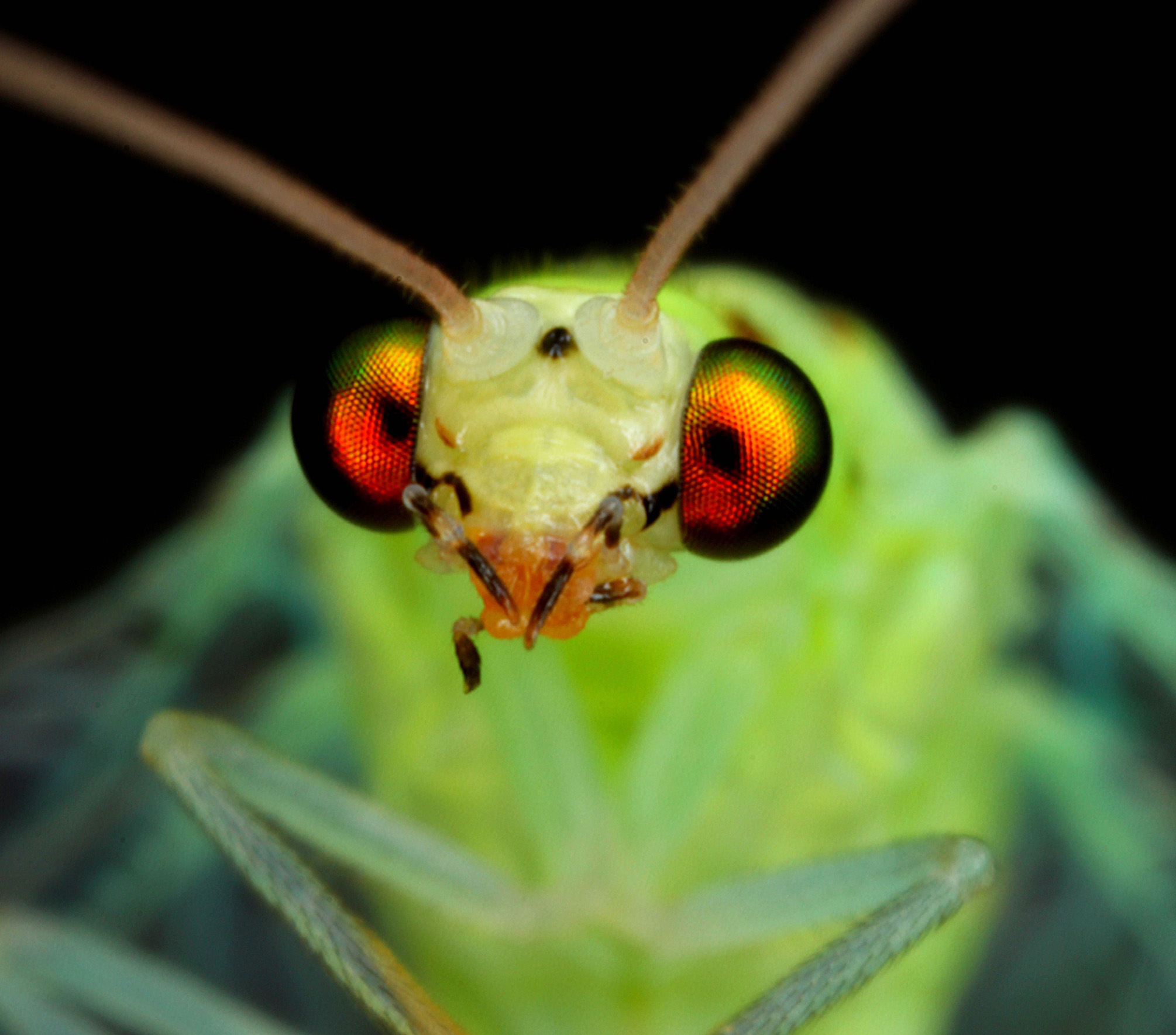
General diagnostic features of prasinoid morphotypes. A medium to large sized grass-green or bluish green lacewing ( Fig. 2 View FIGURE 2 ). Frons and clypeus green or pale. Vertex from green to yellow, sometimes with two reddishbrown spots. Black spot between antennae oval, pear-shaped, bell-shaped or triangular. Genae with black stripes. Scape sometimes with caudal black spots. Prothorax dorsally with one or two pairs of reddish-brown, brown, or black spots, laterally with three brown or black spots. One pair of brown and/or black spots on mesothorax. Abdomen green dorsally, in females sometimes whitish on ventral side. Tergites often with a pair of reddish-brown, brown, or black spots. Dark longitudinal spots or stripes along lateral pleural sutures on abdomen, Setae can vary from blond to black in different morphotypes. Claws dilated. Forewings in females 12.5–18.0 mm long, in males 10.0– 14.5 mm. Black mark at wing base. Costal cross veins dark at both ends, more or less green in the center. Gradates pale or dark. Longitudinal veins mostly green, cross veins variably dark. Genitalia typical for all species of the prasinus group, only gonapsis shape of diagnostic relevance for separating them from other Pseudomallada groups ( Duelli et al. 2017).
Collecting and rearing. Females of the prasinus group showing the above basic features of prasinoid morphotypes were either netted at forest edges or from bushes, or collected at light sources and reared singly in the lab at 22 ±2°C under long day conditions (16L:8D). The rearing containers consisted of plastic cups with a volume of 300 ml and were covered with a paper towel, on which the females deposited their eggs. Moisture was provided with a moist cotton pad. The adults were fed a mixture of yeast, honey and sugar. For each female the mode of oviposition (single eggs vs. bundled eggs) was recorded. For three easily recognizable morphotypes a running culture was kept for crossing experiments. The larvae were fed eggs of Ephestia kühniella , and at later stages also different species of aphids. Mature larvae were kept singly in glass vials and sexed after eclosion.
Morphological assessment. Museum specimens, field collected adults, reared adults, and eclosed hybrids were analyzed with a standard protocol sheet for 26 morphological and biological traits ( Table 1). The general aim was to cover all European species of the prasinus group, but in the present paper the focus is on traits that separate the main European morphs (here called “prasinoid morphs”) that all key out as P. prasinus according to current literature ( Aspöck et al.1980; Plant 1997; San Martin 2004; Mazel et al. 2006). Several of the measured traits were only visible in living specimens. Therefore, the mode of preservation (live, dried, alcohol) was also recorded.
Location: Morpho-Species:
Date: Sex:
Collector: Live, dried, alcohol
Criteria Measurements
Body coloration grass-green, bluish green, bright green, very pale green
Forewing length mm:
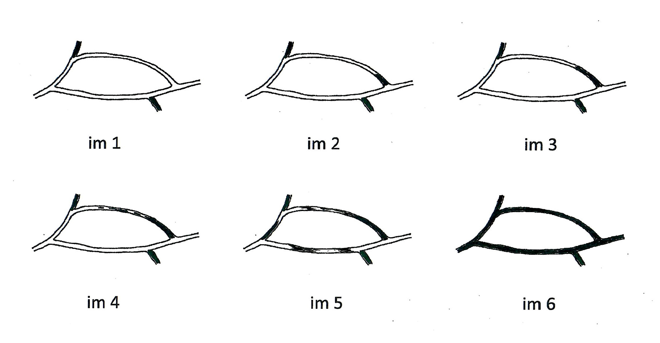
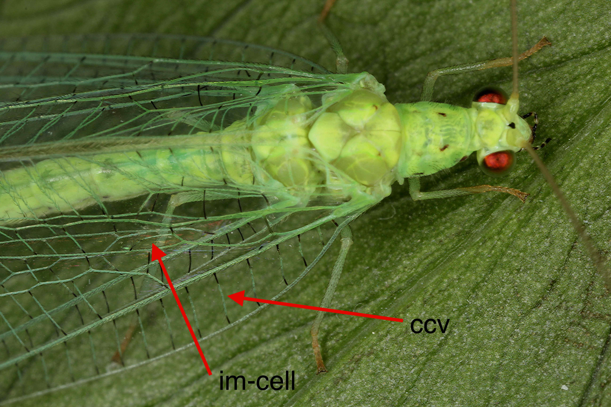
Costal cross veins % black ccv darkness class 1, 2, 3, 4, 5 ( Fig.3 View FIGURE 3 )
Costal cross vein, setae pairs
im cell, % dark veins Intramedian cell type 1, 2, 3, 4, 5, 6 ( Fig. 5 View FIGURE 5 )
Outer gradates black, intermediate, pale
Vertex color green, yellow, intermediate
Dark spots on scape none, caudal, lateral
Interantennal spot oval, pear, triangle or bell-shaped, Y
Subantennal suture above broad black genal band pale (as clypeus), red, brown, black
Two spots on vertex none, reddish, brown, black
Spots on dorsal prothorax (pairs) none, one, two, two in a brown band; reddish, brown, black; large, small Spots on lateral prothorax (one side) 2, 3; small, large; reddish, brown, black
Spots on mesothorax (pairs) 1, 2, twin spots over suture; brown, black
Spots on abdominal tergites (pairs) none, 1/segment, 2/segment; on most segm., few segm.; brown, black Abdominal lateral stripes on suture none, few, most segments; large, small
Ventral side of abdomen green, whitish, black
Eggs deposited in bundles, groups, singly
Further comments:
Explanation for the traits listed in Table 1 and Annex 1, if not self-evident:
Costal cross veins % black ( Fig.3 View FIGURE 3 ): The percentage of black in the costal cross veins (ccv) varies among prasinoid morphs, but there are always some ccv with a green or pale center part. Specimens with entirely black ccv were considered to belong to other species in the prasinus group, such as P. abdominalis ( Brauer, 1856) or P. zelleri ( Schneider, 1851) .
Costal cross veins had a number of paired setae. The number of pairs was counted on the longest ccv of the forewings. In preserved or old specimens, the number of setae was often reduced, so the ccv with the highest number of setae pairs counted on a wing was used. Principi (1956) realized that in P. prasinus not only the shape of the pronotum differed between males and females, but also the pilosity of the veinlets on the wings of males was much denser than that of females. Aspöck et al. (2001) found that in a large part of the distribution range of P. prasinus two types of pilosity occur, often in the same region or even the same habitat ( Fig. 4 View FIGURE 4 ). They cautioned, however, that even if that difference in pilosity turned out to reflect separate species, it would be difficult to find the correct species name among the synonymized species, because most of the type specimens are females.
The intramedian cell (im) was surrounded by three veins that could be more or less green, speckled, or black ( Fig. 5 View FIGURE 5 ).
The subantennal suture above the broad black genal band could be the same color as the clypeus, i.e. pale ( Fig. 6 View FIGURE 6 ), or it is red ( Fig. 7 View FIGURE 7 ), or brown ( Fig. 8 View FIGURE 8 ).
The number, color, and size of spots on the prothorax (dorsal and lateral), as well as of the spots on the mesothorax, were assessed.
The spots on the abdominal tergites are a main discriminant in present keys for the prasinus group. Five traits were tabulated: presence of spots, one or two pairs of spots per segment, spots present on most or only a few of the segments, spot color brown or black, spot size large or small.
The lateral abdominal stripes were usually black. They were often not visible, especially in dried specimens.
Statistical treatment. The initial dataset contained 172 specimens, of which 7 had to be dropped due to missing values. Of the 31 protocolled parameters, 23 could be considered of diagnostic value. Three of these were continuous parameters, measured or counted, that could be subjected to an ANOVA with subsequent TukeyHSD tests and a Quadratic Discriminant Analysis (QDA). Two of them could additionally be coded as factors in three levels each, to be included in a Multiple Correspondence Analysis (MCA). Of the remaining factorial variables, 12 had to be dropped due to many missing or non-discriminating values, leaving a total of 10 for the MCA. All statistical analyses were performed in R 3.5.2 (R Core Team, 2018) using package ‘MASS’ ( Venables & Ripley 2002) for QDA and package ‘FactoMineR’ ( Le et al. 2008) for MCA.
Hybridization. Based on the above morphological and biological (oviposition) assessment, two prasinoid morphs depositing single eggs were chosen to perform experiments in which they were hybridized with the morph depositing bundled eggs. Stocks from different locations in and around Switzerland were reared to yield virgin offspring.
1. The “ marianus ” morph ( Figs. 2 View FIGURE 2 , 12 View FIGURE 12 ): A large species depositing bundled eggs. Frons and vertex green. No red or brown suture below the antennal base. The second instar with obligatory diapause in about half of the larvae, i.e. even under long day conditions. The larval diapause in that morph extended the hybridization experiments over more than one year.
Material: Follatères VS, Switzerland, 31.VIII.2016, on Carpinus betulus , 6♀♀, 3♂♂; Agarone TI, Switzerland, September 2016, at light, 3♀♀, 1♂ .
2. The “greenhead” morph: A small or medium sized species depositing single eggs. Frons pale, Vertex pale green. Red or brown suture below the antennal base. No obligatory diapause in the second larval stage under long day conditions. All males of the strain used for crossing experiments had “furry” wings, i.e. having double the number of setae per costal cross vein. We call them “furwing males” ( Fig.4 View FIGURE 4 ).
Material: Follatères VS, Switzerland, 1.X. 2016, on oak, willows and riparian bushes, 11♀♀, 4♂♂; Agarone TI, Switzerland, September 2016, at light, 13♀♀, 6♂♂ ; La Spezia LI, Italy, 7.V.2017, riparian bushes, 4♀♀, 2♂♂ . Follonica TO, Italy, 6.V.2017, 2♀♀, 1♂ .
3. The “sulfurhead” morph: A medium sized species depositing single eggs. Frons pale, vertex bright yellow ( Fig. 9 View FIGURE 9 ). Subantennal suture red or brown. Mainly Mediterranean. Males of bred stock never with “furry” wings.
Material: Le Mui VAR, France, 15. Sept 2016, 3♀♀, 1♂; Olivetta IM, Italy, Sept. 2016, 1 ♀ ; Follonica TO, Italy, 6.V.2017, 3♀♀, 2♂♂ .
The main goal of the hybridization experiments was to see whether the “ marianus ” morph that is characterized by bundled eggs and obligatory diapause in about half of the larvae in the second stage was reproductively isolated from the two other morphs that are characterized by single eggs and no obligatory diapause.
Single virgin female offspring from females with known egg deposition (bundled or single) were paired with males of one of the other two morphs, or as a control with males of the same morph. Crosses between the two morphs with single eggs are part of an ongoing project and data are not yet ready for publication.
The forced couples were kept in cups of 300 ml and fed a standard Wheast diet. The total number of eggs deposited by each of the females within four weeks was recorded, as well as the number of hatched larvae.
Comparison with type specimens. The type specimen of P. prasinus Burmeister was examined at the Zoological Museum in Halle, Germany, using the standard protocol sheet. The description of the type specimen at the Museum for Natural History in Vienna of C. coerulea Brauer , with mention of eggs and larvae, was studied. The type specimen of C. aspersa Wesmael was examined at the Institut Royale des Science Naturelles de Belgique, Brussels. For the other type specimens, information was gleaned from the original descriptions or reassessments (Legrand et al. 2007).


| TI |
Herbarium of the Department of Botany, University of Tokyo |
| IM |
Indian Museum |
No known copyright restrictions apply. See Agosti, D., Egloff, W., 2009. Taxonomic information exchange and copyright: the Plazi approach. BMC Research Notes 2009, 2:53 for further explanation.
|
Kingdom |
|
|
Phylum |
|
|
Class |
|
|
Order |
|
|
Family |
|
|
Genus |
Pseudomallada prasinus (Burmeister)
| Duelli, Peter & Obrist, Martin K. 2019 |
Chrysopa sachalinensis
| Matsumura 1911 |
Chrysopa mariana Navás, 1905
| Navas 1905 |
Chrysopa coerulea
| Brauer 1850 |
Chrysopa aspersa
| Wesmael 1841 |
Chrysopa prasina
| Burmeister 1839 |