
Parotoplana jondelii, Delogu, Valentina & Curini-Galletti, Marco, 2007
|
publication ID |
https://doi.org/ 10.5281/zenodo.177648 |
|
DOI |
https://doi.org/10.5281/zenodo.6242264 |
|
persistent identifier |
https://treatment.plazi.org/id/039FC129-1E03-FFE2-FF61-6FAE88B9FBE7 |
|
treatment provided by |
Plazi |
|
scientific name |
Parotoplana jondelii |
| status |
sp. nov. |
Parotoplana jondelii sp. n.
( Figs. 3 View FIGURE 3 A–B; Figs. 7 View FIGURE 7 A–B)
Holotype: one whole mount ( SMNH 6666).
Type locality: Apulia, Italy: Santa Maria di Leuca (Lecce), cave ‘la Principessa’(lat. 39°47’58.50”N, long. 18°22’27.86”E), about 5 m deep in medium to coarse sand, May 2005.
Additional material: two specimens from the type locality studied alive, one prepared as whole mount (CZM-55) and one sagittally sectioned (CZM-56)
Etymology: This species is dedicated to Prof. Dr. Ulf Jondelius ( Sweden) in recognition of his contribution to the study of Platyhelminthes, and for his kind patience in dealing with the enthusiasm of the first author in identifying meiofaunal taxa.
Description. Holotype about 1 mm long in fixed conditions. External morphology similar to the other species of the genus, with clearly marked anterior end and fan-shaped caudal end. Limited observations of the internal anatomy could be retrieved from the poor-quality sectioned specimen. The encapsulated oval shaped brain (60 µm long) abuts on the statocyst. Rhabdoids (up to 9 µm in length) are present dorsally and ventrally at both ends of the body. The creeping sole (cilia up to 6,5 µm long) extends from the anterior end to the genital pore. The ciliated epithelium is distinctly higher than the surrounding, non-ciliated epithelium.
The subepidermal longitudinal musculature is particularly well developed ventrally.
The holotype showed a moderately elongate, tubiform pharynx, horizontally oriented. The other two specimens, on the contrary, showed a more typical collar-shaped pharynx.
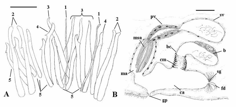
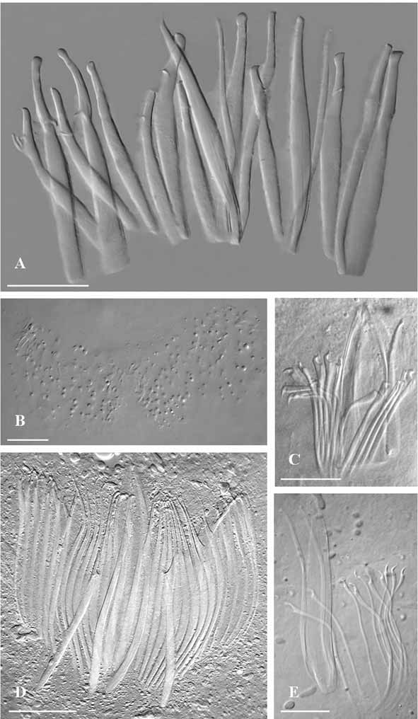
Male genital organs. With two rows of about eight testes each in front of the ovaries. The male copulatory organ consists of a sacciform seminal vesicle, a comparatively short prostatic vesicle ( Fig. 3 View FIGURE 3 B) lined by a ciliated epithelium, a sclerotized apparatus ( Figs. 3 View FIGURE 3 A; 7 A) consisting of 18 spines in both the whole mounts.
Five types of spines, arranged symmetrically into a girdle, can be recognized:
1) two very narrow and straight spines, 75–79 µm long in the holotype;
2) four broad spines, 75–83 µm long, with slightly recurve, bulbous apices provided with a tooth placed at
the basis of the distal third of the spine;
3) four broad spines, 70–80 µm long, narrowing distally into acute apices without any subterminal tooth;
4) two broad spines (62–65 µm long), with sickle shaped apices and marked subterminal tooth;
5) six spines similar in morphology to the previous, but distinctly smaller (53–55 µm long).
In specimen CZM 55, the morphology of the spines was essentially similar, although sizes were somewhat reduced (group 1 = 60 µm; group 2 = 65 µm; group 3 = 65–67 µm; group 4 = 59–60 µm; group 5 = 52– 57 µm). In this specimen, most spines appeared feebly sclerotized basally, presumably due to an early stage of maturity. According to the position of spines in the whole mounts, spines of group 1 seem to be median, and probably act as a functional stylet; spines of group 3 appear to flank the previous ones, and the other spines are symmetrically arranged at their sides.
The male antrum has a nucleated epithelium, and opens into the common atrium.
Female genital organs. With two ovaries anterior to the pharynx. Two rows of numerous vitellaria are present posterior to the ovaries. In preparations of semi-squashed living specimens, a bursa provided with a distinct bursal canal was observed. In sections, the morphology of the bursal canal is obscured by the strong circular musculature that enwraps it entirely. The ovoid bursa shows dorsally a resorbiens portion. In the whole mount, the presence of numerous bursal spines ( Fig. 7 View FIGURE 7 B), arranged into two blocks, could be seen. These spines are sharply triangular in shape (up to 4–5 μm long), feebly sclerotized, and appear as basal lamina derivates. No bursal spines were found in CZM-55 or in the sectioned specimen (CZM-56).
Karyotype. Chromosome number: n = 6; FN = 12. All chromosomes are isobrachial; the first three pairs are distinctly larger than the remaining pairs. The only plate suitable for karyometric analysis yielded the following data: Chrom. I = r.l.: 25.55; c.i.: 47.47 (m); Chrom. II = r.l.: 23.99; c.i.: 49.52 (m); Chrom. III = r.l.: 23.65; c.i.: 41.26 (m); Chrom. IV = r.l.: 10.4; c.i.: 33.33 (sm); Chrom. V = r.l.: 9.47; c.i.: 37.12 (sm); Chrom. VI = r.l.: 6.93; c.i.: 38.19 (m).
Remarks. The limited sample of P. jondelii sp. n. appears heterogeneous. The holotype, which is presumably a mature specimen, showed an elongated pharynx, and the presence of bursal spines, characters that were absent in the other two specimens. However, CZM 55, given the incomplete sclerotization of copulatory spines, was clearly at an early phase of maturity. Furthermore, due to the contraction of the different muscles of the pharynx, a collar shaped pharynx can be held horizontally, at least for a short time (pers. obs.), and may thus not be a specific character. P. jondelii sp. n. appears nonetheless unique in the genus, for the details of its sclerotized apparatus. None of the known species, has spines as broad, or with markedly blunt apices, as the ones found in the new species. P. jondelii sp. n. is the second species of Parotoplana known with a tubiform pharynx (but see above comment). The other species, P. pacifica , was described by Ax & Ax (1967) and belongs to the species group with a central stylet, which is absent in the new species. The new species also shows the presence of bursal spines. This character is shared with two species only: P. capitata (type of the genus Parotoplana ) and P. procerostyla ( Ax, 1956) . Both these species have sickle-shaped, slender spines; the latter, in addition, is provided with an elongate, tubular stylet. It should be however mentioned that the presence of bursal spines is not easily appreciable, except on well-squeezed mounts and the character might be more widespread than presently acknowledged.
The karyotype of P. jondelii sp. n., albeit basic in number (n = 6), is nonetheless distinct for the presence of three pairs of large chromosomes. All the species with the same haploid number have only two distinctly larger pairs ( Curini-Galletti et al., 1984; present paper).
Diagnosis: Parotoplana provided with triangular bursal spines and with a sclerotized apparatus consisting of a girdle of 18 spines: two narrow median spines, four submedian broad spines, with slightly hooked apices, and 12 broad spines, differing for the development of subterminal teeth and the shape of the apex (blunt to hooked). Karyotype with n = 9, with three chromosome pairs markedly larger than the others.
| SMNH |
Saskatchewan Museum of Natural History |
No known copyright restrictions apply. See Agosti, D., Egloff, W., 2009. Taxonomic information exchange and copyright: the Plazi approach. BMC Research Notes 2009, 2:53 for further explanation.