
Parotoplana spathifera, Delogu, Valentina & Curini-Galletti, Marco, 2007
|
publication ID |
https://doi.org/ 10.5281/zenodo.177648 |
|
DOI |
https://doi.org/10.5281/zenodo.6242262 |
|
persistent identifier |
https://treatment.plazi.org/id/039FC129-1E06-FFE0-FF61-68248916F858 |
|
treatment provided by |
Plazi |
|
scientific name |
Parotoplana spathifera |
| status |
sp. nov. |
Parotoplana spathifera sp. n.
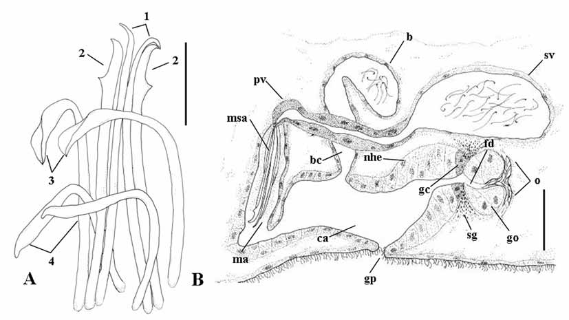
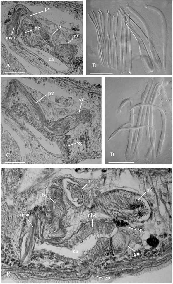
( Figs. 2 View FIGURE 2 A–B; Figs. 6 View FIGURE 6 D–E)
Holotype: one whole mount ( SMNH 6663).
Type locality: Apulia, Italy: Porto Cesareo (Lecce), Torre Scianuli (lat. 40°14’3.72”N, long. 17°54’35.77”E), about 7 m deep in coarse sand among rocks, May 2005.
Additional material: Paratype ( SMNH 6664): one specimen from the type locality, sagittally sectioned. Seven specimens sagittally sectioned (CZM-44/50); one karyological slide (CZM-51); three whole mounts (CZM-52/54), all from the type locality.
Etymology: the specific epithet refers to the shape of one pair of spines in the sclerotized organ, the apex of which somewhat resembles the spathe of an arum lily (fam. Araceae ).
Description. The holotype is an adult worm, about 1.5 mm long in fixed condition. The anterior and the fan-shaped posterior end are provided with numerous adhesive papillae. Short tactile bristles are present at the cephalic end. Encapsulated, oval shaped brain 55 µm long in the paratype; statocyst about 20 µm in diameter. The nucleated epithelium is ciliated ventrally (with cilia about 6 µm long), forming a creeping sole, which runs from the anterior end to behind the genital pore.
Numerous rhabdoids (up to 20 µm long) are scattered all over the body. Subepidermal longitudinal musculature well developed ventrally. The collar shaped pharynx (about 80 µm long) is located in the second half of the body. It is entirely ciliated (cilia about 3,5 µm long), with the exception of its distal tip.
Male genital organs. Two lateral rows of testes (about 34 in each row) are present anterior to the ovaries. The copulatory organ consists of a large sacciform seminal vesicle (76 µm in length in the paratype), which is distally connected to an elongate prostatic vesicle ( Figs. 2 View FIGURE 2 B; 6 E). The seminal vesicle has a nucleated epithelium and it is provided with a layer of well-developed circular musculature. The prostatic vesicle, lined by a glandular, non-ciliated epithelium, is distally connected to the sclerotized apparatus ( Figs. 2 View FIGURE 2 B; 6 E) which consists of four pairs of spines, symmetrically arranged ( Figs. 2 View FIGURE 2 A; 6 D):
1. two median spines with pointed apices (100 µm long in the holotype, ranging 100–102 µm);
2. two spines (94–95 µm long; range: 93–95 µm) with sickle shaped apices, with pointed distal end and a subterminal tooth;
3. two spines (90–92 µm long; range: 89–92 µm) with recurve apices, provided with a feebly sclerotized, distal subtriangular lamina;
4. two spines (70–72 µm long; range 70–74 µm) with slightly sickle shaped apices and obtuse distal tip.
The male antrum is lined with a nucleated epithelium; it opens into the anterior part of the common genital atrium.
Female genital organs. Two ovaries anterior to the pharynx. Two rows of vitellaria extend from posterior to the ovaries to anterior of the copulatory organ. About five vitellarium vesicles are present anterior to the pharynx and 17 behind it. The ovoid bursa is lined by a nucleated epithelium ( Figs. 2 View FIGURE 2 B; 6 E). It opens into the common atrium through a wide, straight canal with a low, nucleated epithelium.
The oviducts are ciliated. They join distally into an extremely short, ciliated, female duct. This female duct is partly surrounded by few, very large glandular cells (the ‘glandular organ’) and by the female glands ( Figs. 2 View FIGURE 2 B; 6 E). It opens into the common genital atrium. The epithelium of the atrium is high and nucleated. Adjacent to the ‘glandular organ’, the epithelium is formed by glandular cells, with very fine, basophilous secretion.
Karyotype. Chromosome number: n = 9; FN = 16. Chromosome pairs differ noticeably in size and centromeric index. Chrom. I is a large metacentric; Chrom. II is medium sized, at the border between submetacentric and subtelocentric. The remaining pairs can be arranged in a decreasing series; Chrom. IX is very small, about 1/9th the size of the largest pair. The only plate suitable for karyometric analysis yielded the following data: Chrom. I = r.l.: 27.9; c.i.: 45.07 (m); Chrom. II = r.l.: 16.13; c.i.: 25.4 (sm); Chrom. III = r.l.: 12.6; c.i.: 28.87 (sm); Chrom. IV = r.l.: 10.22; c.i.: 45.12 (m); Chrom. V = r.l.: 9.3; c.i.: 41.05 (m); Chrom. VI = r.l.: 7.92; c.i.: 17.14 (st); Chrom. VII = r.l.: 7.26; c.i.: 14.83 (st); Chrom. VIII = r.l.: 6.72; c.i.: 28.34 (sm); Chrom. IX = r.l.: 3.28; c.i.: 33.33 (sm).
Remarks. Except for the new species, species of Parotoplana lacking a central stylet include P. capitata Ax 1956 , P. primitiva Ax 1956 , P. multispinosa Ax 1956 , P. m o y a Marcus 1949, P. turgida Ax & Ax 1974 , P. bermudensis Ax & Sopott-Ehlers 1987 , P. l a t a Ax & Sopott-Ehlers 1987, P. subtilis Ax & Sopott-Ehlers 1987 , P. mollis Ax & Sopott-Ehlers 1987 , and P. bicupa Sopott-Ehlers 1976 . Only P. spathifera sp. n. however has a sclerotized apparatus consisting of four pairs of spines, with each pair markedly different in shape from each other. Furthermore, it has the lowest number of spines (eight) known for the genus Parotoplana . Only P. t u r - gida has a sclerotized apparatus of less than 10 spines. In this species, however, the spination consists of two larger and seven distinctly smaller spines; all spines have sickle shaped apices ( Ax & Ax, 1974).
The ‘glandular organ’ seen in the female genital system of the new species is similar to that described for P. c a p i t a t a ( Ax, 1956, fig. 150, pg. 220) and for P. uncinata ( Lanfranchi 1978, fig. 7, pg. 254). In P. capitata , however, a much longer female duct is present. The distal fusion of the oviducts, as seen in the new species, is a rare character in the genus Parotoplana , and only reported from P. macrostyla and P. uncinata ( Lanfranchi, 1978) . Furthermore, in both these species the epithelium lining the female portion of the common atrium is high and glandular, similarly to P. spathifera sp. n. The morphology of the sclerotized structures in the species above is however quite distinct, as both P. macrostyla and P. uncinata have a central stylet, and a girdle of numerous spines with the same morphology. Any phylogenetic inference based on the shared presence of the ‘glandular organ’ in these species should therefore be deferred until a thorough revision of the genus is accomplished.
P. spathifera sp. n. is the only species of Parotoplana so far known with n = 9. The small size of many chromosome pairs and their low centromeric index suggest that the high haploid number is derived through a series of fissioning processes. Robertsonian mechanisms of chromosome fission and fusion have already been documented in Proseriata, and appear to be a widespread pattern of karyological evolution in the group (Curini-Galletti et al., 1989).
Diagnosis: Species of Parotoplana with a glandular organ surrounding the outlet of the extremely short female duct in the common atrium. The sclerotized apparatus consists of two median spines with pointed apices (100–102 µm long); two spines (93–95 µm) with sickle shaped apices, with pointed distal end and a subterminal tooth; two spines (89–91 µm) with spathe-shaped apices; two spines (70–74 µm) with sickle shaped apices and rounded distal tip. Karyotype with n = 9, with chromosome pairs markedly differing in size.
| SMNH |
Saskatchewan Museum of Natural History |
No known copyright restrictions apply. See Agosti, D., Egloff, W., 2009. Taxonomic information exchange and copyright: the Plazi approach. BMC Research Notes 2009, 2:53 for further explanation.