
Lestagella penicillata (Barnard, 1940)
|
publication ID |
https://doi.org/ 10.11646/zootaxa.3750.5.2 |
|
publication LSID |
lsid:zoobank.org:pub:06F7524F-2ABB-412B-A9FA-91B8A059196E |
|
DOI |
https://doi.org/10.5281/zenodo.6149743 |
|
persistent identifier |
https://treatment.plazi.org/id/03A087A6-FFB5-9F34-FF08-1CEDFEB0DD54 |
|
treatment provided by |
Plazi |
|
scientific name |
Lestagella penicillata (Barnard, 1940) |
| status |
|
Lestagella penicillata (Barnard, 1940) View in CoL
Lithogloea harrisoni Barnard, 1932: 253 , pro parte young nymphs, Fig. 43 a–c Lithogloea penicillata Barnard, 1940: 637
Ephemerellina penicillata Allen & Edmunds, 1963: 12 Lestagella penicillata Demoulin, 1970: 130
Material examined. SOUTH AFRICA: Lectotype, ♂ imago, Table Mt. slopes, XI-1932, K.H.B., approximately 33°58′S, 18°25′E. Condition: head, thorax and abdomen mostly disintegrated (kept in ethanol with syntype series). Slide preparations (EPH-A000411): genitalia, wings, mid- and hindlegs (forelegs missing), SAMC.
Paralectotypes. 5 ♀ nymphs, slide preparations of 3 ♀ nymphs (EPH-A000411): Gills (Slide 27), mouthparts (Slide 23, Slide 25, Slide 27) and legs (Slide 27), Table Mt. slopes, XI-1932, K.H.B., SAMC. 1 ♀ subimago, 1 ♂ subimago, 1 ♀ imago, 19 nymphs, (EPH-A000411), Table Mt. slopes, XI-1932, K.H.B., SAMC; 3 ♂ adults (BMNH(E) 1201855–BMNH(E) 1201857), 3 ♀ adults (BMNH(E) 1201858–BMNH(E) 1201860), 1 ♂ subimago (BMNH(E) 1201861), 29 nymphs (BMNH(E) 1201862–BMNH(E) 1201866; BMNH(E) 1201872–BMNH(E) 1201891; BMNH(E) 1239038–BMNH(E) 1239041), Table Mt. slopes, XI-1932, K.H. Barnard, BMNH.
Other material examined. 3 ♂ adult, 1 ♀ adult, (BMNH(E) 1201851–BMNH(E) 1201854) Orange Kloof Table Mt, 28-XI-1934, K.H.B. 2 ♀ nymphs, whole specimen photographed (HMJ 181A), slide preparations: HMJ 60A, whole specimen. 1 ♂ nymph, slide preparations: HMJ 60A, whole specimen. 2 ♂ imagos, slide preparations: genitalia, wings and legs (HMJ 181A), genitalia and wings (HMJ 181A), Table Mt. slopes, Window Stream 18-II- 2012, 14-XII-2012, AMGS. 2 ♂ nymphs, slide preparations: gills (HMJ 118A) and mouthparts (HMJ 118A), Table Mt. slopes, Skeleton Ravine, 29-X-2012, AMGS.
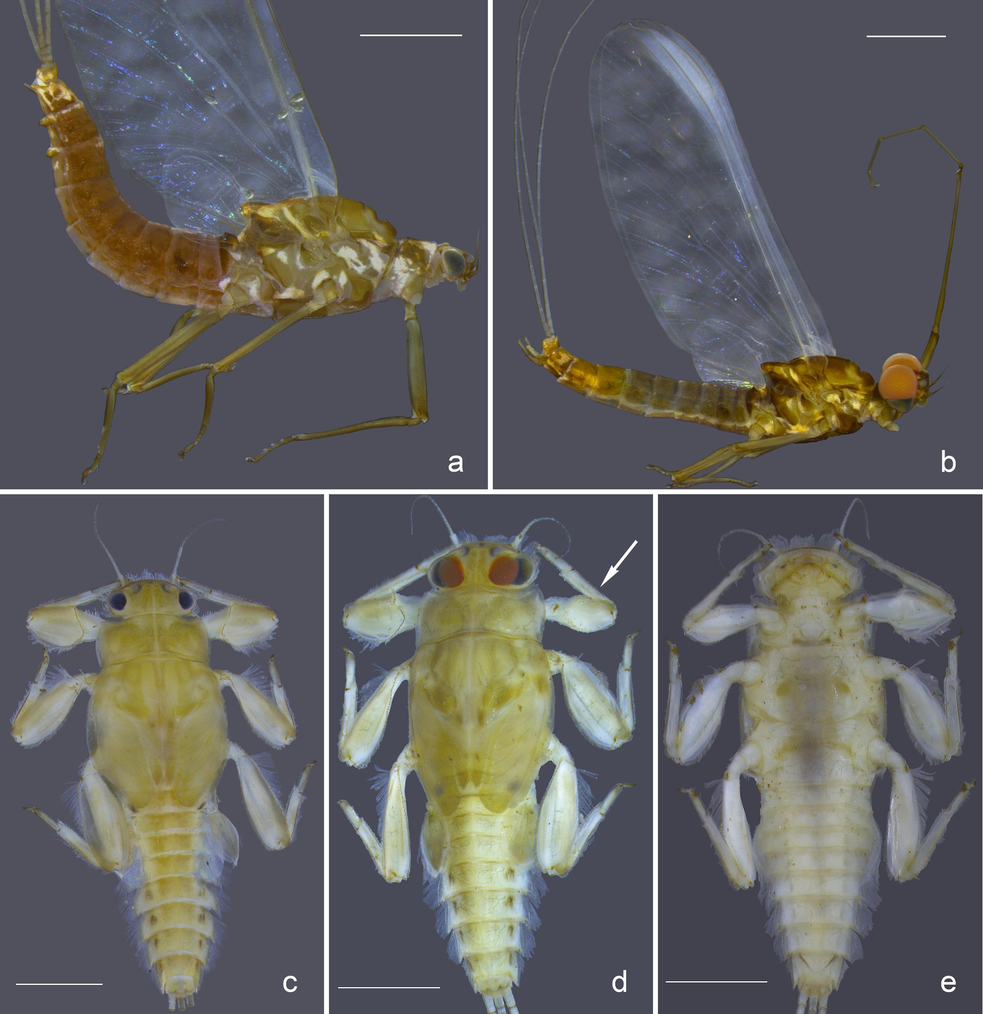
Male Imago (Lectotype and paralectotypes). Head ( Fig. 2 View FIGURE 2 b). Antennae length half the width of the head capsule; dorsal portion of compound eyes large, reddish-brown and spherical; compound eyes only slightly separated dorsally ( Fig. 2 View FIGURE 2 b).
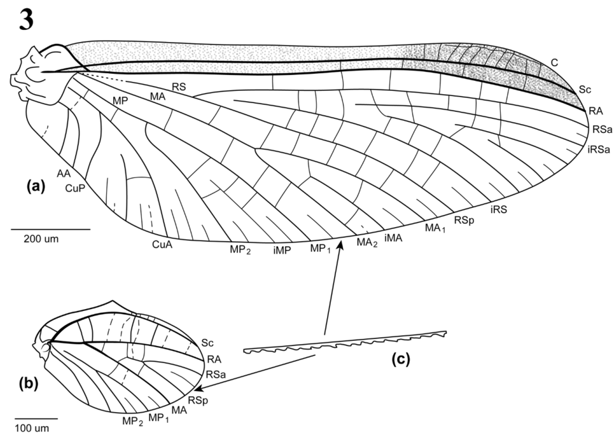
Thorax. Forewings ( Fig. 3 View FIGURE 3 a) narrow with distinct pterostigma present with 8–10 crossveins, variations in intercalaries denoted by dashed lines. RS basally faint. Marginal intercalaries present, sometimes paired, but not present between RA and RSa. Hindwings ( Fig. 3 View FIGURE 3 b) ovoid with costal elevation and slight medial depression in costal margin. Ventral margins of both fore- and hindwings jagged, possibly as a result of shedding of subimaginal falcate microtrichia.
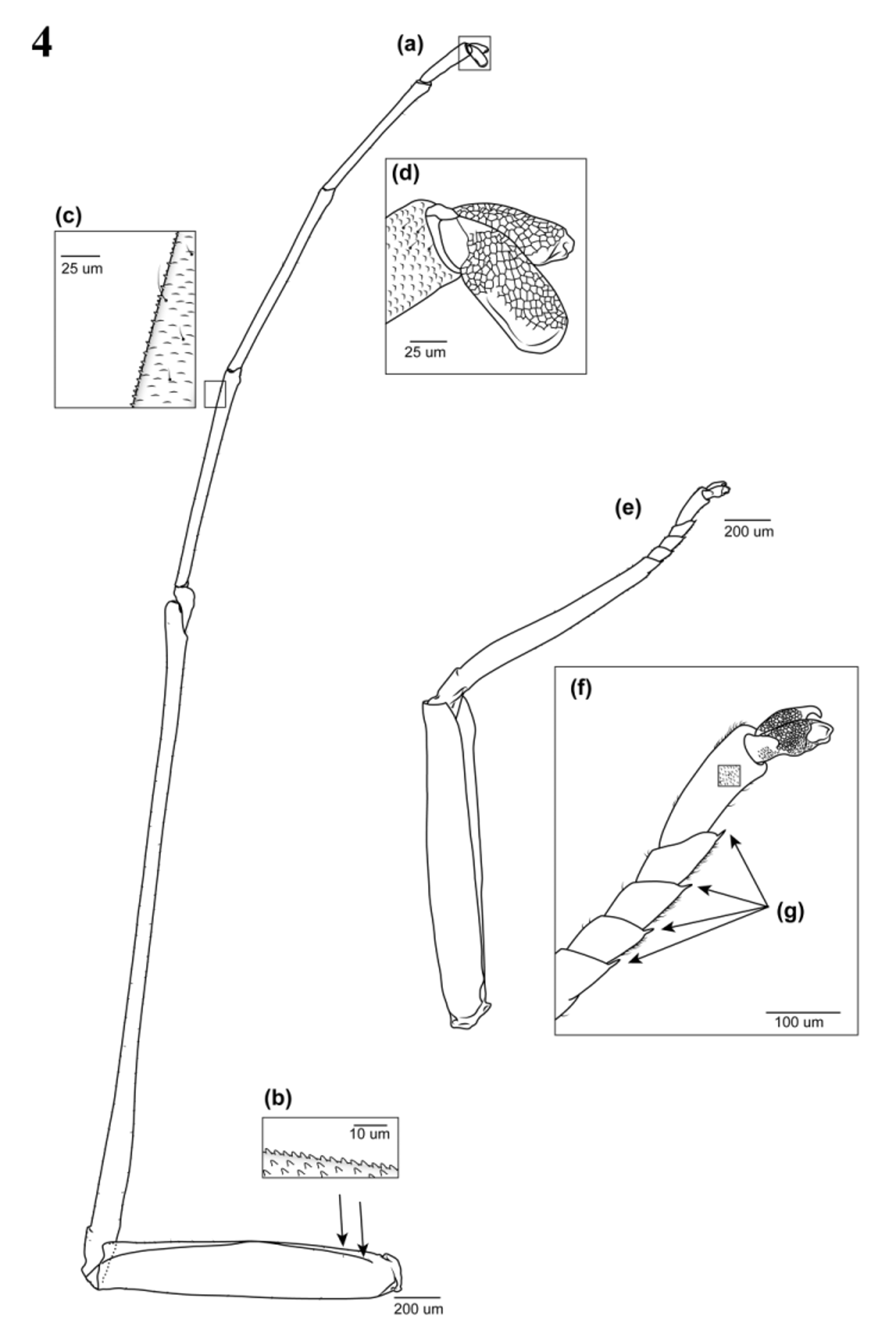
Forelegs ( Fig. 4 View FIGURE 4 a–d) with tibiae just over twice the length of femora and slightly longer than tarsi, segments I and II of tarsi subequal, segment III half-length of segment II and segment IV is ca. one third length of segment III. Femora with subtriangular, scale-like projections and few scattered setae ( Fig. 4 View FIGURE 4 b). Tibiae and tarsi with smaller scale-like protrusions and scattered setae ( Fig. 4 View FIGURE 4 c). Claws modified into two pad-like lobes with flat scale-like dorsal surface and smooth, slightly grooved ventral surface ( Fig. 4 View FIGURE 4 d). Mid- and hindlegs ( Fig. 4 View FIGURE 4 e) with tibiae ca. 0.8 times femora length, few setae scattered on tibia becoming slightly more frequent nearer the tarsi ( Fig. 4 View FIGURE 4 f). Tarsi one third the length of tibiae, tarsal segments I–III subequal in length with many short simple setae, segment IV twice the length of segment III. Distal end of tibia and tarsal segments I–III with distinct spike on ventral surface ( Fig. 4 View FIGURE 4 g). Claws with one pad-like lobe and one hook, dorsal surface with flat scales as in foreleg.
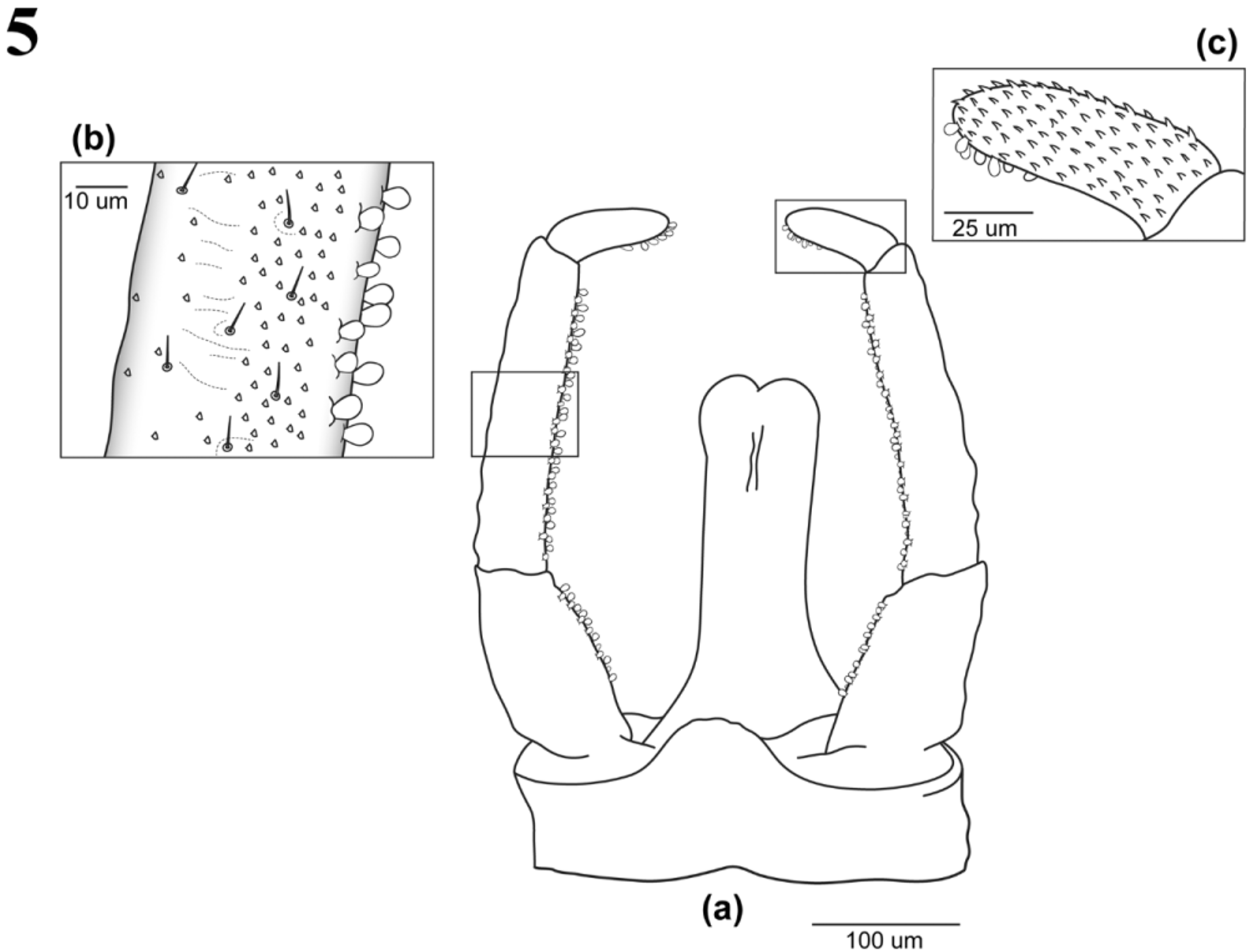
Abdomen with gill socket vestiges present on segments II - IV. Male abdomen ca. two thirds thickness of female abdomen. Male genitalia (lectotype ( Fig. 5 View FIGURE 5 a–c)), genital forceps three-segmented, segment I more broad than other segments and ca. two-thirds the length of segment II ( Fig. 5 View FIGURE 5 a). Segment III reduced, bending inwardly, nearly forming a right angle, inner lateral margin of genital forceps with ovoid scale-like processes on all three segments ( Fig. 5 View FIGURE 5 a). The ventral, dorsal and outer margin surfaces of segment I and II covered with few simple setae scattered and small subtriangular scales ( Fig. 5 View FIGURE 5 b) while the third segment has many larger, slightly more elongate subtriangular scale-like processes ( Fig. 5 View FIGURE 5 c). Penis lobes elongated but shorter than genital forceps, fused with a small medial notch or indentation, slightly broadened distally, longitudinal groove vestigial. Styliger plate distinctly convex. Three caudal filaments ca. subequal in length and one third longer than body length, few setae scattered all over caudal filament surface.
Female Imago. Head ( Fig. 2 View FIGURE 2 a). Antennae as in male; without dorsal portion of compound eye, compound eyes relatively small and laterally situated.
Thorax. Forewings as in male; fore-, mid- and hindlegs similar in structure to mid- and hindlegs of male.
Abdomen with gill socket vestiges present. Caudal filaments as in male.
Nymph. Colouration of immature nymphs pale, straw-coloured and slightly darker as nymphs mature with darker abdominal markings ( Fig. 2 View FIGURE 2 c–d). Body dorso-ventrally flattened.
Head with well-developed marginal fringe of setae extending to lateral margin of head with setae longer anteriorly and shorter laterally ( Fig. 2 View FIGURE 2 c–d). Male nymphs show developing dorsal compound eyes ( Fig. 2 View FIGURE 2 d).
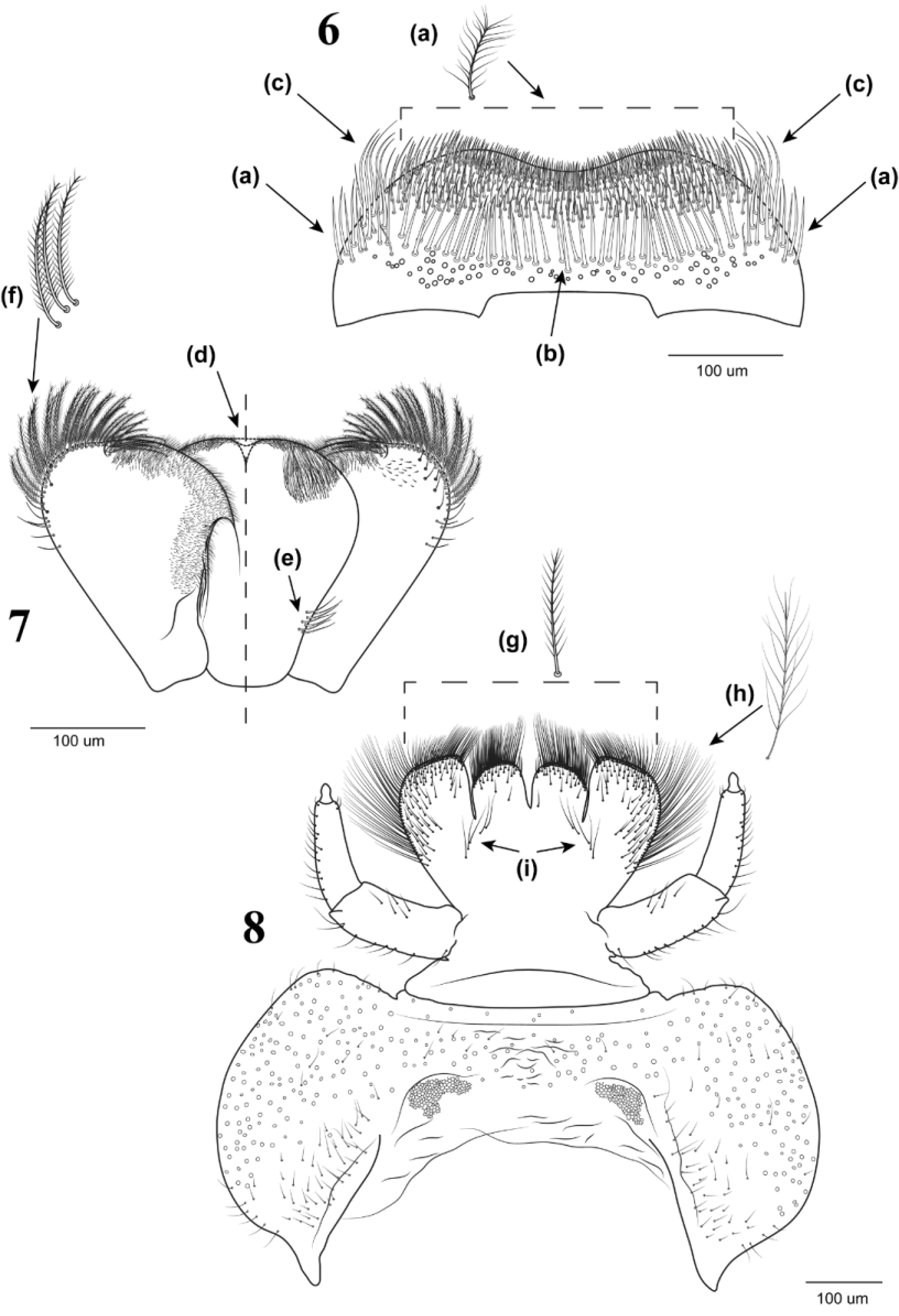
Labrum ( Fig. 6 View FIGURE 6 – 8 ) width 3 times length. Lateral margins rounded, anterior with slight emargination. Basal margin with elongated, squared notch medially. One third of apico-dorsal margin with densely clustered, feathered setae; similar feathered setae scattered along lateral margin ( Fig. 6 View FIGURE 6 – 8 a). Dorso-medial transverse row of setae 3–4 times larger than apico-dorsal setae and not feathery and possible remnant setal bases situated towards dorso-basal margin ( Fig. 6 View FIGURE 6 – 8 b). Dorso-lateral margin with single cluster of setae 1.5 times length of dorso-medial transverse setae ( Fig. 6 View FIGURE 6 – 8 c).
Hypopharynx ( Fig. 7 View FIGURE 6 – 8 ), lingua and superlinguae of similar size, both rounded apically, narrowing towards the base. Lingua with variable distomedial notch, ranging from deep indentation to none ( Fig. 7 View FIGURE 6 – 8 d). Dorsal surface with tuft of fine setae antero-laterally; very short brush setae covering dorsal to ventral anterior surface and a small cluster of larger setae postero-laterally ( Fig. 7 View FIGURE 6 – 8 e). Superlinguae with long, feathery setae on outer antero-lateral margin ( Fig. 7 View FIGURE 6 – 8 f) becoming shorter and non-feathery on outer lateral margin closest to base. Small, short setae sparsely covering inner antero-lateral surface extending from antero-dorsal to inner latero-ventral surface, covering two thirds toward base.
Labium ( Fig. 8 View FIGURE 6 – 8 ) with partly divided glossae and paraglossae. Paraglossae slightly falciform, larger than glossae which are rounded apically. Apical surfaces of glossae and paraglossae with short-feathered setae ( Fig. 8 View FIGURE 6 – 8 g). Lateral outer surface of paraglossae with long, thin-feathered setae 2 times the length of apical setae ( Fig. 8 View FIGURE 6 – 8 h). Dorsal surface of paraglossae and glossae with short, scattered setae and 2 rows of longer setae situated obliquely from paraglossal base towards base of glossae ( Fig. 8 View FIGURE 6 – 8 i). Labial palps three-segmented, segment I as long as segment II and III combined. Articulation between segment II and III distinct. Segment II narrows slightly distally, segment III small with slight medial constriction and tapered distally. Lateral surfaces of segment II and III with scattered setae. Prementum and postmentum as seen in Fig. 8 View FIGURE 6 – 8 ; postmentum covered with sparsely scattered simple setae and disc-shaped sensory pits or setal bases.
Mandibles ( Fig. 9–11 View FIGURE 9 – 13 ) elongate, length almost 4 times width. Few long socketed-setae (<10) from middle of outer lateral margin towards the base. Incisors single, prominent, with small tuft of very fine setae on outer edge of incisor near distal end ( Fig. 10–11 View FIGURE 9 – 13 a). Ventral surface of mandibles ( Fig. 9 View FIGURE 9 – 13 ) with many disc-shaped sensory pits or setal bases, medio-transverse groove with small triangular scales or sclerotized projections ( Fig. 9 View FIGURE 9 – 13 b). Left mandible ( Fig. 10 View FIGURE 9 – 13 ) with prostheca close to incisor, well-developed into long sclerotized setae with the longest seta feathered or brush-like ( Fig. 10 View FIGURE 9 – 13 c). Molar region prominent with 2–3 long thin setae below mola. Right mandible ( Fig. 11 View FIGURE 9 – 13 ) with prostheca well-developed, with branched sclerotized setae, with a single feathery or brush-like seta protruding out towards the mola ( Fig. 11 View FIGURE 9 – 13 d); molar region with 3 sclerotized seta-like projections distally ( Fig. 11 View FIGURE 9 – 13 e) and an elongated, thumb-like proximal ridge ( Fig. 11 View FIGURE 9 – 13 f); row of setae (<10) below mola.
Maxillae ( Fig. 12–13 View FIGURE 9 – 13 ) uniform with maxillary palp absent. Canines fused into a single elongate canine, with a depression along inner margin containing a single seta ( Fig. 12 View FIGURE 9 – 13 g). Cluster of long, thin setae at base of canine on outer face. Apex of galea-lacinia with two dentisetae ( Fig. 12 View FIGURE 9 – 13 h, 13h) and 6–7 long socketed setae. Proximal dentiseta slightly serrated on the inner margin. One large seta present on inner dorso-lateral surface below galealacinia apex and 5 thin simple setae on inner lateral surface below large single seta as seen in Fig. 12 View FIGURE 9 – 13 . A few large and thin, simple setae present on lower, outer lateral margin.
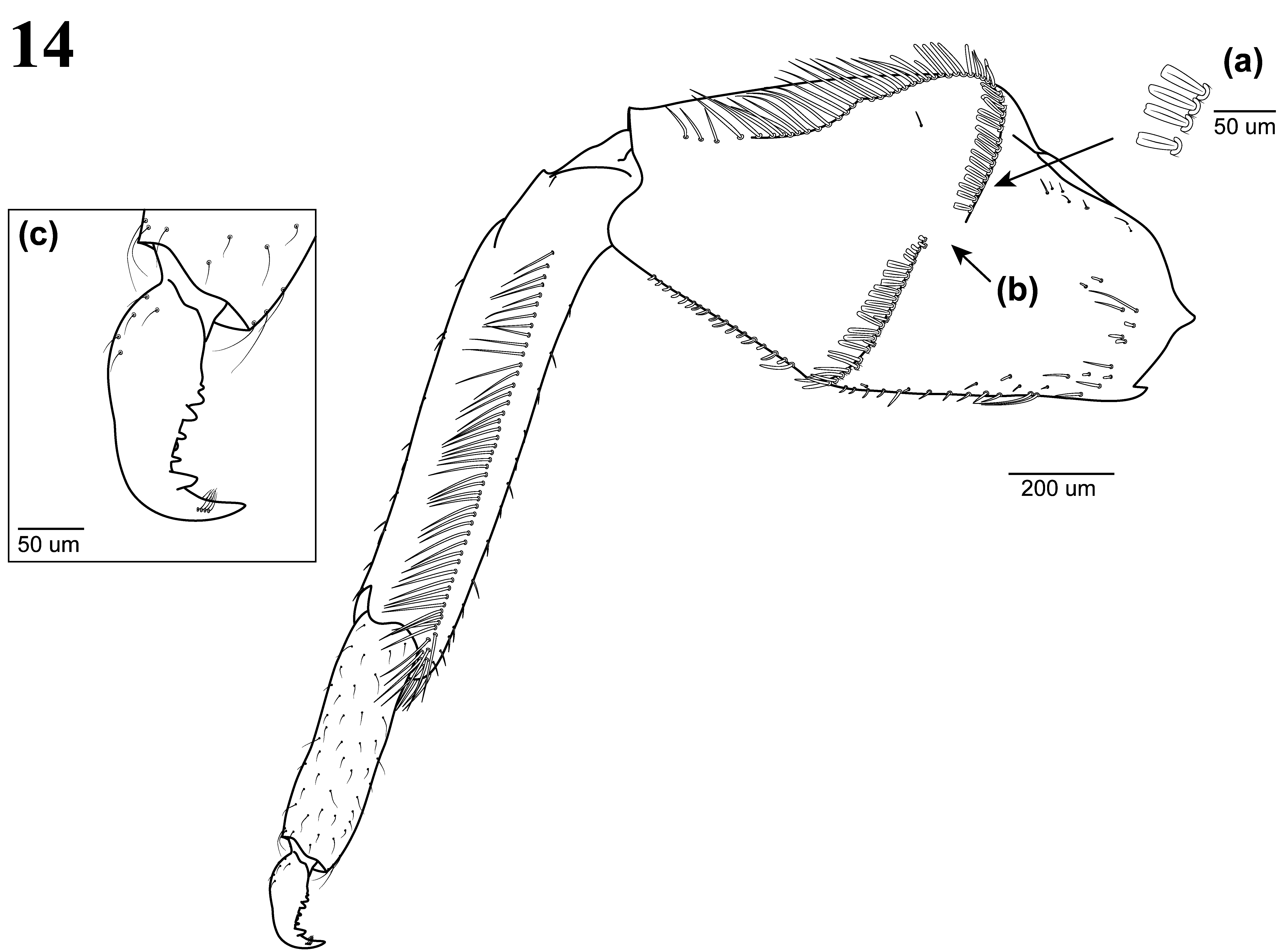
Thorax. Outer edges of prothorax lined with spatulate setae similar to spatulate setae shown in Figure 14 View FIGURE 14 a on forefemur. Foreleg ( Fig. 14 View FIGURE 14 ), femur subequal to tibia in length, medially broadened, narrowing toward distal and proximal ends. Medio-transverse spatulate setae ( Fig. 14 View FIGURE 14 a) present along transverse ridge on dorsal surface of forefemur; size of medial gap between transverse ridges of spatulate setae variable ( Fig. 14 View FIGURE 14 b). This ridge extends distally along the postero-lateral margin where the setae become longer (over 3 times length of spatulate setae). Short spatulate and lanceolate setae are present along antero-lateral margin with few long setae scattered near proximal base. Dorsal surface of tibia with single row of long, perpendicular setae, lateral margins interspersed with small, simple setae. Tarsus ca. half the length of tibia, entire surface interspersed with small, simple setae. Tarsal claw ( Fig. 14 View FIGURE 14 c) elongate with a single row of 4–6 variably sized smaller denticles followed by one large apical tooth. A single row of 4 small, subapical setae present apicolaterally on each side of claw. Ventral base of tarsal claw with small sparsely scattered setae. Mid- and hindlegs as seen in Fig. 2 View FIGURE 2 c–e, femora slightly more elongate and without transverse ridge of stout setae, antero-lateral margins with long setae and postero-lateral margins with spatulate or stout setae. Tibia, tarsi and claws similar to foreleg.
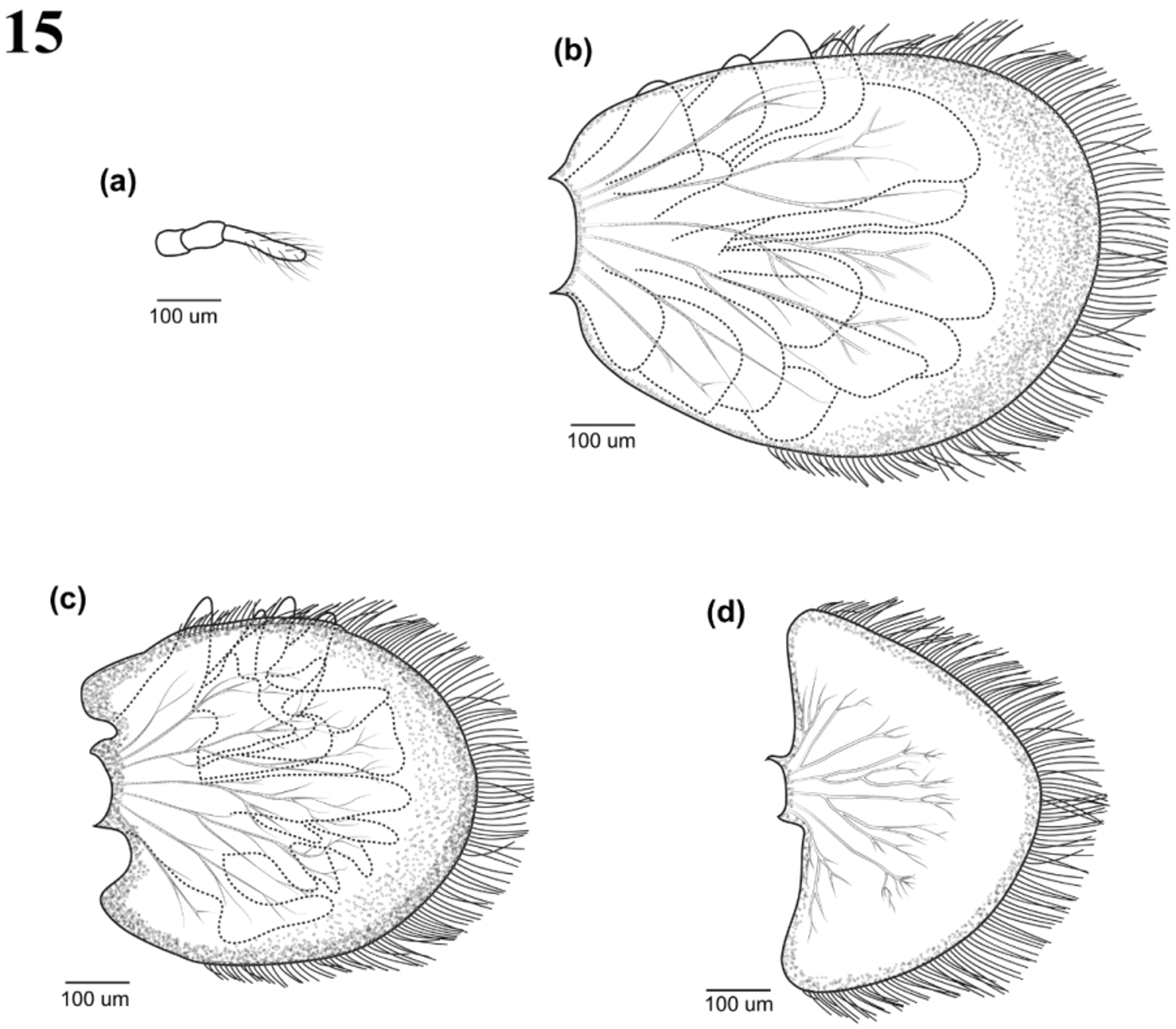
Abdomen with many fine setae covering the ventral surface, becoming more hirsute closer to the lateral margins. Abdominal segments with series of dark spots dorso-laterally, largest occurring on segment II ( Fig. 2 View FIGURE 2 c–d). Posterolateral processes subtriangular, moderately developed and slightly separated from base of following segment, processes with single row of long setae. Filamentous gill I ( Fig. 15 View FIGURE 15 a) present on abdominal segment I; threesegmented with long, thin, fine setae scattered around segment III. Lamellate gills present on segments II to IV. Gill II ( Fig. 15 View FIGURE 15 b) fully operculate, upper lamella ovoid with thin setae present starting ca. two thirds from base, setae longer anteriorly and shorter laterally; lower lamella bifid and highly lobed. Gill III ( Fig. 15 View FIGURE 15 c) upper lamella more circular in shape with thin setae present starting ca. three quarters from base, setae longer anteriorly and shorter laterally, lower lamella not bifid, singular and highly lobed. Gill IV ( Fig. 15 View FIGURE 15 d) almost semicircular, lower lamella absent, thin setae present along lateral margin to apex, setae longer anteriorly and shorter laterally. All three caudal filaments banded at base, sparsely setose with setae becoming slightly longer distally, medial caudal filament well-developed, cerci ca. two thirds length of body.
Discussion
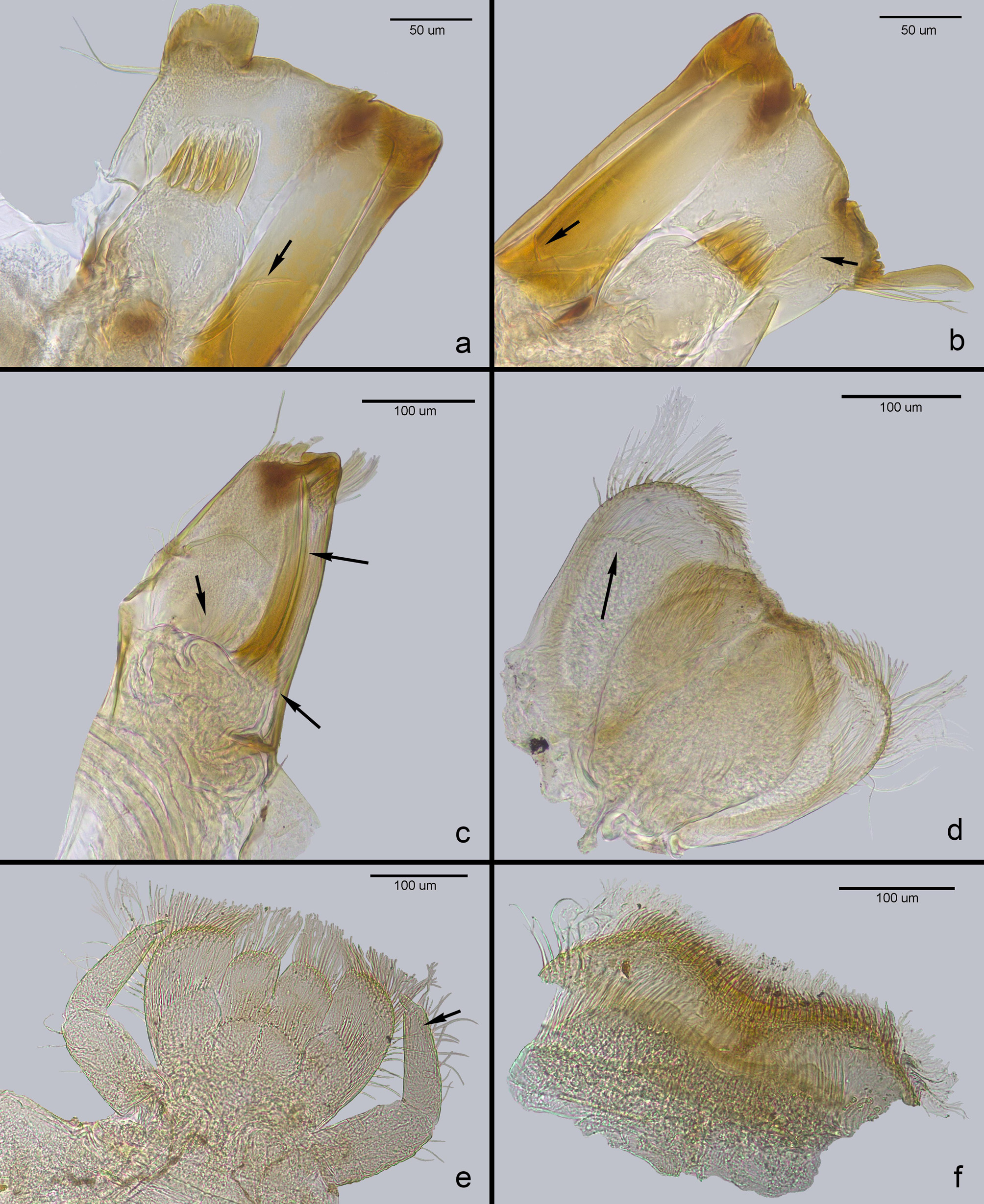
L. penicillata shows a high degree of variability in the lingual medial notch of the hypopharynx ( Fig. 7 View FIGURE 6 – 8 ), other factors such as wear-and-tear of mouthparts ( Fig. 16 View FIGURE 16 ), regeneration of limbs ( Fig. 2 View FIGURE 2 d, arrowed) and different moulting stages can affect the interpretation of morphological characters and identification of the species. However, the imago stage is less variable than the nymphal stage and differences have been seen in the adult stages of potential new and undescribed species of Lestagella . Edmunds (1959) attributed the differences in distinctiveness between life stages to different rates of evolution between the nymphs and short-lived adults and emphasised the importance of knowing the adult and nymphal stages when recognising genera and species groups (Edmunds 1962).
McCafferty & Wang (1997) stated that the presence of a medial notch on the lingua of the hypopharynx was diagnostic of the genus. However, in the material examined for the species L. penicillata alone the hypopharynx was found to be variable and not a suitable character for generic or species diagnosis.
Demoulin (1970) and McCafferty & Wang (1997) both described the mandibles of L. penicillata as being atrophied. The use of the term “atrophy” in both studies is ambiguous and unclear. Demoulin (1970) noted that the nymphal mandibular and maxillary canines underwent “progressive atrophy” with age. McCafferty & Wang (1997) suggested that the mandibles are apically atrophied while the maxillae have modified apices, which could refer to either the dentition being reduced, or the thickness and width of the mandibles. In this particular case, it is assumed that McCafferty & Wang (1997) were referring to the reduction in dentition, as the width of the mandibles were already mentioned as being narrow, and both left and right mandibles clearly show worn-down dentition in the illustrations.
Demoulin (1970) examined two nymphs at a younger and older stage, and it is by chance that the younger nymph had moulted more recently than the older nymph, therefore having less worn-down mandibular and maxillary canines, thus making it appear that there is a process of “progressive atrophy” in these mouthparts. It is therefore important to ensure that freshly moulted specimens are used for slide mounting when used for identification purposes, as stressed in other taxonomic studies (e.g. Kluge 2004, Sartori et al. 2008).
By the aforementioned definition of atrophy in the introduction section, neither of these reported “atrophies” are true for Lestagella ; this “atrophy” is due to wear-and-tear and is seen and well-documented in various other groups [ Baetidae (Muller-Liebenau 1973) ; Ephemeroidea (Elpers 1997); and other stream animals such as Plecoptera, Coleoptera, Diptera, an isopod and snail (Arens 1990)]. When the Lestagella nymph approaches the next moult, the mouthparts are well worn down (appearing atrophied), but the new mouthparts are evident ( Fig. 16 View FIGURE 16 ). As McCafferty & Wang (1997) only described the ultimate nymphal stage, it is most likely that the nymphal mouthparts were worn down and they would not be replaced due to final nymphal moult to the subimaginal stage.
Figure 16 View FIGURE 16 (a–f) shows the mouthparts of a L. penicillata nymph approaching the next moult. New, welldeveloped incisors and molars are evident in the left and right mandibles ( Fig. 16 View FIGURE 16 a and b respectively, arrowed), which appear to be initially distinctly separated; new setae and molar projections are arrowed. The maxilla ( Fig. 16 View FIGURE 16 c) regrowth shows a very well-developed, fused incisor and the dentisetae and other setae are clearly visible (see arrows). Newly developed mouthparts can also be seen for the hypopharynx, labium and labrum as seen in Figure 16 View FIGURE 16 (d–f) respectively.

No known copyright restrictions apply. See Agosti, D., Egloff, W., 2009. Taxonomic information exchange and copyright: the Plazi approach. BMC Research Notes 2009, 2:53 for further explanation.