
Zoropsis pekingensis Schenkel, 1953
|
publication ID |
https://doi.org/ 10.11646/zootaxa.3981.3.10 |
|
publication LSID |
lsid:zoobank.org:pub:0A4A181C-B0B9-4CF5-9A0B-9B808F225241 |
|
DOI |
https://doi.org/10.5281/zenodo.6116366 |
|
persistent identifier |
https://treatment.plazi.org/id/03A087AD-FFBA-FFE8-2AF3-A5A7FCEF6387 |
|
treatment provided by |
Plazi |
|
scientific name |
Zoropsis pekingensis Schenkel, 1953 |
| status |
|
Zoropsis pekingensis Schenkel, 1953 View in CoL
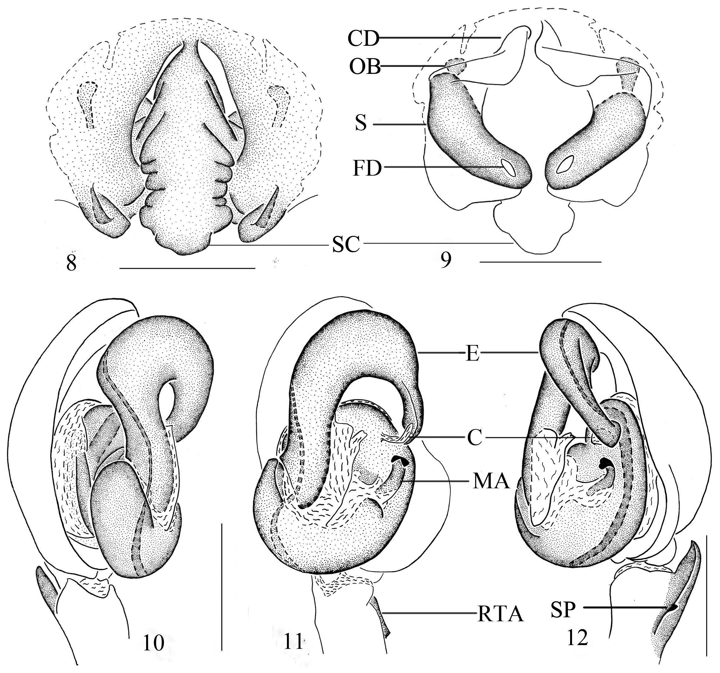
( Figs 1–12 View FIGURES 1 – 7 View FIGURES 8 – 12 )
Zoropsis pekingensis Schenkel, 1953: 11 View in CoL , fig. 6.
Diagnosis. The male can be distinguished from other Zoropsis species by the strong embolus that originates prolaterally from the tegulum, extending along the cymbial margin to the distal side of the tegulam and forming an arc in ventral view. The female can be distinguished by the epigyne with an unfolded and slender scape with a narrow stalk and the vulva with the ovoid bodies hidden below the copulatory ducts.
Description. Male (from Xiaolongmen Forestry Park, Beijing). Total body length 10.46: prosoma 4.83 long, 3.61 wide; opisthosoma 5.65 long, 2.94 wide. Carapace ( Fig. 1 View FIGURES 1 – 7 ) longer than wide, yellowish-brown. Median furrow longitudinal, yellowish-brown. Eight eyes in two rows. Diameters and interdistances: AME 0.18, ALE 0.23, PME 0.22, PLE 0.13; AME–AME 0.13, AME–ALE 0.11, PME–PME 0.15, PME–PLE 0.15. MOA 0.65 long, front width 0.49, back width 0.60. Clypeal height 0.30. Labium yellowish-brown. Sternum yellowish, with sparse black dots. Chelicerae yellow-brown with three promarginal and three retromarginal teeth. Metatarsus IV with a short calamistrum retrolaterally. Measurements of legs: I 22.60 (4.78, 2.10, 7.70, 6.07, 1.95), II 20.74 (4.75, 1.94, 6.60, 5.87, 1.58), III 18.26 (4.46, 1.81, 4.85, 5.44, 1.70), IV 24.77 (5.94, 1.90, 6.86, 7.87, 2.20). Leg formula: 4123. Spination of palp and legs: palp 1, 3; femora I lost, II p5, d3, r4, III p4, d3, r4, IV p4, d3, r3; patellae I lost, II p1, r1, III p1, r1, IV p1, r1; tibiae I lost, II p2, d1, r3, v14, III p2, d1, r2, v6, IV p2, d1, r2, v8; metatarsi I lost, II p3, d2, r2, v5, III p3, d4, r3, v6, IV p3, d2, r3, v6. Opisthosoma oval, with several separated chevron-like markings posteriorly.
Male palp ( Figs 5–7 View FIGURES 1 – 7 , 10–12 View FIGURES 8 – 12 ). Cymbium with dorsally arising dense patch of short setae, fine retrolateral marginal bump, and striated concavity. Tibia with strong blackish retrolateral apophysis and subdistal process. Bulb relatively enlarged. Embolus long, strongly sclerotised, arising prolaterally from bulb and extending along cymbial margin, with tip reaching retrolateral side of tegulum, with slim, translucent tip. Median apophysis sclerotised, hooked. Conductor small, narrow and membranous, rising from narrow stalk near distal bulb.
Female (from Mt. Xiaowutai) total length 14.47–15.30. One of them total length 14.47: Prosoma 6.50 long, 4.79 wide; Opisthosoma 8.29 long, 4.81 wide. Prosoma with similar pattern as male ( Fig. 2 View FIGURES 1 – 7 ), but darker in color. Diameters and interdistances of eyes: AME 0.22, ALE 0.28, PME 0.28, PLE 0.31; AME–AME 0.20, AME–ALE 0.14, PME–PME 0.16, PME–PLE 0.27. MOA 0.80 long, front 0.64 wide, back 0.64 wide. Clypeal height 0.20. Measurements of legs: I 21.84 (5.30, 2.60, 7.06, 5.17, 1.71), II 19.31 (5.15, 2.36, 5.41, 4.88, 1.51), III 16.29 (4.42, 2.16, 4.07, 4.08, 1.56), IV 22.33 (5.62, 2.14, 5.91, 6.81, 1.85). Leg formula: 4123. Spination of palp and legs: palp 5, 5, 1; femora I lost, II p4, d3, r4, III p4, d3, r4, IV p2, d3, r3; patellae I lost, II –IV p1, r1; tibiae I lost, II p2, d1, r3, v14, III–IV p2, d1, r2, v6; metatarsi I lost, II p3, d1, r2, v2, III–IV p3, d1, r3, v7.
Epigyne ( Figs 3–4 View FIGURES 1 – 7 , 8–9 View FIGURES 8 – 12 ). Plate flanked laterally by dark, narrow and sclerotised folds in ventral view. Stalk short, with a large median scape that projecting beyond epigastric furrow posteriorly. Spermathecae large, robust, with uniform texture. Ovoid bodies small, connected by projecting tube hidden below copulatory ducts.
Type material. The female holotype collected from ‘pai t'a, n. von peking’ (today the White Pagoda Temple in Beijing), presumably lost ( Marusik & Li 2011), not examined.
Material examined. CHINA: Beijing: Xiaolongmen Forestry Park, Nangou Valley (N 39.95°, E 115.42°), 1 male, 28 July 2014, Chi Jin leg.; Hebei: Yu County, Mt. Xiaowutai (N 39.97°, E 114.82°): 3 females, Zhengjia Valley, 14 July 2007, Feng Zhang leg.; 2 females, Zhangjiayao Village, 2 July 2014, Feng Zhang leg.; Laiyuan County, Mt. Baishi (N 39.21°, E 114.71°), 1 male and 3 females, Zhaojia Valley, 13 August 2014, Xiaoyu Dong leg.
Distribution. China (Beijing, Hebei) ( Fig. 25 View FIGURE 25 ).
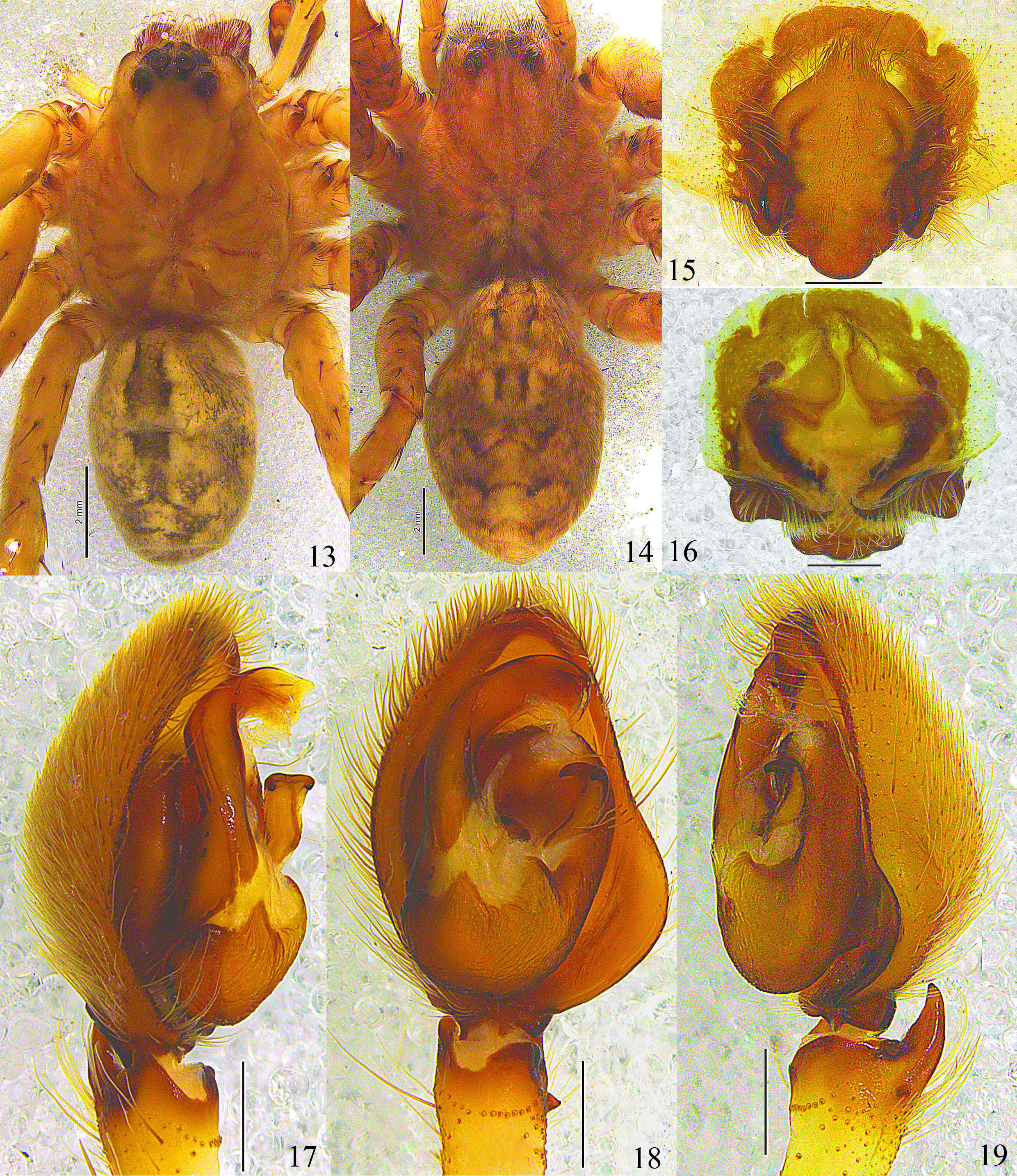
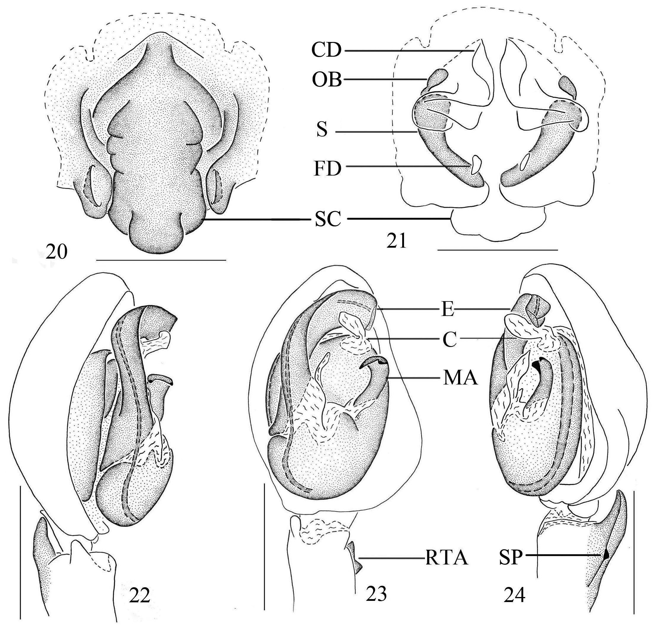
Zoropsis tangi sp. nov. ( Figs 13–24 View FIGURES 13 – 19 View FIGURES 20 – 24 )
Zoropsis pekingensis: Tang, Kim & Song, 1999: 38 View in CoL , figs 1–8; Song, Zhu & Chen,1999: 398, figs 13J, 232V–W, 233B–D; Song, Zhu & Chen, 2001: 300, figs 191A–H. (Misidentified)
Type material. Holotype male, Helan Mountains (38.60°N, 105.90°E), Alxa League, Inner Mongolia, 30 July 1991, Guiming Tang leg. (Inner Mongolia Normal University, Hohhot, China). Paratypes: 2 females, with same data as holotype (Inner Mongolia Normal University, Hohhot, China). These are the specimens that Tang et al. (1999) misidentified and described as the male of Zoropsis pekingensis . Song et al. (1999, 2001) based their illustrations on Tang et al. (1999).
Etymology. This specific name is a patronym in honour of Prof. Guiming Tang of lnner Mongolia Normal University, China, who collected the type specimens.
Diagnosis. Females of the new species are similar to Z. pekingensis ; both have a median scape and small ovoid bodies on epigyne ( Figs 15–16 View FIGURES 13 – 19 , 20–21 View FIGURES 20 – 24 ). Males of both species have a small and membranous conductor, hook-like median apophysis and RTA on male palp ( Figs 17–19 View FIGURES 13 – 19 , 22–24 View FIGURES 20 – 24 ), but can be distinguished by the smaller, narrower and shorter embolus and the large median apophysis on the male palp. Females can be differentiated by the wider median scape and the smaller ovoid bodies.
Description. Male (holotype). Total length 12.05; prosoma 6.21 long, 4.47 wide; opisthosoma 5.87 long, 3.38 wide. Carapace ( Fig. 13 View FIGURES 13 – 19 ) longer than wide, yellowish-orange. Median furrow longitudinal, brown. Eight eyes in two rows, anterior eye row slightly recurved, posterior eye row strongly recurved. Diameters of eyes: AME 0.20, ALE 0.29, PME 0.28, PLE 0.28. Interdistances of eyes: AME–AME 0.19, AME–ALE 0.16, PME–PME 0.19, PME–PLE 0.27. MOA 0.89 long, front 0.59 wide, back 0.86 wide. Clypeal height 0.16. Labium yellowish-brown. Sternum yellowish-brown. Chelicerae reddish-brown, with three promarginal and three retromarginal teeth. Metatarsus of leg IV with a short calamistrum retrolaterally. Measurements of legs: I 25.84 (6.45, 2.44, 7.42, 7.34, 2.19), II 24.51 (6.11, 2.63, 6.93, 6.74, 2.11), III 19.64 (5.08, 2.12, 5.07, 5.63, 1.69), IV 26.17 (7.10, 1.79, 6.95, 8.28, 2.05). Leg formula: 4123. Spination of palp and legs: palp 1, 4; femora I p1, d8, r2, II p4, d3, r4, III p1, r1, IV lost; patellae I r2, II lost III p1, r1, IV lost; tibiae I–II lost, III p2, d1, r2, v6, IV lost; metatarsi I–IV lost. Opisthosoma oval, spotted; with several separate dark chevronlike markings on posterior half, sides pale brown.
Palp ( Figs 17–19 View FIGURES 13 – 19 , 22–24 View FIGURES 20 – 24 ). Cymbium with dorsal, dense patch of short setae, fine retrolateral bump and striated concavity. Tibia with strong blackish retrolateral apophysis and subdistal process. Bulb relatively small. Embolus short and strongly sclerotised, arising from prolateral side of bulb, and its tip reaching apical part of tegulum. Median apophysis sclerotised and hook-like, with tip pointed at base of embolus, membranous tegular apophysis, and basal part unsclerotised. Membranous conductor rising near apex of bulb.
Females. Total length 16.21–17.61; one paratype measured, total length 16.21, prosoma 7.38 long, 6.13 wide; opisthosoma 8.82 long, 5.21 wide. Carapace ( Fig. 14 View FIGURES 13 – 19 ) colour pattern as in male, but with deeper colour. Diameters of eyes: AME 0.22, ALE 0.39, PME 0.36, PLE 0.32. Interdistances of eyes: AME–AME 0.30, AME–ALE 0.22, PME– PME 0.24, PME–PLE 0.56. MOA 0.93 long, front 0.73 wide, back 1.03 wide. Clypeal height 0.22. Labium deep yellowish-brown. Sternum yellowish-brown. Chelicerae deep reddish-brown, with three promarginal and three retromarginal teeth. Metatarsus of leg IV with short calamistrum retrolaterally. Measurements of legs: I 26.50 (6.25, 3.21, 8.26, 6.84, 1.94), II 23.98 (6.14, 3.14, 6.94, 5.90, 1.86), III 21.70 (5.96, 2.80, 5.78, 5.56, 1.60), IV 27.17 (6.99, 2.96, 7.25, 7.80, 2.17). Leg formula: 4123. Spination of palp and legs: palp 5, 5, 1; femora I p2, d3, r4, II p5, d2, r4, III p3, d3, r4, IV p4, d3, r2; patellae I–II0, III p1, r1, IV p1, r1; tibiae I p3, r3, v14, II p4, r4, v14, III p2, d1, r3, v6, IV p2, d1, r3, v6; metatarsi I p3, r2, v6, II p4, r4, v8, III p2, r2, v4, IV p3, d6, r2, v3.
Epigyne ( Figs 15–16 View FIGURES 13 – 19 , 20–21 View FIGURES 20 – 24 ) with broad median scape attached by narrow stalk. Far sides of plate flanked by dark, narrow sclerotised folds in ventral view. Short stalk attached to wide and long median scape. Spermathecae thin, with uniform texture throughout. Small ovoid bodies connected by a tube project and observed above the copulatory ducts.
Distribution. Known from the type locality only ( Fig. 25 View FIGURE 25 ).


No known copyright restrictions apply. See Agosti, D., Egloff, W., 2009. Taxonomic information exchange and copyright: the Plazi approach. BMC Research Notes 2009, 2:53 for further explanation.
|
Kingdom |
|
|
Phylum |
|
|
Class |
|
|
Order |
|
|
Family |
|
|
Genus |
Zoropsis pekingensis Schenkel, 1953
| Li, Zhiyue, Hu, Lanlan & Zhang, Feng 2015 |
Zoropsis pekingensis:
| Song 2001: 300 |
| Tang 1999: 38 |
| Song 1999: 398 |
Zoropsis pekingensis
| Schenkel 1953: 11 |