
Parapsectra
|
publication ID |
https://doi.org/ 10.5281/zenodo.193438 |
|
DOI |
https://doi.org/10.5281/zenodo.6195906 |
|
persistent identifier |
https://treatment.plazi.org/id/03A087F6-423B-FFAC-8F9F-FA93FA08AE63 |
|
treatment provided by |
Plazi |
|
scientific name |
Parapsectra |
| status |
|
Key to males of Parapsectra View in CoL View at ENA
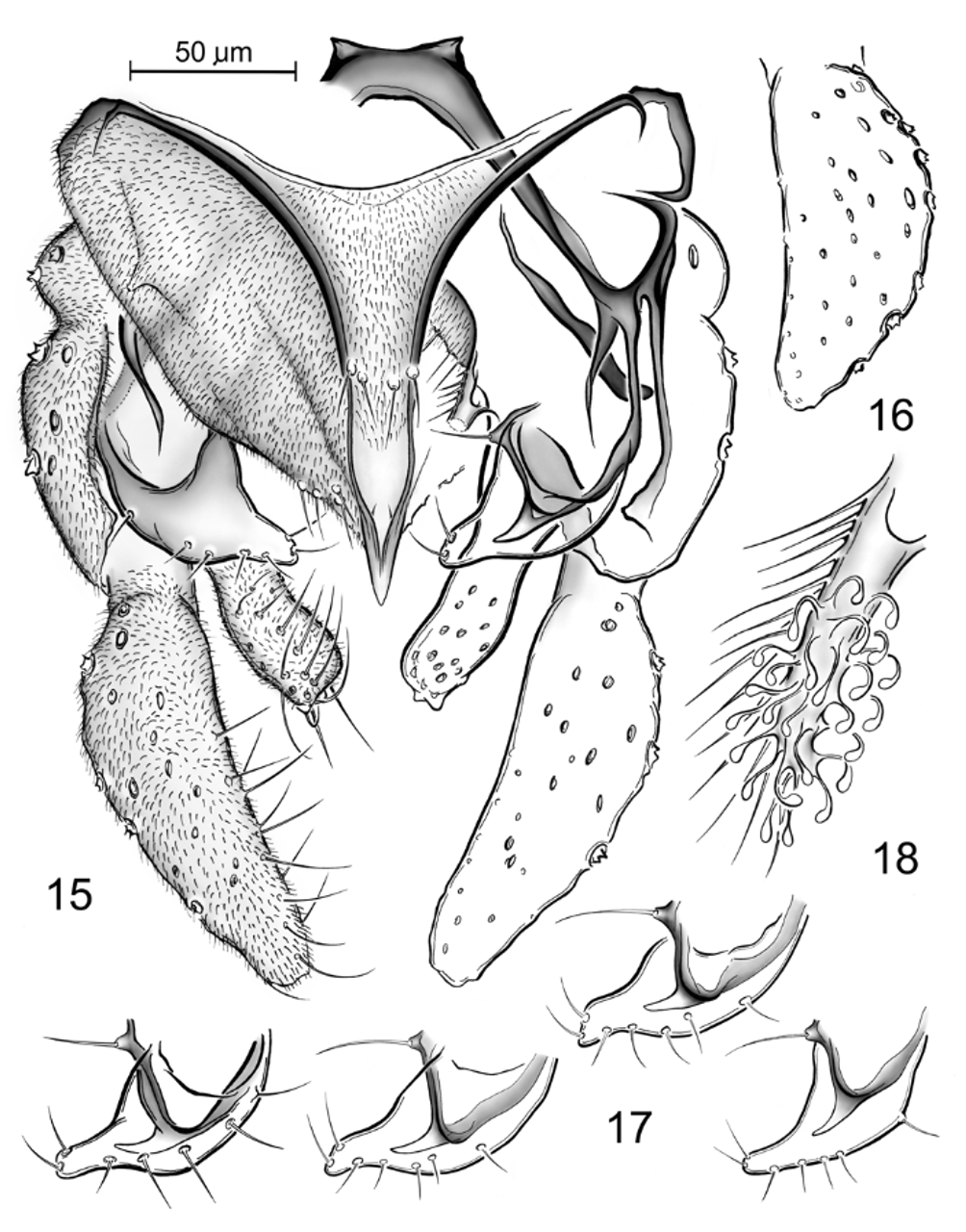
1. Anal point acute ( Fig. 15 View FIGURES 15 – 18 ). Superior volsella with slender apical prolongation and concave anteromedian margin ( Figs 15, 17 View FIGURES 15 – 18 ) ..................................................................................................................................................... P. mendli View in CoL
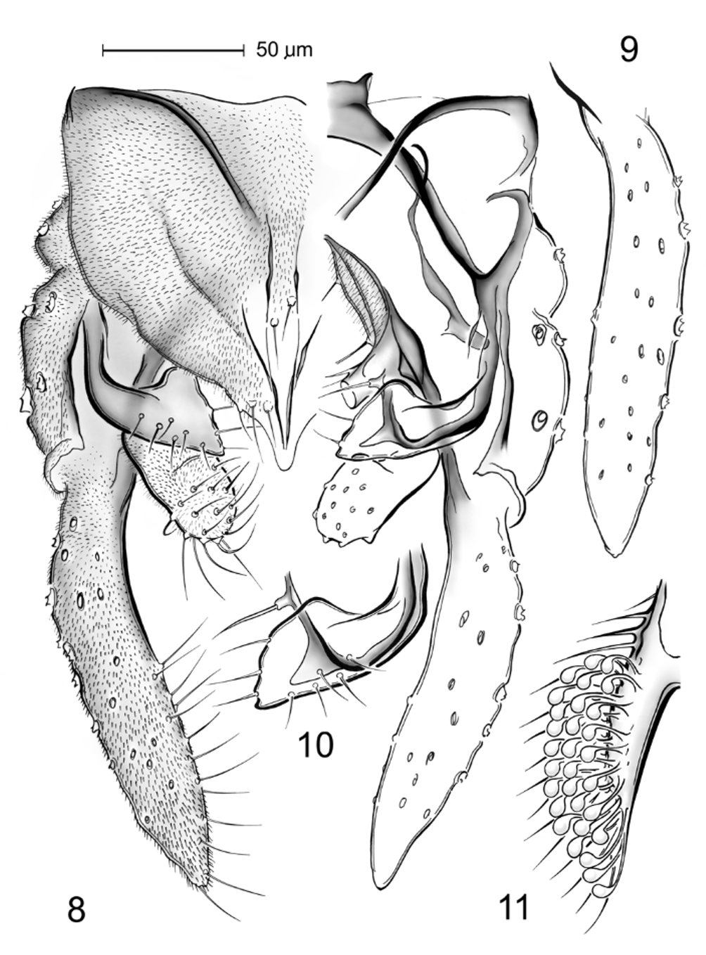
-. Anal point with rounded, transversely cut or slightly concave apex ( Figs 8 View FIGURES 8 – 11 , 12 View FIGURES 12 – 14 , 19 View FIGURES 19 – 21 , 22 View FIGURES 22 – 24 , 26 View FIGURES 25 – 28 , 30 View FIGURES 29 – 31 ). Superior volsella without apical prolongation and/or with straight or convex anteromedian margin ( Figs 10 View FIGURES 8 – 11 , 13 View FIGURES 12 – 14 , 20 View FIGURES 19 – 21 , 23 View FIGURES 22 – 24 , 27 View FIGURES 25 – 28 , 30 View FIGURES 29 – 31 ). ....... 2
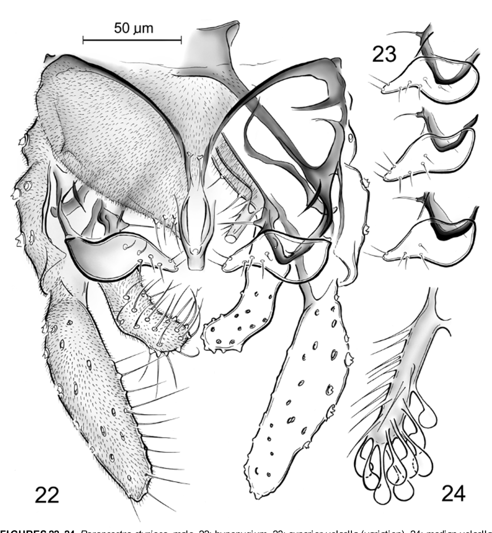
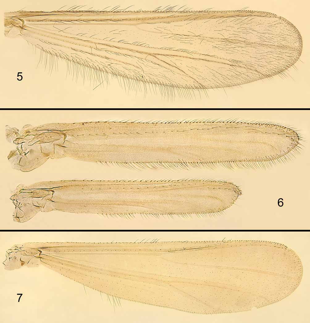
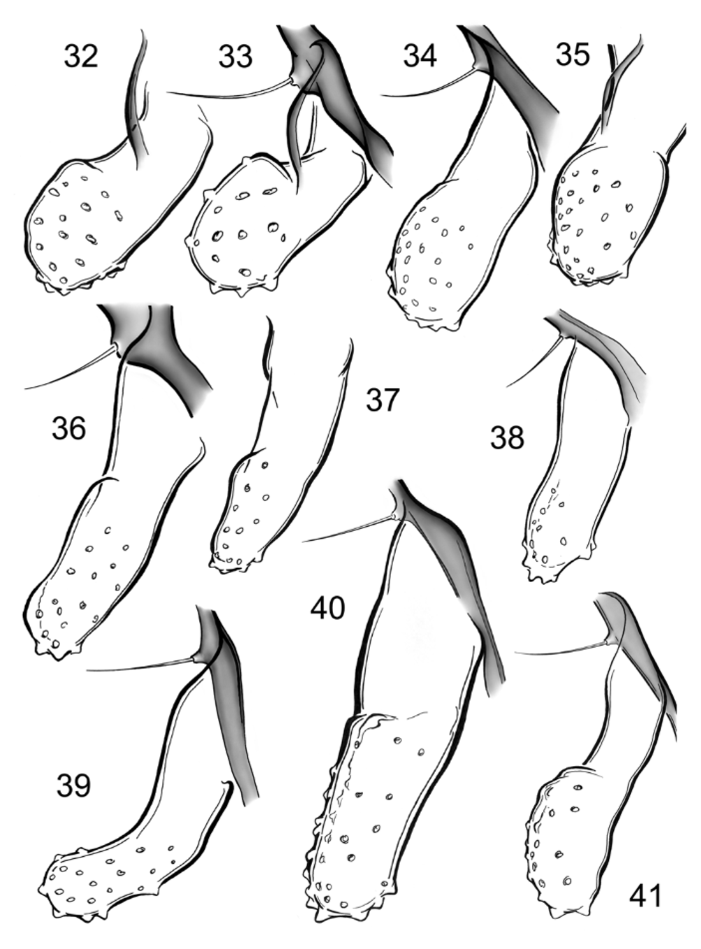
2. Wing with FCu well distal of RM ( Fig. 5 View FIGURES 5 – 7 ). Lateral teeth of anal tergite present ( Fig. 22 View FIGURES 22 – 24 ). Superior volsella sinuous, digitus absent ( Figs 22, 23 View FIGURES 22 – 24 ). Median volsella with sparse and large, spoon-shaped lamellae ( Fig. 24 View FIGURES 22 – 24 ). Inferior volsella long and strongly curved, L-shaped ( Figs 22 View FIGURES 22 – 24 , 39 View FIGURES 32 – 41 ). .............................................................................. P. styriaca View in CoL
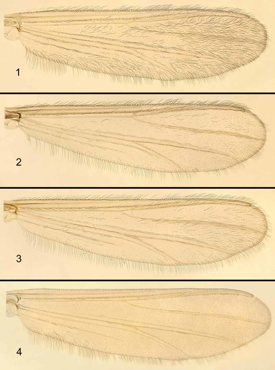
-. Wing with FCu very slightly distal of RM ( Fig. 1 View FIGURES 1 – 4 ), under RM ( Figs 2, 4 View FIGURES 1 – 4 , 7 View FIGURES 5 – 7 ) or proximal of RM ( Fig. 6 View FIGURES 5 – 7 ). Lateral teeth of anal tergite absent. Superior volsella never sinuous, digitus present. Median volsella with lamellae never as above ( Figs 11 View FIGURES 8 – 11 , 14 View FIGURES 12 – 14 , 21 View FIGURES 19 – 21 , 28 View FIGURES 25 – 28 , 31 View FIGURES 29 – 31 ). Inferior volsella never L-shaped, short when bent ( Figs 32–35, 38, 40, 41 View FIGURES 32 – 41 ). ...................... 3
3. Median volsella with falciform lamellae ( Figs 28 View FIGURES 25 – 28 , 31 View FIGURES 29 – 31 )................................................................................................. 4
-. Median volsella with spoon-shaped lamellae ( Figs 11 View FIGURES 8 – 11 , 14 View FIGURES 12 – 14 , 21 View FIGURES 19 – 21 ).................................................................................... 5
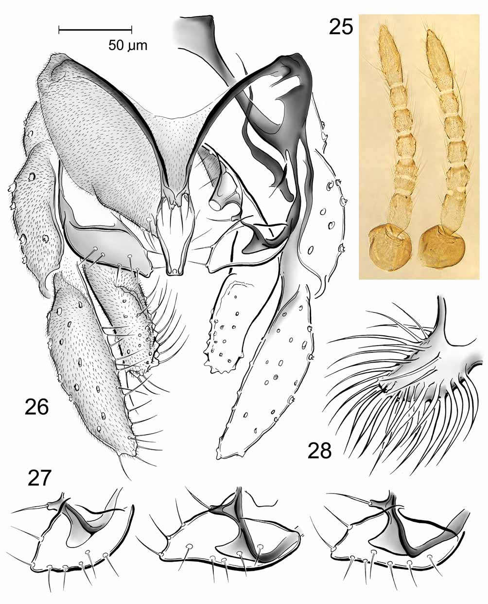
4. Antenna with 6 or 7 flagellomeres ( Fig. 25 View FIGURES 25 – 28 ). Wing reduced ( Fig. 6 View FIGURES 5 – 7 ). Superior volsella elongated ( Figs 26, 27 View FIGURES 25 – 28 ). Median volsella with slender falciform and fusiform lamellae ( Fig. 28 View FIGURES 25 – 28 ). Inferior volsella reaching beyond half length of gonostylus, evenly tapering to apex ( Figs 26 View FIGURES 25 – 28 , 40 View FIGURES 32 – 41 ). ............................................................................... P. uliginosa View in CoL
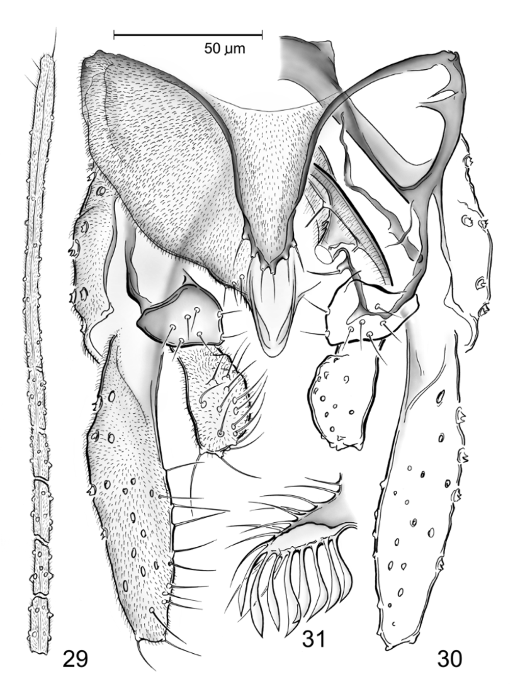
-. Antenna with 13 flagellomeres, with distal flagellomeres slightly separated ( Fig. 29 View FIGURES 29 – 31 ). Wing well developed ( Fig. 7 View FIGURES 5 – 7 ). Superior volsella short ( Fig. 30 View FIGURES 29 – 31 ). Median volsella with broad falciform and lanceolate lamellae ( Fig. 31 View FIGURES 29 – 31 ). Inferior volsella reaching at most half length of gonostylus, with abruptly swollen head-like distal half ( Figs 30 View FIGURES 29 – 31 , 41 View FIGURES 32 – 41 ) ............ ...................................................................................................................................................................... P. wagneri View in CoL
5. Anal lobe of wing strongly reduced, RM very short, membrane below An with sparse macrotrichia ( Fig. 2 View FIGURES 1 – 4 ). Dark
brown to fuscous body ............................................................................................................................. P. chionophila View in CoL -. Anal lobe of wing well developed, RM long, membrane below An with dense macrotrichia ( Figs 1, 4 View FIGURES 1 – 4 ). Brownishgreen body..................................................................................................................................................................... 6 6. Stem of median volsella with large lamellae placed posteromedially ( Fig. 11 View FIGURES 8 – 11 ). Inferior volsella bent at base, with transverse protrusion ( Figs 8 View FIGURES 8 – 11 , 32, 33 View FIGURES 32 – 41 ) ................................................................................................ P. b u m a s t a sp. n.
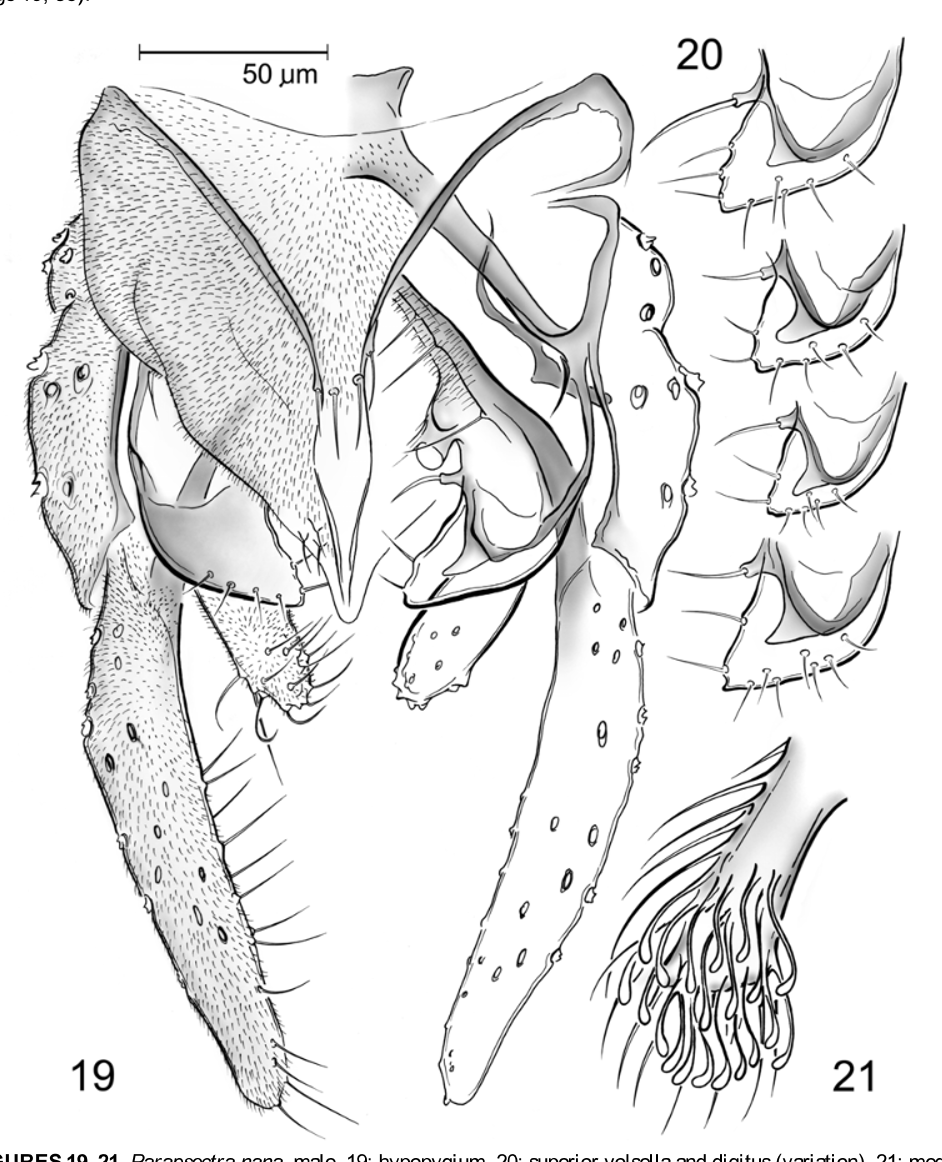
-. Stem of median volsella with small lamellae placed apically ( Fig. 21 View FIGURES 19 – 21 ). Inferior volsella short and straight, without transverse protrusion ( Fig. 19 View FIGURES 19 – 21 , 38 View FIGURES 32 – 41 ) .................................................................................................................... P. nana View in CoL

No known copyright restrictions apply. See Agosti, D., Egloff, W., 2009. Taxonomic information exchange and copyright: the Plazi approach. BMC Research Notes 2009, 2:53 for further explanation.