
Epigomphus rufus, Bota-Sierra & Novelo-Gutiérrez, 2020
|
publication ID |
https://doi.org/10.11646/zootaxa.4896.2.7 |
|
publication LSID |
lsid:zoobank.org:pub:71E90DA6-7595-4010-A849-0EBA20D15921 |
|
DOI |
https://doi.org/10.5281/zenodo.4382787 |
|
persistent identifier |
https://treatment.plazi.org/id/03A087FA-231E-FF8A-FF3D-615AFDF0B82B |
|
treatment provided by |
Plazi |
|
scientific name |
Epigomphus rufus |
| status |
sp. nov. |
Epigomphus rufus View in CoL sp.nov.
( Figs. 1–4 View FIGURE 1 View FIGURE 2 View FIGURE 3 View FIGURE 4 )
Material examined: Three specimens were collected (1♂, 2♀♀), we assigned them as conspecific due to their share of the same habitat in the same forest patch, their conspicuous coloration, and mainly the correspondence of male cerci and epiproct with the posterior portion of the female head. All three specimens are teneral, and the two females were reared from the last larval instar to the adult stage (one of them was kept alive and feed with small insects for five days, which allowed the coloration pattern to get very close to the mature pattern). CEUA: Holotype. ♀ teneral, CEUA 113500, 19 February 2019, COLOMBIA, Antioquia Department, La Forzoza Reserve, Anorí Municipality, El Roble Township, 7.001010, -75.130792; 1,644 m a.s.l., Leg: R. Novelo-G. and C. Bota. Paratypes, 1♀, teneral, same as holotype but CEUA 113490 . 1 ♂, teneral, CEUA 99305 , 27 May 2012, Arrierito Antioqueño Reserve , 6.982318, -75.111666; 1,687 m a.s.l., Leg: J.A. Cogollo and C. Bota.
Etymology: The color of this species is unique among the species in the genus, since its dark ground coloration is reddish-brown instead of the common dark brown or black. Thus we named it rufus from Latin, which means red.
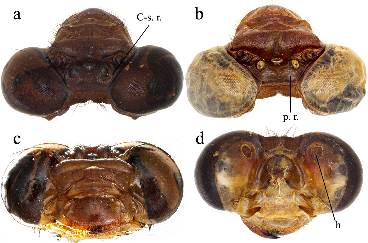
Description of holotype. Head reddish-brown, eyes when alive greenish-gray. Posterior ocelli over a tumid area, bordered by a C-shaped thick rim ( Figs. 1a, c View FIGURE 1 ); occipital posterior border sinuous with a small medial concave incision ( Fig. 1a View FIGURE 1 ); posterior eye margin tumid, with a conic hole located under it ( Fig. 1d View FIGURE 1 ).
Thorax. Reddish brown,the following greenish-gray areas were visible when the specimen died, but unfortunately dispel with time ( Fig. 2a View FIGURE 2 ): two medial spots and two lateroapical spots on posterior lobe of pronotum, mesepisternum with a longitudinal line, a transversal line on anterior border, the second pale antehumeral stripe reduced to a single small posterior spot, the rest of the pleura with three longitudinal lines: one on mesepimeron, one on metepisternum, and one on metepimeron. The antealar crest yellow. Legs brown except distal portion of femora, tibiae, and tarsi which are black. Metafemora approximately 1 ½ the length of pro- and mesofemora. All femora with an external row of spines. Tibiae with long, slender spines on external border, except protibiae with a row of eleven scale-like spines on the external side. Tarsal claws brown with a subapical tooth. Wings. Hyaline with amber tinge on base, veins black and Pt brown ( Fig. 2a View FIGURE 2 ). Pt covering 7 cells. Left FW with 20 Ax, right FW with 21, FWs with 15 Px; left HW with 14 Ax, right HW with 15 Ax; HWs with 14 Px; supratriangles, triangles and subtriangles without crossevins. Second reinforced antenodal the 8th in left FW, the 7th in right FW, in both HW the 7th; ( Fig. 4a View FIGURE 4 ). Cubito-anal crossveins in addition to inner side of subtriangle, 3 in FWs, 2 in left, and 3 in right HW. Five crossveins in space between sectors of arculus and point of branching of RP on HW.
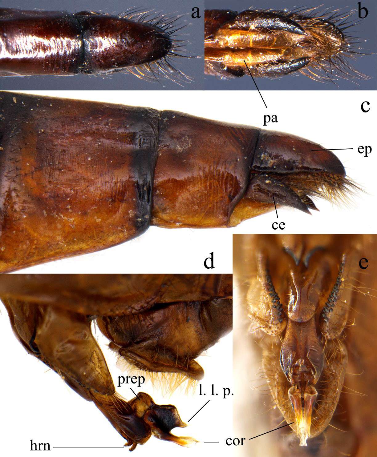
Abdomen. Club-like, narrowing slightly on S3 and widening again on S8, S9 widest. Brown, but S1–S2 darker, the distal part of S3 to S7 light brown, S8–S10 dark brown dorsal and light brown laterally, black distal rings on S2–S7 ( Fig. 2a View FIGURE 2 ). Auricles rounded and smooth. Epiproct smooth, long with convex apex, almost as long as S10 ( Figs. 3 a, c View FIGURE 3 ). Cerci approximately ¾ of epiproct length, cylindrical with pointed tips slightly curved ventrad, their apices convergent ( Figs. 3 View FIGURE 3 a–c). Paraprocts conical, straight, with pointed tips ( Fig. 3b View FIGURE 3 ).
Measurements (mm): TL 58, Ab 44, FW 43, HW 41.
Male paratype: Teneral, probably do not bear the final coloration pattern. As female holotype but with following differences: Head. With greenish-gray spots on antefrons and in the tumid area behind ocelli, the vertex lacks the C-shaped thick rim, instead it presents a complete transverse postocellar ridge, emarginate medially ( Fig. 1b View FIGURE 1 ). The posterior part of head behind eyes is not tumid and lacks the conic groove on the occipital region.
Thorax. Without greenish gray lines on mesepisternum. Metatibia and first two tarsomeres with a row of peglike spines on internal side. Wings. Pt covering 5 ½ cells. Left FW with 20 Ax, left FW with 14 Px; left HW with 16 Ax, left HW with 13 Px; supratriangles, triangles and subtriangles without crossevins. Second reinforced antenodal the 6th in left FW, in left HW the 7th. Cubito-anal crossveins in addition to inner side of subtriangle, 3 in left FW, 2 in left HW ( Fig. 2b View FIGURE 2 ).
Abdomen. The width of abdominal segments is more uniform, just slightly wider on S8–10 ( Fig. 2b View FIGURE 2 ). Auricles subquadrangular, each with rows of 1–4 black denticles on posterior border ( Figs. 4b, d View FIGURE 4 ). Anterior lamina of genital fossa swollen and rounded, densely covered by long golden setae ( Figs. 4b, d View FIGURE 4 ); anterior hamuli with pointed black apical margins ( Figs. 3 View FIGURE 3 d–e, 4b, d); posterior hamuli long, approximately twice the length of anterior hamuli, subtriangular in lateral view, their mesial side covered by black tubercles ( Figs. 3 View FIGURE 3 d–e, 4b, d). Vesica spermalis reddish brown: segment 1 large, concave in ventral view, in lateral view with a ventroapical rounded process directed distally ( Figs. 3 View FIGURE 3 d–e, 4b, d); segment 3 widening towards its distal portion, two ventro apical pointed horns directed ventrally; segment 4, in lateral view, with prepuce subquadrangular, lateral lobes strongly bulging dorsally, in ventral view, forming a nozzle-like slender projection directed posteriorly and parallel to cornua; cornua short ( Figs. 3 View FIGURE 3 d–e); Cercus and epiproct subequal in length, less than half the length of S10 ( Figs. 4a, c View FIGURE 4 ). Cerci concave mesially, in lateral view strongly curved ventrally with a pointed dark tip ( Figs. 4a, c View FIGURE 4 ). Epiproct divided in two lateroapical processes, in ventral view widely V-shaped forming an angle close to 90°, each lateroapical process with two black sharp teeth, one apical and another one subapical ( Figs. 4a, c View FIGURE 4 ).
Measurements (mm): TL 54, Ab 42, FW 40, HW 38.
Variation of female paratype: Ax in FW 19, in HW 16; Px in FW 13–14, in HW 12–14.
Measurements (mm): TL 59, Ab 44, FW 44, HW 42.
Diagnosis: The typical color pattern of species of this genus usually alternates black or dark brown with yellow, green and light blue; this is the only known species that has reddish brown coloration as dark background color making it easily recognizable ( Figs. 2 View FIGURE 2 a–b). Epigomphus rufus shares with a group of 16 species, including most of the South American and some Central American species, its second pale antehumeral stripe reduced to a single small posterior spot ( Table 1 View TABLE 1 ). Among them, the males of E. camelus , E. compactus , E. subsimilis , E. tumefactus , and E. verticicornis share with E. rufus cercus length shorter than S10 length ( Figs. 4a, c View FIGURE 4 ), but males of E. rufus can be easily distinguished from all these species by cercus strongly curved downwards and with a sharp tip which is unique among all 33 known Epigomphus species ( Figs. 4a, c View FIGURE 4 ), and so is the shape of male epiproct by having two teeth, one apical and one subapical, on each lateroapical process ( Figs. 4a, c View FIGURE 4 ). As pointed out by Calvert (1920), these caudal structures of males perfectly match with structures on vertex, occiput, and posterior area of females’ head, including the C-shaped tubercles behind the posterior ocelli and the conical holes where cerci match on rear of head ( Figs. 1a View FIGURE 1 , c–d), the females of only ten species in this group of Epigomphus are known ( Table 1 View TABLE 1 ), unfortunately, the descriptions of the posterior area of the head are not meticulous for all of them, but in all the cases where the structures are well described E. rufus females can be easily distinguishable since no other has C-shaped tubercles behind the posterior ocelli or conical holes in the rear area and this combination of characteristics is expected to be unique as the shape of the male cercus and epiproct among the species in the genus.
Distribution and habitat: This species is so far only known to occur in two close localities, which are actually part of the same forest patch at the northern portion of the Colombian Central Cordillera, close to first and second order streams in the interior of the forest.


| R |
Departamento de Geologia, Universidad de Chile |
No known copyright restrictions apply. See Agosti, D., Egloff, W., 2009. Taxonomic information exchange and copyright: the Plazi approach. BMC Research Notes 2009, 2:53 for further explanation.
|
Kingdom |
|
|
Phylum |
|
|
Class |
|
|
Order |
|
|
Family |
|
|
Genus |