
Tumidopteris astra NAUGOLNYKH, 2020
|
publication ID |
https://doi.org/ 10.37520/fi.2020.022 |
|
persistent identifier |
https://treatment.plazi.org/id/03A187D4-CA6E-FFE9-FF3E-A55C9237F7A0 |
|
treatment provided by |
Diego |
|
scientific name |
Tumidopteris astra NAUGOLNYKH |
| status |
sp. nov. |
Tumidopteris astra NAUGOLNYKH sp. nov.
Text-figs 2a–e View Text-fig , 3a–e View Text-fig , 4a–f View Text-fig , 5a, b View Text-fig
H o l o t y p e. GIN 4851/343h; the holotype is figured here in Text-figs 2a, d View Text-fig , 3a–e View Text-fig , 4a–c, e View Text-fig , 5a–c. View Text-fig
P l a n t F o s s i l N a m e s R e g i s t r y N u m b e r.
PFN001847.
R e p o s i t o r y. Monographic Department of the State Darwin Museum (Moscow, Russian).
During study, the collection is kept in the Geological Institute of Russian Academy of Sciences (Moscow, Russia)
D e r i v a t i o n o m i n i s. From “ astra ” (Latin for “star”; as in: “Per aspera ad astra ”); after the star-like shape of the sori in this new species.
T y p e l o c a l i t y. Borehole IK-675, depth 961.7 m,
Pechora coal-basin; Middle Permian, Roadian.
D i a g n o s i s. Fronds at least bipinnate. Fertile pinnules from pecopteroid to sphenopteroid, of subtriangular shape, with acute apices. Venation pinnate, with thin undulating midvein and three to five pairs of lateral veins. Lateral veins simple to dichotomizing up to three times. Terminations of lateral veins on fertile pinnules bear rosette-like sori. Each sorus consists of six to seven sporangia fused by their basal parts. Sporangia of short-clavate or prolonged ovoid shape, with small shallow sinus at apex, with wide indistinct annulus and vertical split for releasing spores. Sterile pinnules similar to fertile ones, but lacking sporangia. Associated spores round to ovoid, with small trilete mark.
Measurements of morphological structures are as followed: largest pinnule size: 7 mm long and 4 mm wide; average size of sporangia: 500 μm long, 300 μm wide; size of sporangial cells: 150 μm long, 60–70 μm wide; number of rows of cells in the annulus: presumably 1.
D e s c r i p t i o n. Four specimens of well-preserved frond fragments are attributed to this new species. Two of them are fertile, and two are sterile.
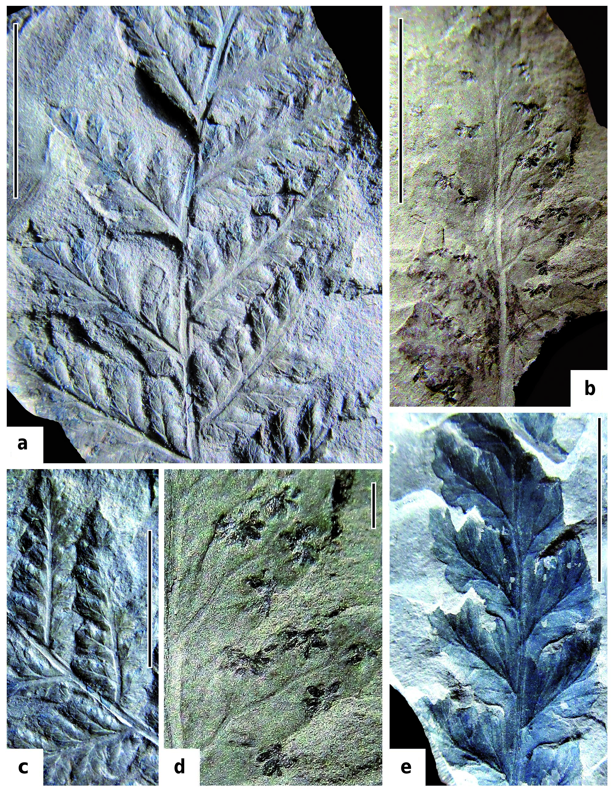
The most complete frond fragment is a bipinnate sterile pinna of penultimate (last-but-one) order ( Text-fig. 2a View Text-fig ). Although this specimen is sterile, the details of venation are completely identical to venation on the fertile pinna selected as the holotype ( Text-fig. 2b, d View Text-fig ).
The pinna rachis of penultimate order is slightly curved presumably towards the frond apex. Maximal observed width of the pinna rachis does not exceed 1 mm. The rachis has a well-defined prolonged axial furrow located on its adaxial surface ( Text-fig. 2c View Text-fig ). Similar but smaller furrows are present on the rachises of the ultimate pinnae (= pinnae of the last order or segments of the last-but-one order). The ultimate pinnae (pinnae of the last order) is attached to the rachis of the penultimate order in regularly alternate order, at an angle of 45–50°. The most well-developed penultimate pinnae possess five to six pairs of pinnules (segments of the last order). The pinnules are attached to the pinna rachis at an angle of 55–65°. The largest pinnules are 7 mm long and 4 mm wide. The size of the pinnules gradually decreases towards the pinna apex. The shape of the pinnules varies from subtriangular (apical pinnules) to ovoid (proximal or basal pinnules). The pinnule apex is acute; pinnule margin is lobate. The lobes are of subtriangular shape, with acute to round apices. The pinnules are fused by their bases and form a wing (= limb) of the last-order pinna rachis ( Text-fig. 2c, e View Text-fig ). Venation is pinnate. The midvein is thin, undulating variably. The lateral veins are thin, dichotomizing only once. The anterior branch of the lateral vein usually dichotomizes once more.
Gross morphology of the fertile pinnae in general is the same as that of sterile pinnae, but the venation of the fertile pinnules is somewhat simpler.
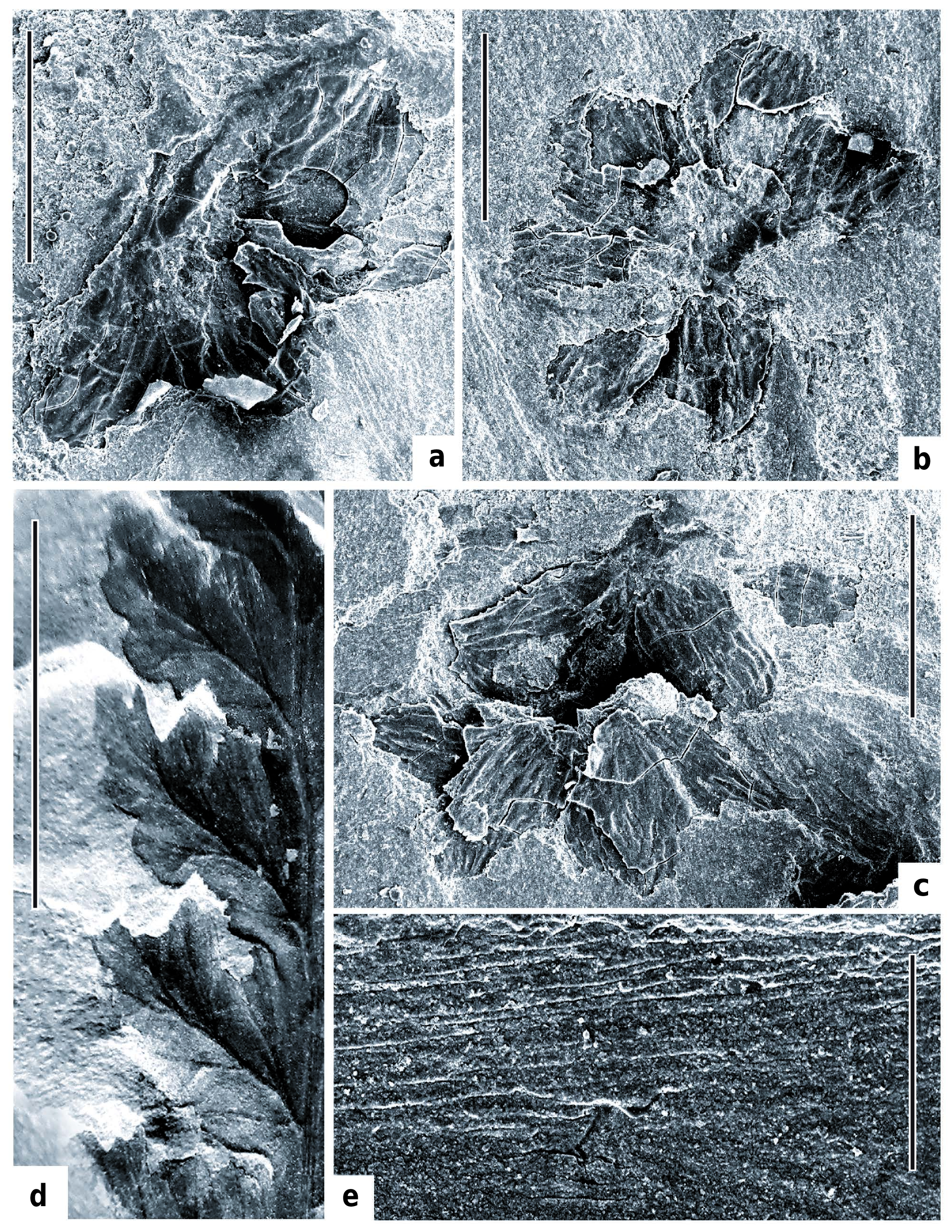
The best-preserved fertile pinna, which was selected as the holotype, shows a clear pattern of arrangement and structure of the sori and sporangia ( Text-figs 2b, d View Text-fig , 3a–e View Text-fig , 4a, b, d View Text-fig ). Each fertile pinnule has one to two sori (on apical pinnules) extending to seven or eight sori (on proximal or basal pinnules). Each sorus consists of six to seven short-ovoid sporangia. The sporangia are free along almost the whole of their length, but are slightly fused by their bases where they attach to the common base (placenta or receptaculum). The average diameter of the sori is 1 mm. The average length of the sporangium is 500 μm, and width of the sporangium is 300 μm. The apex of the sporangia has a small apical sinus or notch. The sporangial wall consists of elongated polygonal cells. The annulus is wide and indistinct having a subequatorial position on the sporangia, most probably consisting of a one cell wide row, situated along both sides of the sporangium split. The vertical split is hardly visible on the sporangia. It can be traced by the small narrow elongated cells surrounding the split ( Text-fig. 3e View Text-fig , uppermost sporangia of both sori). The average size of the cells is 150 μm long and 60–70 μm wide.
Several spores were found on the surface of the holotype fertile pinnules, on the wall of an open sporangium. The spores are round to ovoid ( Text-fig. 5c View Text-fig ), with finely granulate sporoderm and a small trilete scar.
C o m p a r i s o n a n d r e m a r k s. The new species is different from the type species Tumidopteris clavata NAUGOLNYKH in the smaller number of sporangia per sorus (six to seven sporangia per sorus in T. astra compared to nine to eleven in T. clavata), also in the shorter pinnules, and the weak development of the sporangia apical sinuses.
The spores of T. astra are similar to the spores extracted from sporangia of Dichotomopteris lindleyii (ROYLE) MAITHY, the fern from the Lower Gondwana (Lower Permian) deposits of India ( Lele et al. 1981, see for example, text-fig. 8A–F, pl. 2, figs. 12–17), but the exact taxonomic position of the genus Dichotomopteris MAITHY is still unclear. The same or very similar type of in situ spores is known in the presumably gleicheniaceous fern Wingatea plumosa (DAUGHERTY) ASH from the Late Triassic age Chinle Formation, North America ( Litwin 1985, see for example, pl. VI, figs 1–5, pl. VII, figs 1–7). Small round spores with trilete tetrad scar were extracted from the gleicheniaceous fern Oligocarpia lindsaeoides (ETTINGSH.) STUR from the Carboniferous (Westphalian) of the Czech Republic ( Pšenička and Bek 2001: pl. 3) and Lower Permian fern Oligocarpia kepingensis Y.D.WANG et WU from China ( Wang and Wu 1999: figs. 15, 17, Wang et al. 1999: fig. 1E, G).
Similar spores were reported in some other Late Palaeozoic sphenopterid ferns, for instance, Discopteris opulenta DANZÉ from Westfalian C and D (Upper Carboniferous) of Western Europe ( Brousmiche 1979: pl. IX, figs 8–10). The in situ spores with relatively small trilete tetrad scar were obtained from the sporangia of the Upper Carboniferous ferns Doneggia complura G.W.ROTHWELL ( Rothwell 1978: figs 21, 22) and Grambastia goldenbergii (ANDRAE) BOUSMICHE ( Brousmiche 1978: pl. 4, figs 6, 7). Similar spores are characteristic of many geologically younger ferns. Thus, spores practically identical to the spores found on the sporangia and cuticles of Tumidopteris astra , were found in sporangia of leptosporangiate ferns (Aspidiacae and Polypodiaceae ) from Paleogene deposits in the Russian Far-East ( Fedotov 1970: pl. XV, figs 8, 16, 17).
Some remarks comparing T. astra and completely extinct Palaeozoic fern families Sermayaceae ( Sermaya D.A.EGGERT et DELEV. and Doneggia G.W.ROTHWELL ) and Tedeliaceae (e.g., Tedelia glabra D.A.EGGERT et T.N.TAYLOR) should also be made here.
All the sermayacean and tedeliacean ferns (for details see Eggert and Taylor 1966, Eggert and Delevoryas 1967, Rothwell 1978) are characterized by fronds generally similar to those of T. astra . Nonetheless, these taxa vary considerably in important characters such as type and presence of morphologically pronounced annulus and general shape of the sporangia, which are round to obovoid in contrast to the elongated sporangia of representatives of the genus Tumidopteris NAUGOLNYKH. The construction of the sori in sermayacean and tedeliacean ferns is also different. The sori of the two latter fern groups are represented by looser clusters in contrast to the compact sori of Tumidopteris . In addition, the leaf lamina could be strongly reduced in the fertile pinnae of Tedelia glabra, and thus the sori of this fern extend outwards from the fertile pinnule margin.
D i s t r i b u t i o n. Upper part of the Lower Permian (Kungurian) to Ufimian (lower Roadian) stages of the Pechora coal-basin and probably adjacent areas of the Cis- Urals and Russian platform.


| C |
University of Copenhagen |
No known copyright restrictions apply. See Agosti, D., Egloff, W., 2009. Taxonomic information exchange and copyright: the Plazi approach. BMC Research Notes 2009, 2:53 for further explanation.