
Rheotanytarsus
|
publication ID |
https://doi.org/ 10.5281/zenodo.158146 |
|
publication LSID |
lsid:zoobank.org:pub:9E918826-9476-4A0E-9528-ABAC9911BA6C |
|
DOI |
https://doi.org/10.5281/zenodo.5681686 |
|
persistent identifier |
https://treatment.plazi.org/id/03A1A20A-FFBA-333A-C239-FE16FAF4FD91 |
|
treatment provided by |
Plazi |
|
scientific name |
Rheotanytarsus |
| status |
|
Key to males of Rheotanytarsus View in CoL View at ENA of China
1. Antenna with 12 flagellomeres .................................................................................... 2
Antenna with 13 flagellomeres .................................................................................... 3
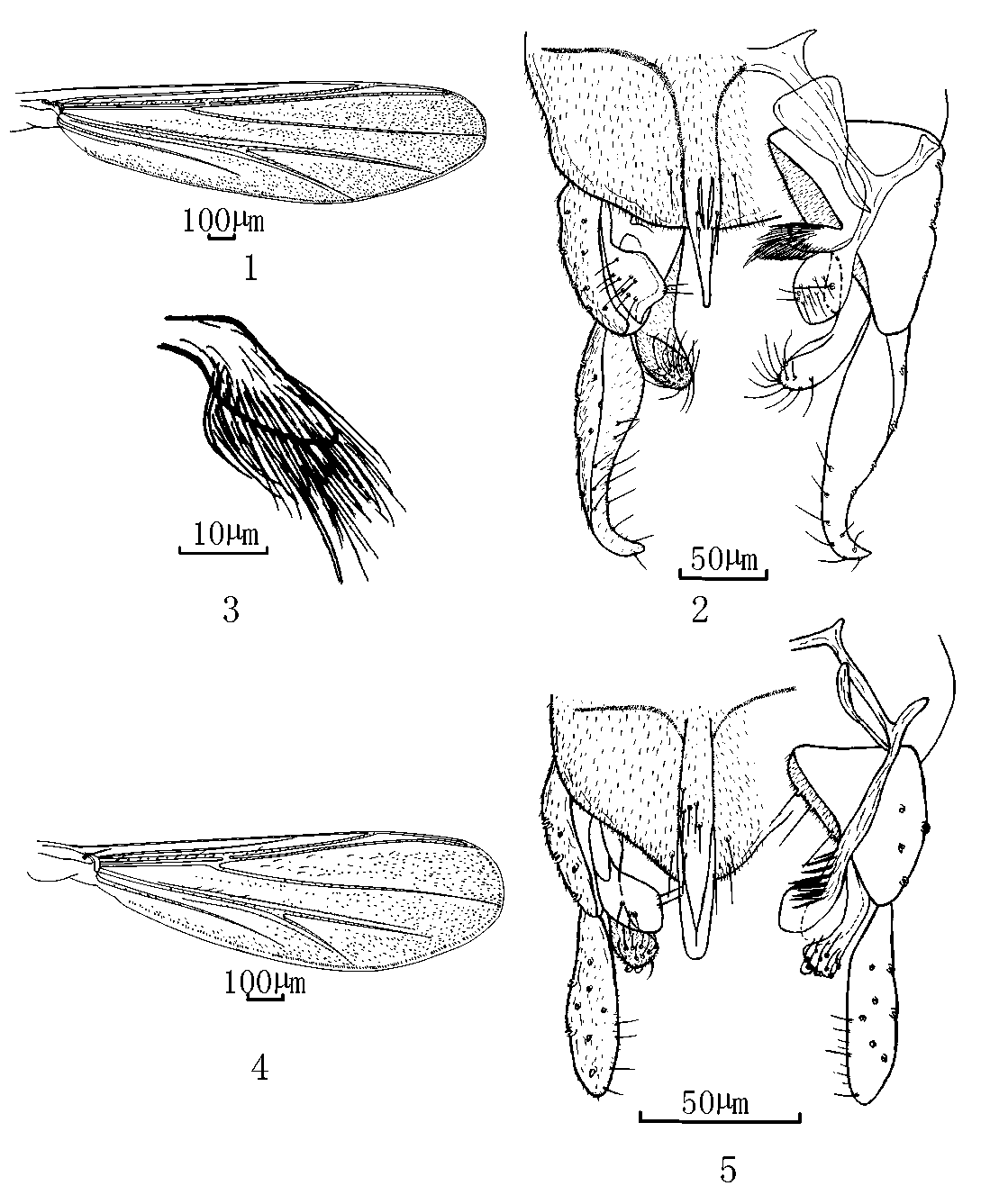
2. Apical lamellae of median volsella bulbous ( Fig. 5 View FIGURES 1 – 5 ) ............................. R. bullus View in CoL sp. n.
Apical lamellae of median volsella not bulbous ( Fig. 7 View FIGURES 6 – 9 ) ............... R. polychaetus View in CoL sp. n.
3. Apex of anal point pointed; tergite IX bare ( Fig. 13 View FIGURES 10 – 13 ) ........................... R. apiculus View in CoL sp. n.
Apex of anal point spatulate, as wide as its basal width, or at least rounded................ 4
4. Abdomen banded. Posterior margin of tergite IX lateral to anal point straight ( Fig. 9 View FIGURES 6 – 9 ). ................................................................................................................... R. liuae View in CoL sp. n.
Abdomen evenly coloured. Posterior margin of tergite IX forming a triangular base for the anal point ................................................................................................................ 5
5. Median volsella long, reaching beyond apex of inferior volsella (Kyerematen & Saether 2000: Figs. 43–46) ................................ R. buculicaudus View in CoL Kyerematen & Saether
Median volsella short, not reaching beyond apex of inferior volsella ......................... 6
6. Base of anal point with a tuft of setae ( Fig. 15 View FIGURES 14 – 18 ) ..................................... R. fundus View in CoL sp. n.
Base of anal point without setal cluster ........................................................................ 7
7. Gonostylus tapering gradually to rounded apex ........................................................... 8
Gonostylus abruptly tapered in apical portion and with narrow apex ........................ 10
8. Superior volsella subtriangular ( Fig. 18 View FIGURES 14 – 18 )....................................... R. brevipalpus View in CoL sp. n.
Superior volsella oblong .............................................................................................. 9
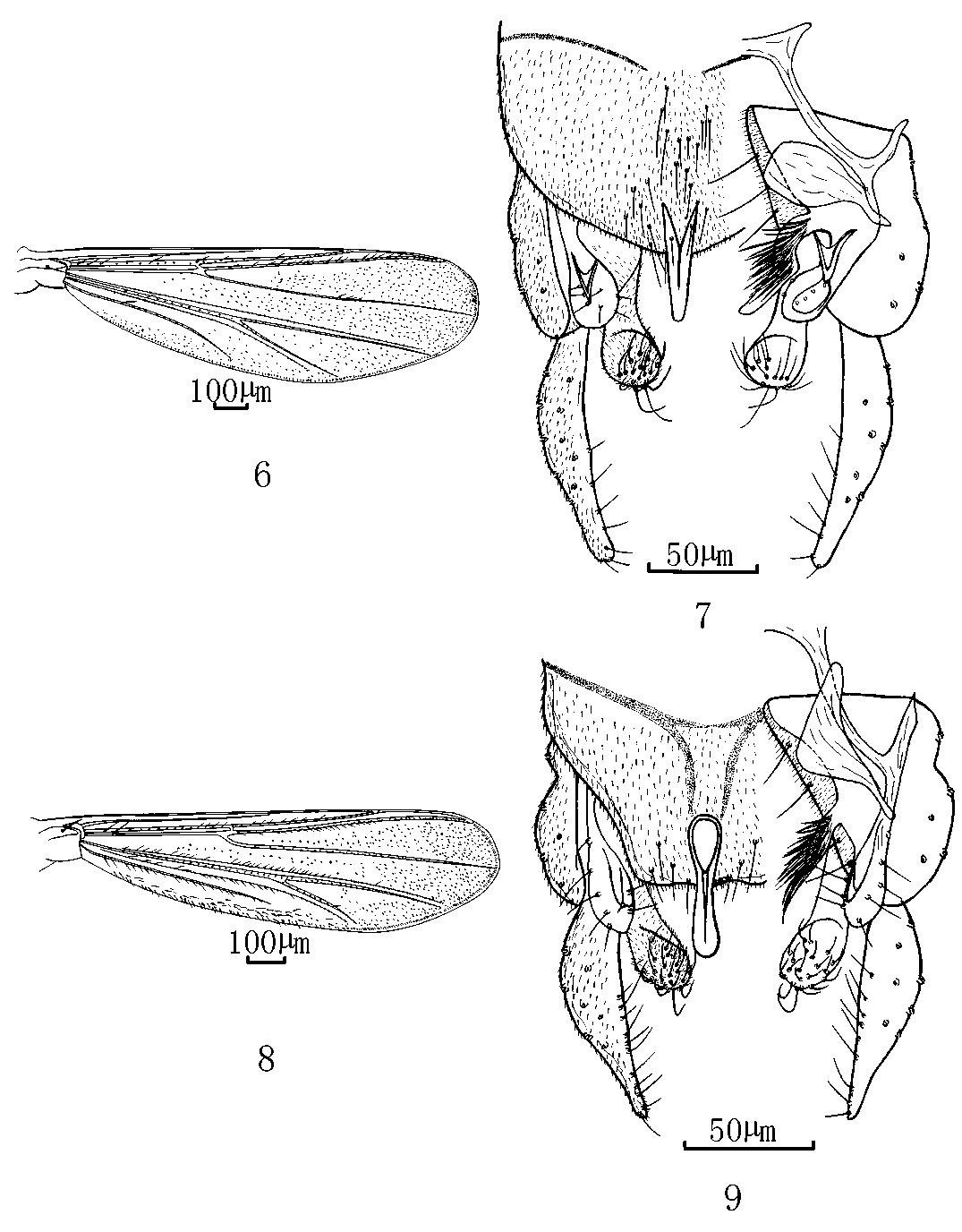
9. Lamellae of median volsella fused into plate, anal point without anal crests ( Sasa 1980: Fig. 11 View FIGURES 10 – 13 ; Wang & Zheng 1993: Figs. 1–2 View FIGURES 1 – 5 ; Kyerematen, Andersen & Saether 2000: Figs. 3 View FIGURES 1 – 5 A–D) .......................................................................... R. tamaquartus View in CoL Sasa
Lamellae of median volsella not fused into plate, anal point with well developed crests ( Tokunaga 1938: Fig. 31; Chaudhuri et al 1994: Figs. 2–3 View FIGURES 1 – 5 ; Kyerematen, Andersen & Saether 2000: Figs. 1 View FIGURES 1 – 5 A–D; Sasa & Kikuchi 1986: Fig. 3 View FIGURES 1 – 5 J; Wang & Zheng 1993: Fig. 4 View FIGURES 1 – 5 ) ................................................................................................. R. aestuarius (Tokunaga) View in CoL
10. Gonostylus recurved at apex ...................................................................................... 11
Gonostylus not recurved at apex ................................................................................ 12
11. Superior volsella with bluntly rounded apical projection ( Johannsen 1932: Fig. 37; Chaudhuri, Datta & Mazumdar 1994: Fig. 1 View FIGURES 1 – 5 ; Kyerematen, Andersen & Saether 2000: Figs. 2 View FIGURES 1 – 5 D–G; Wang & Zheng 1993: Figs. 5–6 View FIGURES 1 – 5 View FIGURES 6 – 9 ) .......................... R. acerbus (Johannsen) View in CoL
Superior volsella rounded ( Fig. 2 View FIGURES 1 – 5 ) ......................................................... R. aphelus View in CoL sp. n.
12. Superior volsella rectangular. Median volsella with almost all lamellae fused into plate ( Fig. 11 View FIGURES 10 – 13 ) .................................................................................... R. quadratus View in CoL sp. n.
Superior volsella subtriangular or beanshaped. Median volsella with apical foliate
setae fused into plate and markedly recurved with sickleshaped apex ..................... 13 13. Superior volsella with posterior margin produced giving a hooklike projection ( Lehman 1970: Figs. 11–13 View FIGURES 10 – 13 ; Albu 1980: Fig. 191; Wang and Zheng 1993: Fig. 3 View FIGURES 1 – 5 ) .............. .............................................................................................. R. muscicola Thienemann View in CoL Superior volsella rectangular and with rounded margin ............................................ 14
14. Anal point spatulate ( Sasa 1980: Fig. 8 View FIGURES 6 – 9 ) .......................................... R. tamatertius View in CoL Sasa
Anal point narrow at apex ( Tokunaga 1938: Fig. 29; Lehmann 1970: Figs. 9–10 View FIGURES 6 – 9 View FIGURES 10 – 13 ) ....... ................................................................................................... R. pentapodus (Kieffer)


No known copyright restrictions apply. See Agosti, D., Egloff, W., 2009. Taxonomic information exchange and copyright: the Plazi approach. BMC Research Notes 2009, 2:53 for further explanation.