
Macrobiotus ariekammensis, ARIEKAMMENSIS WEGLARSKA, 1965
|
publication ID |
https://doi.org/ 10.1093/zoolinnean/zlab101 |
|
publication LSID |
lsid:zoobank.org:pub:B66AA0F0-04D1-4CEE-833A-1C9EE623166E |
|
DOI |
https://doi.org/10.5281/zenodo.6994544 |
|
persistent identifier |
https://treatment.plazi.org/id/03A1D418-FF9A-5A05-FCD0-F9B9FAFBFE27 |
|
treatment provided by |
Plazi |
|
scientific name |
Macrobiotus ariekammensis |
| status |
|
MACROBIOTUS ARIEKAMMENSIS ARIEKAMMENSIS WĘGLARSKA, 1965 View in CoL
( TABLES 2 View Table 2 , 3 View Table 3 ; FIGS 1–4 View Figure 1 View Figure 2 View Figure 3 View Figure 4 )
Material examined: Seven animals and four eggs. Specimens mounted on microscope slides in Hoyer’s medium (four animals + four eggs), processed for DNA sequencing (three animals).
Population locality: 78°40’33’’N, 16°38’49’’E; 208 m a.s.l.: Norway, Svalbard, Fortet ; moss on soil GoogleMaps .
Specimen depositories: Four animals (slides: NO.393.393.2-4) and four eggs (slide: NO.393.01) are deposited at the Institute of Zoology and Biomedical Research , Jagiellonian University , Gronostajowa 9, 30-387, Kraków, Poland .
Description of the Norwegian population from Spitsbergen
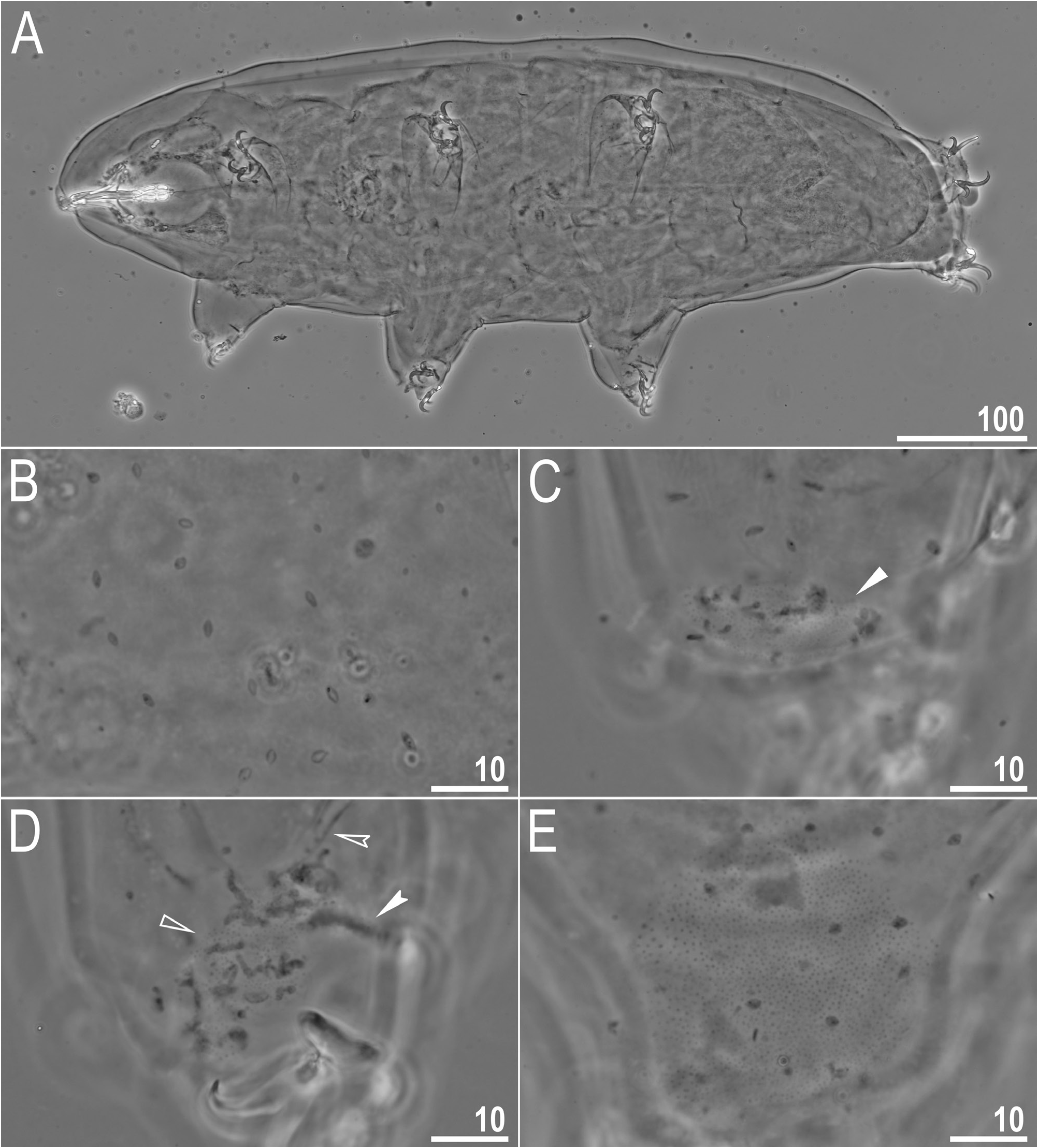
Animals (measurements and statistics in Table 2 View Table 2 ): Body transparent in smaller individuals (juveniles) and whitish in adults, after fixation in Hoyer’s medium transparent ( Fig. 1A View Figure 1 ). Eyes present in all specimens, visible after mounting in Hoyer’s medium. Cuticular pores (0.7–1.2 µm in diameter) present, clearly visible under PCM ( Fig. 1B–E View Figure 1 ) and scattered randomly on the entire body cuticle, including the external and internal surface of all legs. Patches of fine granulation present on internal and external surfaces of all legs I–III, as well as on legs IV and visible clearly in PCM ( Fig. 1C–E View Figure 1 ). A pulvinus present on the internal surfaces of legs I–III ( Fig. 1D View Figure 1 ). Granulation on legs IV is visible as a single large granulation patch on dorsal and lateral leg surfaces ( Fig. 1E View Figure 1 ).
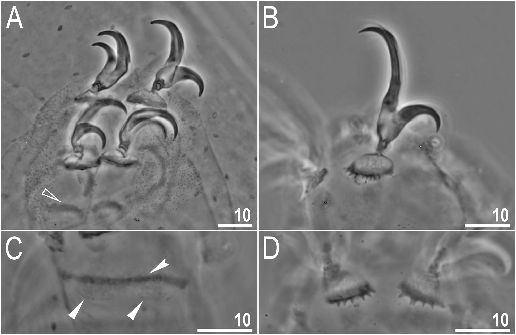
Claws slender, with flat and wide common tract, beginning with a visible stalk that connects the claws to the wide lunulae and ending with elongated branches (especially the primary branch; Fig. 2A, B View Figure 2 ). Primary branches with distinct accessory points, visible in PCM ( Fig. 2A, B View Figure 2 ). Lunulae I–III smooth ( Fig. 2A View Figure 2 ), whereas lunulae IV with clear dentation ( Fig. 2B, D View Figure 2 ). A single continuous cuticular bar and double muscle attachments visible on each leg I–III ( Fig. 2C View Figure 2 ).
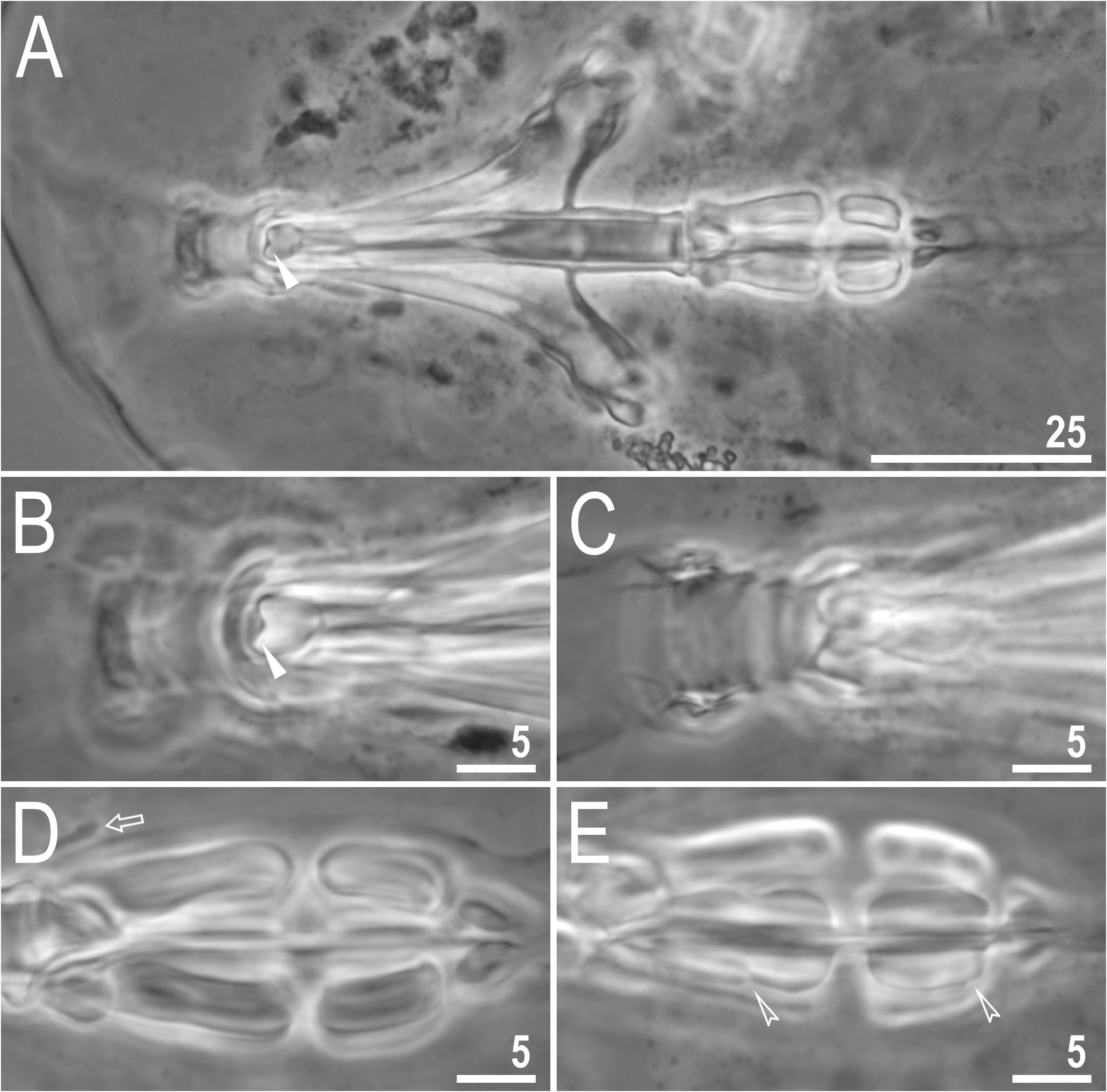
Mouth anteroventral with ten peribuccal lamellae. Bucco-pharyngeal apparatus of the Macrobiotus - type ( Fig. 3A View Figure 3 ). Under PCM, oral cavity armature extremely reduced to only one large tooth present in the dorsal portion of the third band of teeth, whereas other bands of teeth invisible or absent ( Fig. 3A, B View Figure 3 ). Pharyngeal bulb spherical, with triangular apophyses, cuticular spikes, two rod-shaped macroplacoids (macroplacoid sequence: 2 <1) and a triangular small microplacoid ( Fig. 3A, D–E View Figure 3 ). The first macroplacoid with a weak central constriction, whereas the second macroplacoid is weakly subterminally constricted ( Fig. 3D, E View Figure 3 ) .
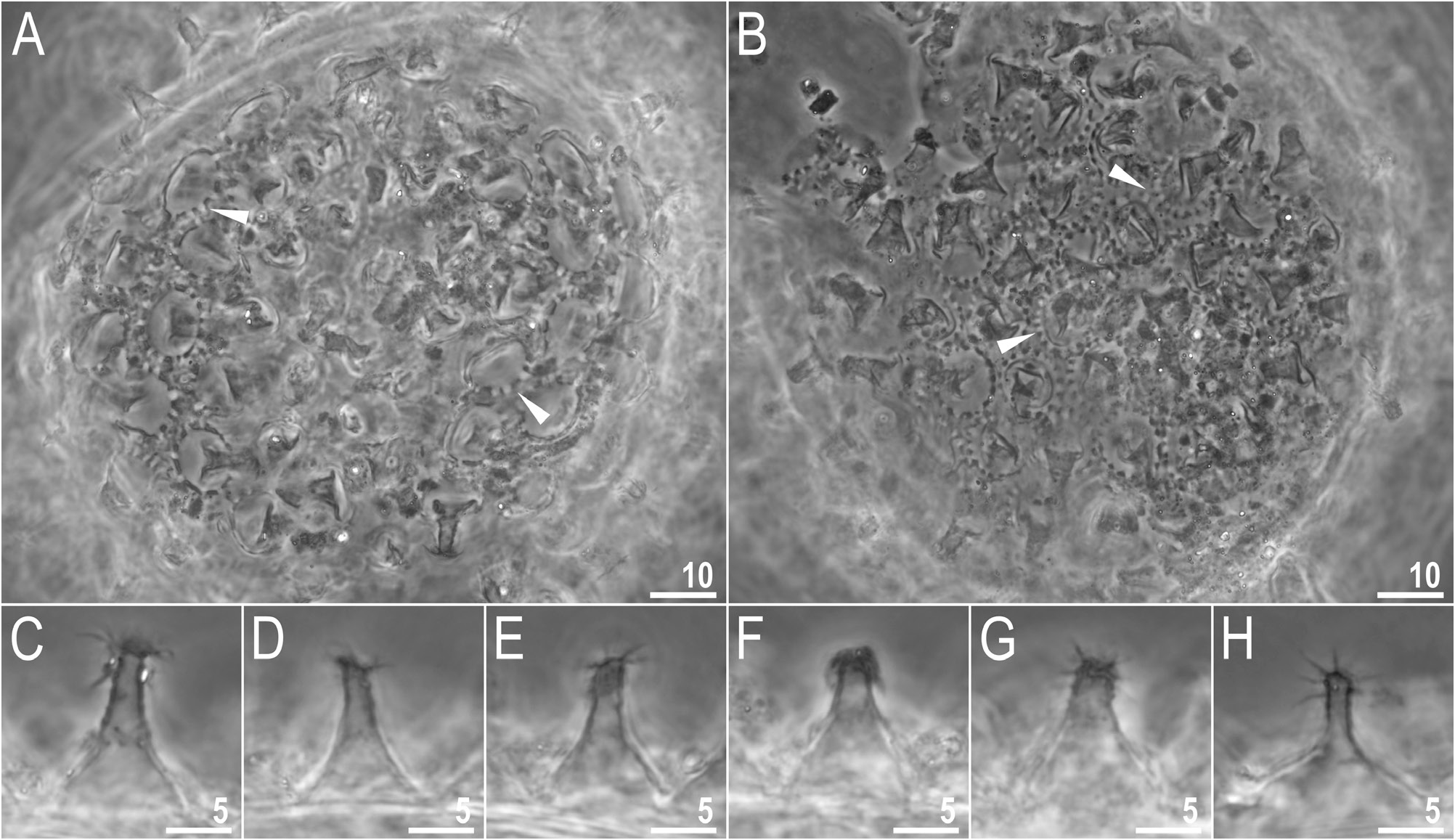
Eggs (measurements and statistics in Table 3 View Table 3 ): Eggs laid freely, whitish, spherical or slightly oval ( Fig. 4A, B View Figure 4 ). The spaces between processes are small, the surface between processes is of the persimilis - type, i.e. with the continuous smooth chorion, with no pores visible ( Fig. 4A, B View Figure 4 ). Egg processes single-walled (without reticulation caused by the labyrinthine layer) with dome-shaped basal part and thinner and elongated distal portions ( Fig. 4C–H View Figure 4 ). Internal septa are sometimes visible between the basal and the distal portion of the process in PCM ( Fig. 4H View Figure 4 ). The basal portions of the processes are pierced by pores that are arranged alternately with dark thickenings and around the process base ( Fig. 4A, B View Figure 4 ). The apical parts of the processes are flat but devoid of terminal discs, and are covered with short, thin and flexible filaments ( Fig. 4C–H View Figure 4 ) .
Reproduction: The population is dioecious (the examination of specimens freshly mounted in Hoyer’s medium revealed testis filled with spermatozoa), but no secondary sexual dimorphism has been observed. DNA sequences: All obtained DNA sequences were represented by a single haplotype per each marker:
18S rRNA: MZ463668 View Materials , MZ463669 View Materials , MZ463670 View Materials . 28S rRNA: MZ463674 View Materials , MZ463675 View Materials , MZ463676 View Materials . ITS2: MZ463656 View Materials , MZ463657 View Materials , MZ463658 View Materials .
COI: MZ460999 View Materials , MZ461000 View Materials , MZ461001 View Materials .
| PCM |
Polish Collection of Microorganisms |
No known copyright restrictions apply. See Agosti, D., Egloff, W., 2009. Taxonomic information exchange and copyright: the Plazi approach. BMC Research Notes 2009, 2:53 for further explanation.
|
Kingdom |
|
|
Phylum |
|
|
Class |
|
|
Order |
|
|
Family |
|
|
Genus |