
Macroplea huaxiensis Lou & Liang
|
publication ID |
https://doi.org/10.5281/zenodo.278456 |
|
DOI |
https://doi.org/10.5281/zenodo.6184805 |
|
persistent identifier |
https://treatment.plazi.org/id/03A28793-FFB0-FFEF-FF4E-57FFAB2F03DB |
|
treatment provided by |
Plazi |
|
scientific name |
Macroplea huaxiensis Lou & Liang |
| status |
sp. nov. |
Macroplea huaxiensis Lou & Liang , sp. nov.
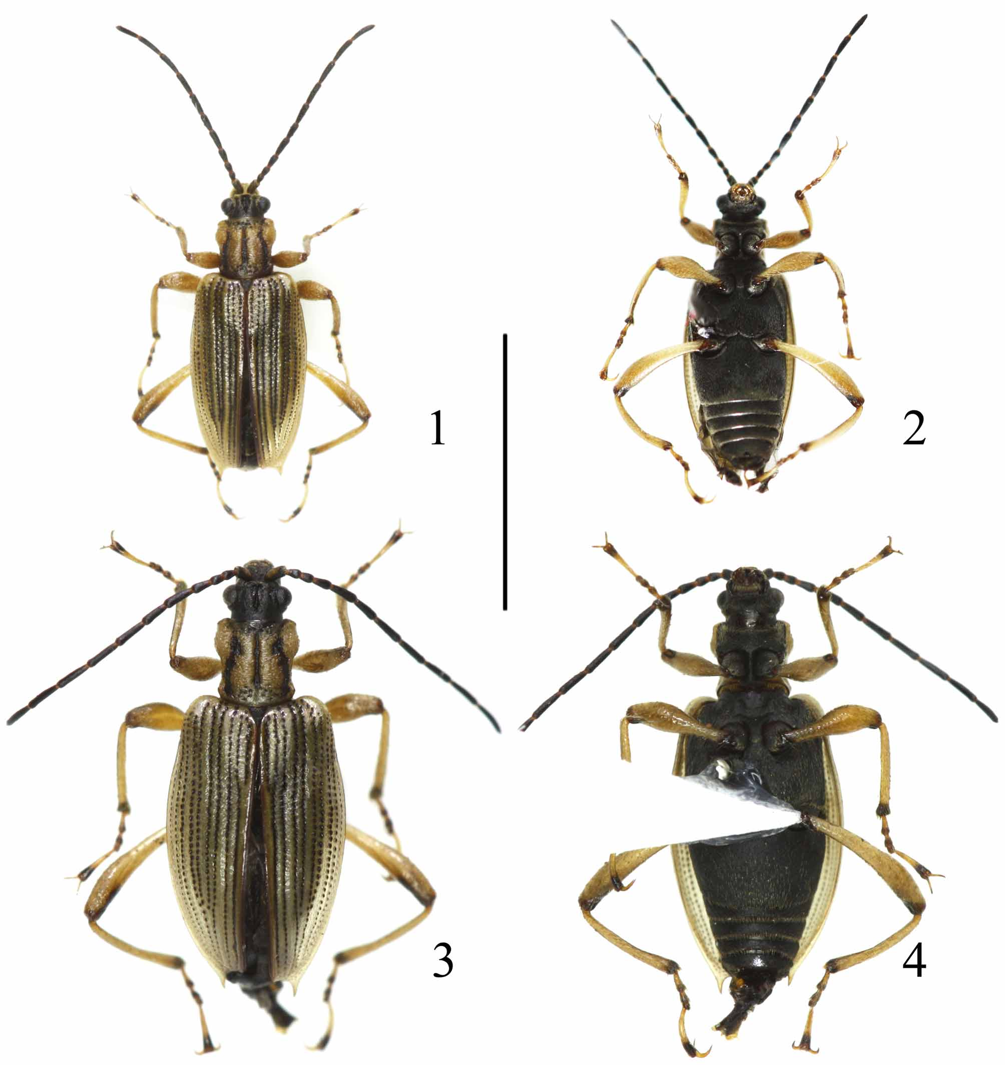
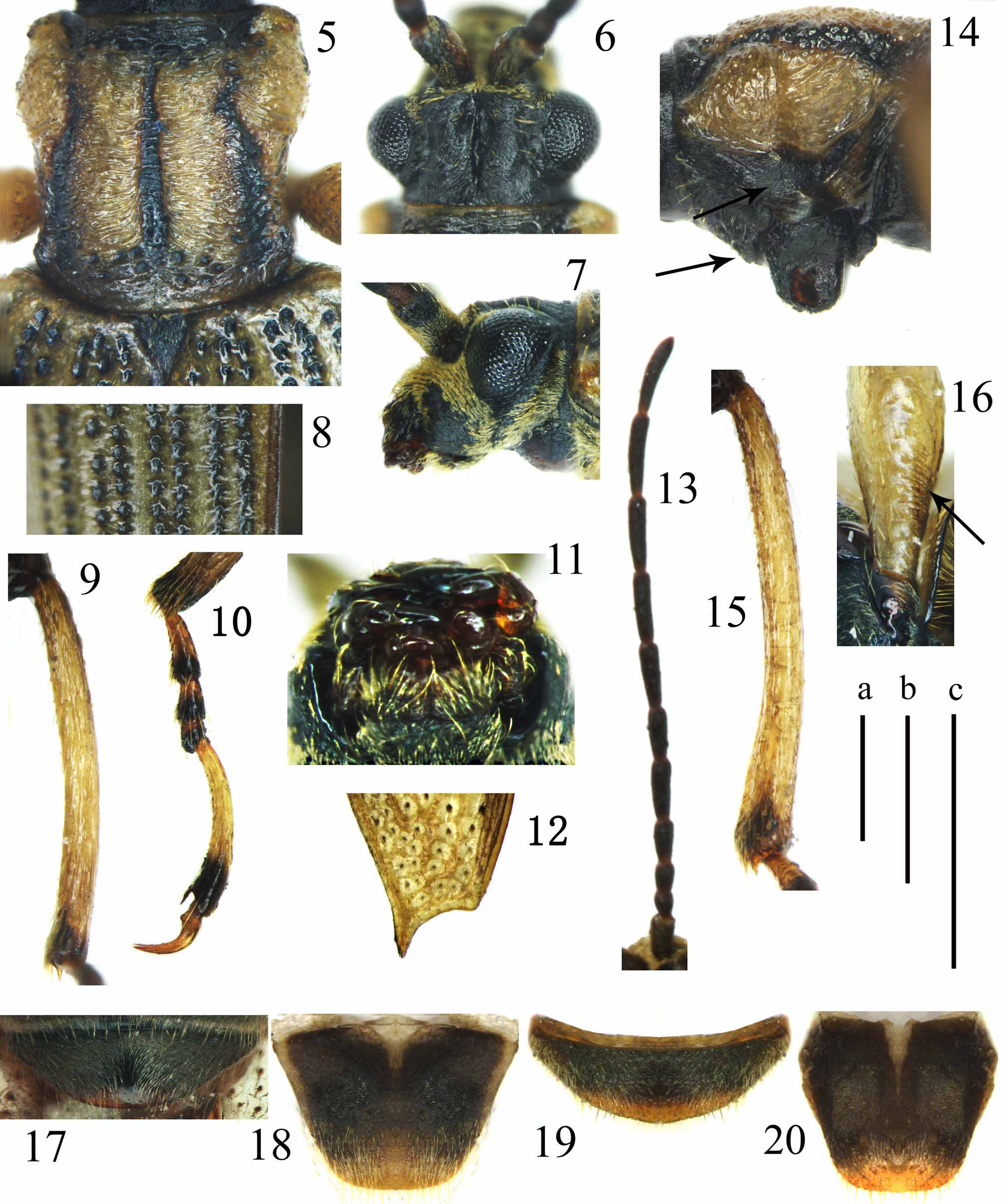
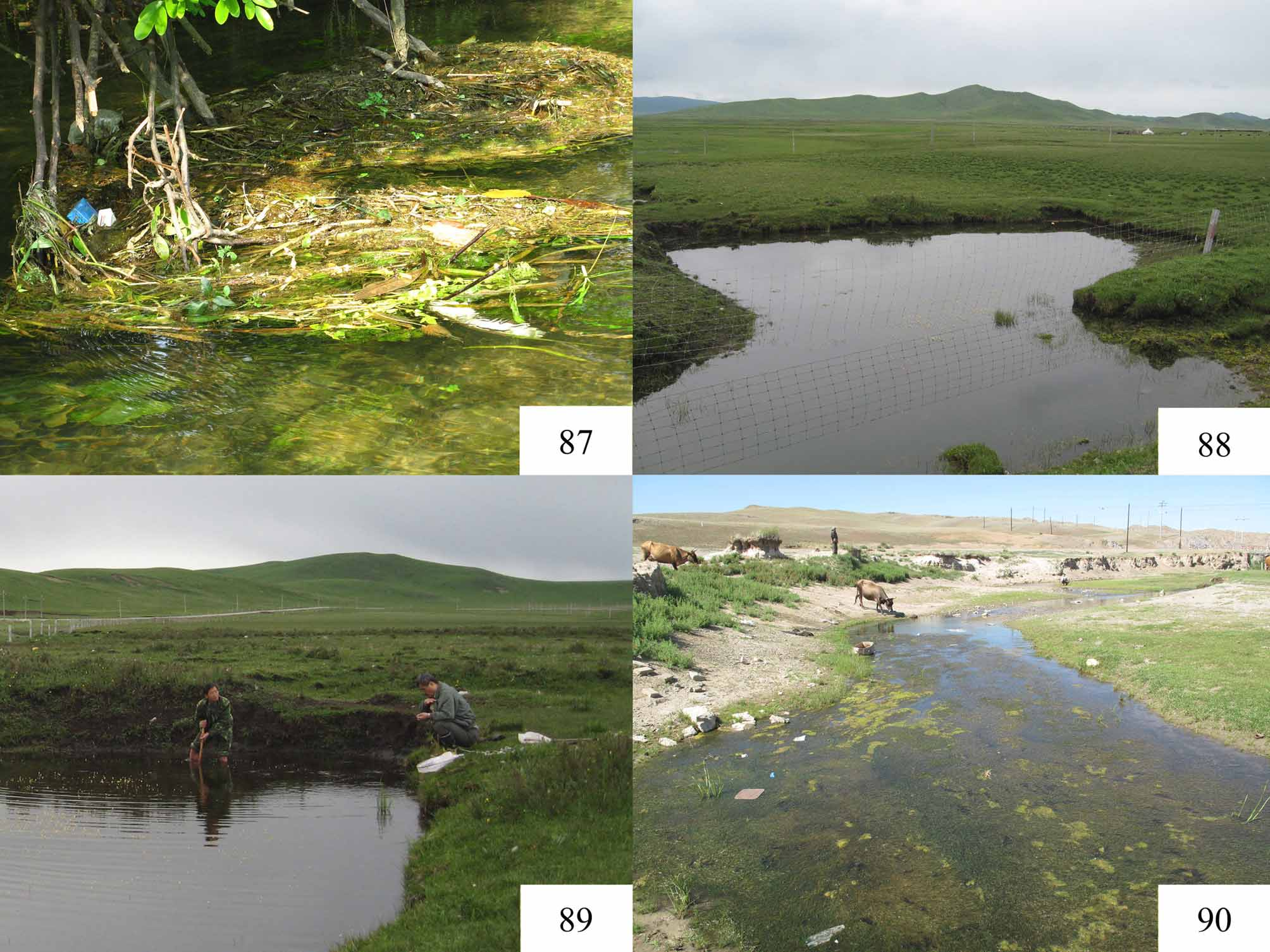
( Figs 1–23 View FIGURES 1 – 4 View FIGURES 5 – 20 View FIGURES 21 – 23 , 86 View FIGURE 86 , 87 View FIGURES 87 – 90 )
Type locality. Huaxi District, Guizhou Province, China, 26.43578°N, 106.65092°E, 1100 m.
Types. Holotype: male, “ CHINA, Guizhou Provin., Guiyang City, Huaxi Distr. Huaxi river. 26.43578°N, 106.65092°E ”/ “ 1100 m, 2009. X.16 D. Ye Liu & Hongliang Shi coll. Institute of Zoology, Chinese Acad. Sci.”/ “ Macroplea huaxiensis NEW SPECIES, HOLOTYPE, designated by Lou and Liang, 2011” [red label]. Paratypes (total 18 specimens): 11 males and 3 females, one pair deposited in the Osaka Museum of Natural History, “ CHINA, Guizhou Provin., Guiyang City, Huaxi Distr., Huaxi river. 26.43578°N, 106.65092°E ”/ “ 1100 m, 2009. X.16 D. Larval host plants: Vallisneria natans and Ottelia acuminata . Ye Liu & Hongliang Shi coll. Institute of Zoology, Chinese Acad. Sci.”/ “ Macroplea huaxiensis NEW SPECIES, PARATYPE, designated by Lou and Liang, 2011” [yellow label]; 2 males and 2 females, “ CHINA, Guizhou Provin., Guiyang City, Huaxi Distr., Huaxi river. 26.43578°N, 106.65092°E ”/ “ 1100 m, 2008. I.06 D. Larval host plants: Vallisneria natans and Ottelia acuminata . Ye Liu coll. Institute of Zoology, Chinese Acad. Sci.”/ “ Macroplea huaxiensis NEW SPECIES, PARATYPE, designated by Lou and Liang, 2011” [yellow label].
Diagnosis. Pronotum and elytra glabrous ( Figs 5, 8 View FIGURES 5 – 20 ); ventral surface covered with dense flat pubescence, mixed with sparse long individually distinguishable setae ( Figs 14, 16, 17, 19 View FIGURES 5 – 20 ); antenna long, extending beyond middle of elytron ( Figs 1–4 View FIGURES 1 – 4 ); fourth antennomere about 1.4 times as long as third ( Fig. 13 View FIGURES 5 – 20 ); mentum with dense pubescence ( Fig. 11 View FIGURES 5 – 20 ); pronotum wrinkled, with three longitudinal black stripes on disc, coarsely punctate near apical and basal margins ( Figs 5, 14 View FIGURES 5 – 20 ); hypomeron with a small triangular supracoxal pubescent patch ( Fig. 14 View FIGURES 5 – 20 ); elytral apex truncate, with triangular spine at outer apical angle, sutural apex angulated ( Fig. 12 View FIGURES 5 – 20 ); punctures along striae arranged in paired rows ( Figs 1, 3 View FIGURES 1 – 4 , 8 View FIGURES 5 – 20 ); first metatarsomere longer than second ( Fig. 10 View FIGURES 5 – 20 ).
Description.
Size. Male: BL = 5.1–5.4 mm, BW = 1.9–2.1 mm; female: BL = 5.4–6.4 mm, BW = 1.9–2.8 mm.
Color. Head and antenna black ( Figs 1–4 View FIGURES 1 – 4 ); mouth parts brown to dark brown; pronotum yellow with three black stripes and black punctures ( Fig. 5 View FIGURES 5 – 20 ); scutellum black; elytra yellow with punctures along striae black and elytral suture brown ( Figs 1, 3 View FIGURES 1 – 4 ); underside black ( Figs 2, 4 View FIGURES 1 – 4 ); legs yellow, apices of femora, tibiae and tarsomeres black ( Figs 1–4 View FIGURES 1 – 4 ).
Head. Eyes prominent, supraocular furrow distinct ( Fig. 6 View FIGURES 5 – 20 ); vertex with distinct tubercles along medial groove, with dense, flat, individually indistinguishable pubescence extending over tubercles ( Figs 6, 7 View FIGURES 5 – 20 ); transverse carina behind antennal base as high as vertex, covered with long yellow setae ( Fig. 6 View FIGURES 5 – 20 ); frontoclypeus with long yellowish setae ( Figs 6, 7 View FIGURES 5 – 20 ); anteclypeus exposed or enclosed; labrum broad, apical margin slightly protruding, with a row of punctures at base and a few punctures at lateral sides; mentum with dense long setae, apex emarginated ( Fig. 11 View FIGURES 5 – 20 ); apical labial palpomere flattened, short, broad; labial and maxillary palpi with setae restricted to mesal surface; antenna long, extending beyond middle of elytron ( Figs 1–4 View FIGURES 1 – 4 ); scape (antennomere 1) with mesal surface flat, smooth, glabrous, rest of scape and antennomeres 2–11 with dense, flat pubescence, most setae individually indistinguishable ( Figs 6, 13 View FIGURES 5 – 20 ); length ratio of scape and antennomeres 2–4 equals 31:16:19:26.
Thorax. Pronotum ( Figs 5, 14 View FIGURES 5 – 20 ) quadrate, PL/PW = 0.99 (0.92–1.03); pronotal angles projecting, each bearing a seta (two or three setae at a single angle in a few specimens); apex slightly wider than base; apical margin convex; basal margin flat; anterolateral tubercles protruded distinctly; lateral margin gradually narrowing backwards from anterolateral tubercle; lateral margin narrowed in middle; no ridge or carina anterior to basal angle; disc slightly shiny, glabrous, with fine transverse wrinkles and fine punctures outside median groove, with scattered coarse punctures near apical and basal margins; median groove deep, wide, with fine transverse wrinkles and fine punctures at bottom; longitudinal black stripes long, exceeding anterior and posterior transverse grooves. Hypomeron ( Fig. 14 View FIGURES 5 – 20 ) with wrinkles and a small triangular supracoxal pubescent patch. Prosternum ( Fig. 14 View FIGURES 5 – 20 ) covered with dense flat pubescence, mixed with sparse long individually distinguishable setae, prosternum slightly convex in central area before procoxae. Scutellum ( Fig. 5 View FIGURES 5 – 20 ) triangular, with sharp apex.
Elytra. EL/EW = 1.85 (1.64–1.94), widest near middle, narrowed backwards; apex truncate with triangular spine at outer angle ( Figs 1, 3 View FIGURES 1 – 4 , 12 View FIGURES 5 – 20 ); spine slightly longer than its width at base; punctures along striae relatively fine, arranged in paired rows; surface of intervals shiny, alternating intervals 2, 4, 6, 8 wider and convex ( Figs 1, 3 View FIGURES 1 – 4 , 8 View FIGURES 5 – 20 ), elytral suture convex with shallow transverse wrinkles; sutural apex with obtuse angulation ( Fig. 12 View FIGURES 5 – 20 ); epipleuron as wide as outermost interval, convex, extending over spine at outer angle ( Fig. 4 View FIGURES 1 – 4 ); groove at shoulder shallow, basal depression shallow, medial depression inconspicuous ( Figs 1, 3 View FIGURES 1 – 4 ).
Abdomen. Sterna with dense, flat pubescence, mixed with sparse long individually distinguishable setae ( Figs 17, 19 View FIGURES 5 – 20 ).
Legs. Profemur with a short linearly arranged brush of setae basally on posterior surface ( Fig. 16 View FIGURES 5 – 20 ); pro- and meso-tibial spurs short, inconspicuous, metatibia slightly curved in both sex ( Figs 9, 15 View FIGURES 5 – 20 ); metatarsus ( Fig. 10 View FIGURES 5 – 20 ) with markedly reduced pubescence, fifth tarsomere elongate, 1.2–1.4 times as long as basal three combined, first tarsomere longer than second, second longer than third, third tarsomere cylindrical, slightly bilobed in dorsal view; tarsal claw simple, elongate, with a small tooth at base.
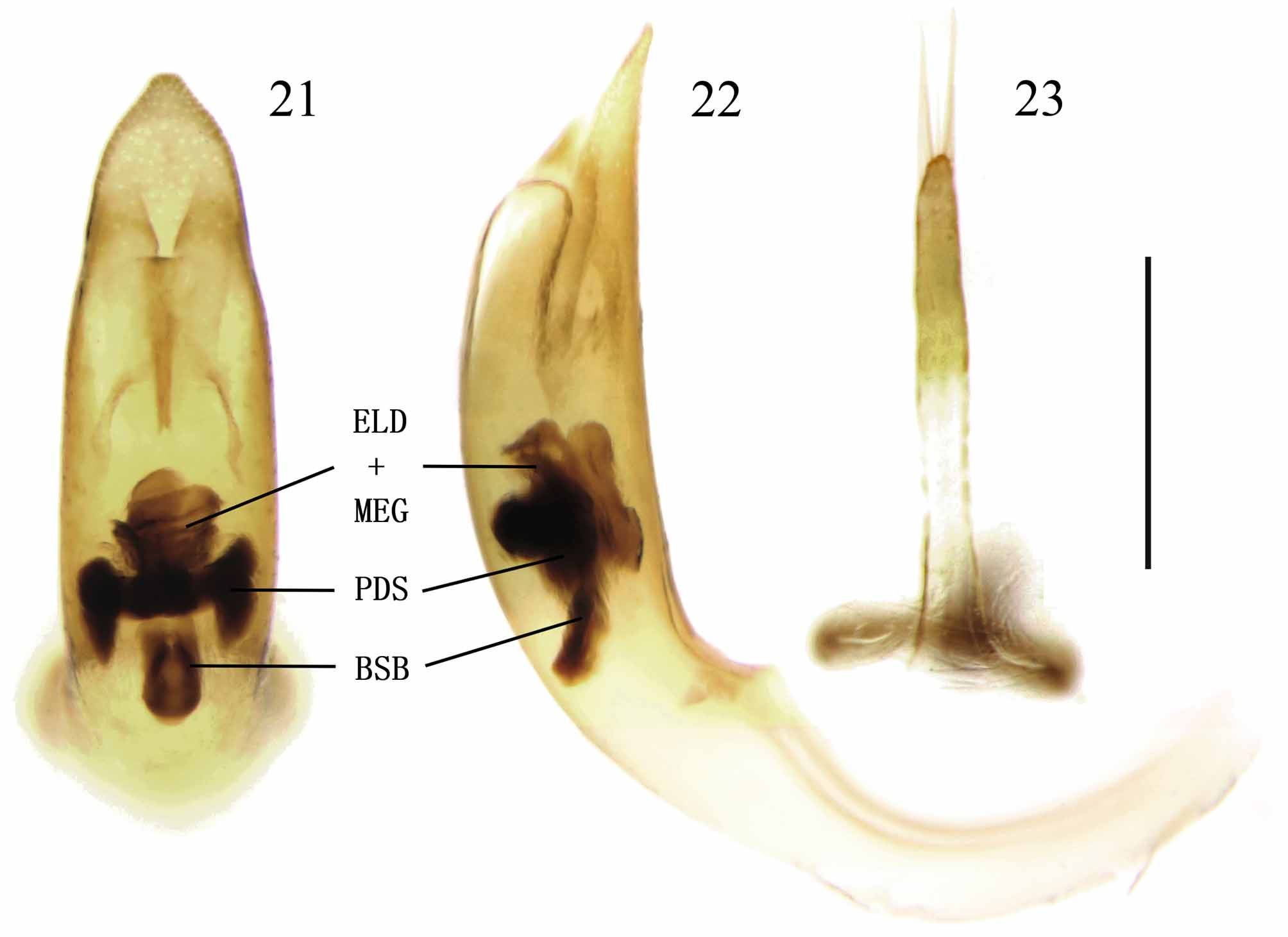
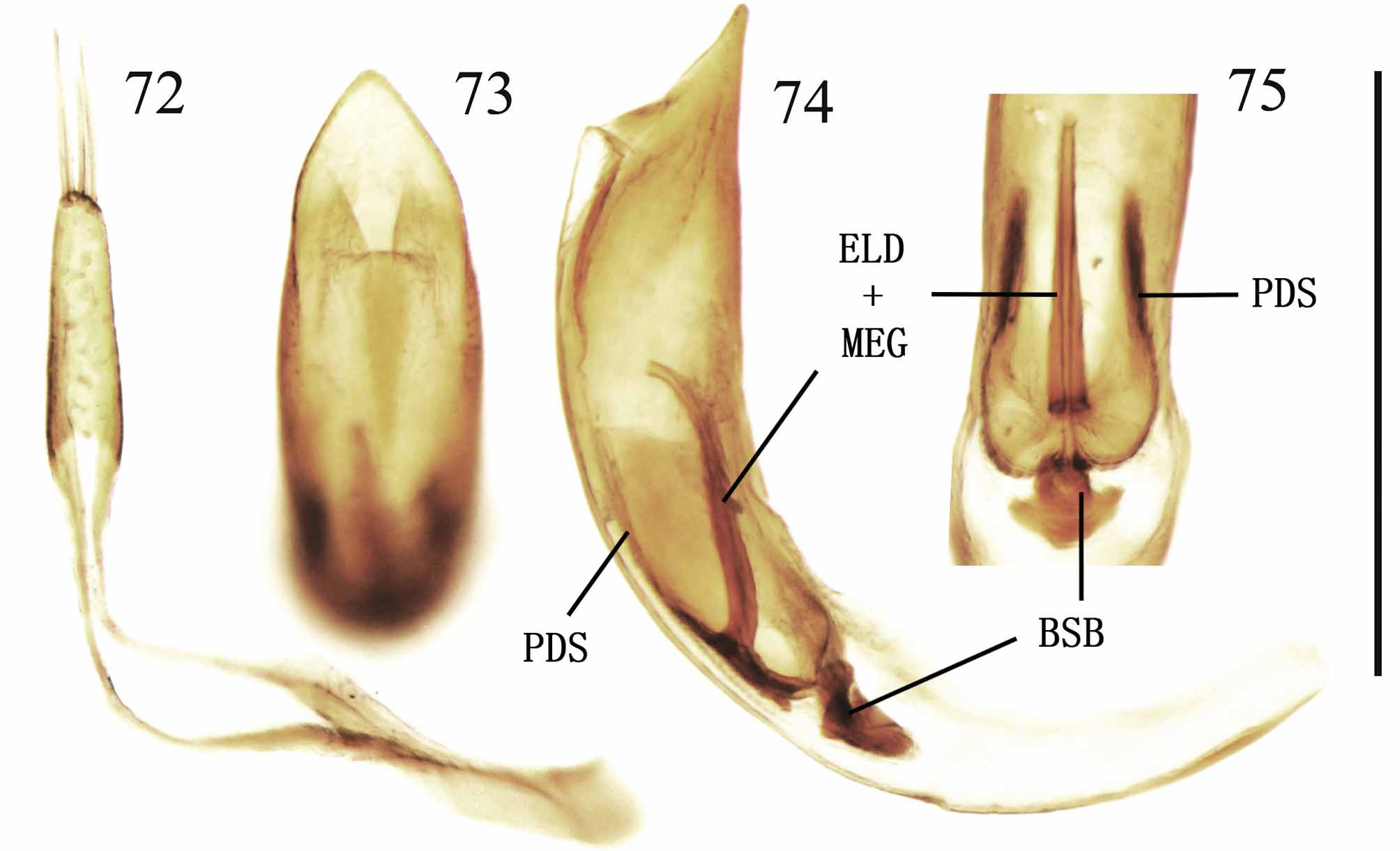
Male genitalia. Median lobe of aedeagus stout, slightly truncate at apex in dorsal view ( Fig. 21 View FIGURES 21 – 23 ); cap of tegmen slender, more sclerotized at apical half, with several long setae at apex ( Fig. 23 View FIGURES 21 – 23 ); endophallus with apex of MEG turned dorsal, two ELDs fused, enclosing MEG, PDS paired, with apex broadly expanded in dorsal view, without pELD ( Figs 21, 22 View FIGURES 21 – 23 ).
Sexual dimorphism. Male generally smaller than female ( Figs 1–4 View FIGURES 1 – 4 ); apical abdominal sternum with a medial depression of variable depth in male ( Fig. 17 View FIGURES 5 – 20 ), such a depression is absent in female ( Fig. 19 View FIGURES 5 – 20 ); hind margin of pygidium slightly rounded in male ( Fig. 18 View FIGURES 5 – 20 ), truncate in female ( Fig. 20 View FIGURES 5 – 20 ).
Distribution. China (Guizhou) ( Fig. 85 View FIGURE 85 ).
Host plants. Vallisneria natans (Lour.) Hara and Ottelia acuminata (Gagnep.) Dandy (both Hydrocharitaceae ) are recorded as larval host plants, with the former being the major host.
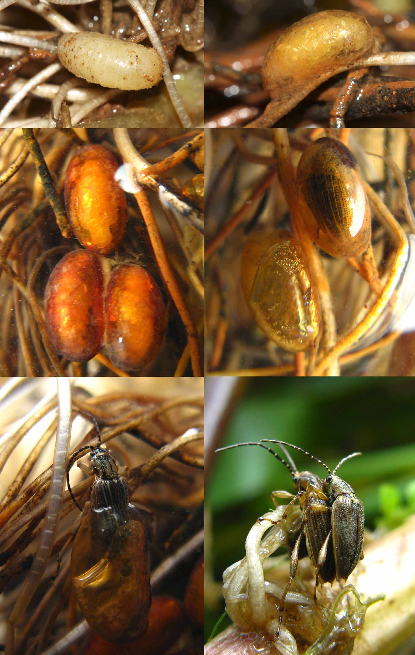
Biology. The type locality of this new species is a clean river running at a moderate speed while harbouring a variety of aquatic vegetation ( Fig. 87 View FIGURES 87 – 90 ). We observed larvae and cocoons (with larvae, pupae or adults inside) adhering to the roots of host plant ( Fig. 86 View FIGURE 86 ). In the laboratory, adults were never observed flying, although they can walk slowly out of water for a long time (more than two hours).
Etymology. The species name huaxiensis refers to the type locality, Huaxi river.
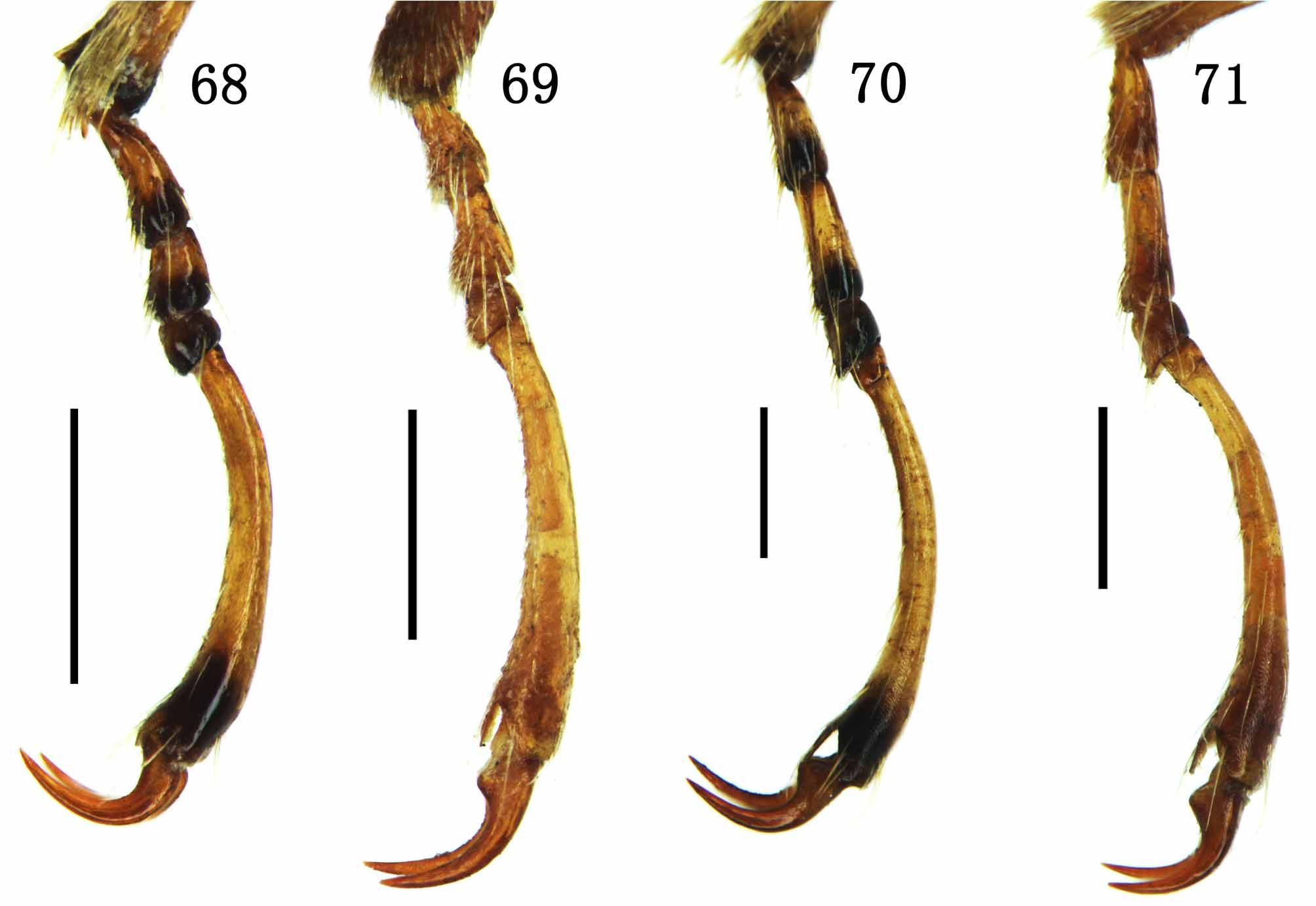
Remarks. This new species was wrongly identified as M. japana by Zhang et al. (2010). It resembles M. japana in many characters (see the key), among which three black stripes on pronotal disc ( Figs 5 View FIGURES 5 – 20 , 56 View FIGURES 54 – 59 ) and a triangular supracoxal pubescent patch on hypomeron ( Figs 14 View FIGURES 5 – 20 , 54 View FIGURES 54 – 59 ) are shared only by these two species in Macroplea . However, M. huaxiensis can be easily separated from M. japana by the characters listed in the key. Other characters useful in separating these two species are the following: mentum is densely pubescent in M. huaxiensis ( Fig. 11 View FIGURES 5 – 20 ), while the same is sparsely pubescent in M. japana ; ventral surface is mixed with sparse long individually distinguishable setae in the former ( Figs 14, 16, 17, 19 View FIGURES 5 – 20 ), while such setae are absent in M. japana ; prosternum in middle part before procoxae is slightly convex in M. huaxiensis ( Fig. 14 View FIGURES 5 – 20 ), however the same is distinctly convex in M. japana ( Fig. 54 View FIGURES 54 – 59 ); elytral apex is truncate in M. huaxiensis ( Fig. 12 View FIGURES 5 – 20 ), and the same is emarginate in M. japana ( Fig. 64 View FIGURES 60 – 67 ); base of metatarsal claw with a small tooth in M. huaxiensis ( Fig. 10 View FIGURES 5 – 20 ), but such a tooth is absent in M. japana ( Fig. 68 View FIGURES 68 – 71 ); tip of median lobe of aedeagus is slightly truncate in M. huaxiensis ( Fig. 21 View FIGURES 21 – 23 ), while the same is more acute in M. japana ( Fig. 73 View FIGURES 72 – 75 ); endophallus with ELD robust and apex of PDS broadly expanded in dorsal view in the former ( Fig. 21 View FIGURES 21 – 23 ), however the same are much more slender in M. japana ( Fig. 75 View FIGURES 72 – 75 ).
The densely pubescent mentum ( Fig. 11 View FIGURES 5 – 20 ) in M. huaxiensis is unique as the same is glabrous or sparsely pubescent in all other members of the genus. Densely pubescent mentum is a feature of Neohaemonia species distributed in the new world ( Askevold, 1988: 403).


| NEW |
University of Newcastle |
No known copyright restrictions apply. See Agosti, D., Egloff, W., 2009. Taxonomic information exchange and copyright: the Plazi approach. BMC Research Notes 2009, 2:53 for further explanation.
|
Kingdom |
|
|
Phylum |
|
|
Class |
|
|
Order |
|
|
Family |
|
|
SubFamily |
Donaciinae |
|
Genus |