
Macroplea ranina Lou & Yu
|
publication ID |
https://doi.org/10.5281/zenodo.278456 |
|
DOI |
https://doi.org/10.5281/zenodo.6184807 |
|
persistent identifier |
https://treatment.plazi.org/id/03A28793-FFB4-FFE2-FF4E-56D1ABFA00E5 |
|
treatment provided by |
Plazi (2016-04-11 02:47:03, last updated 2024-11-29 13:31:37) |
|
scientific name |
Macroplea ranina Lou & Yu |
| status |
sp. nov. |
Macroplea ranina Lou & Yu , sp. nov.
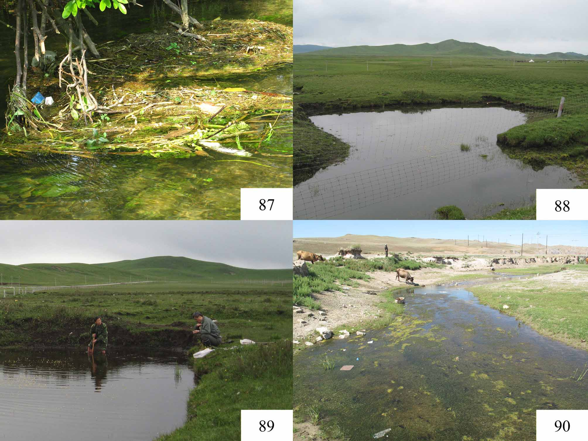
( Figs 24–49 View FIGURES 24 – 27 View FIGURES 28 – 43 View FIGURES 44 – 49 , 88, 89 View FIGURES 87 – 90 )
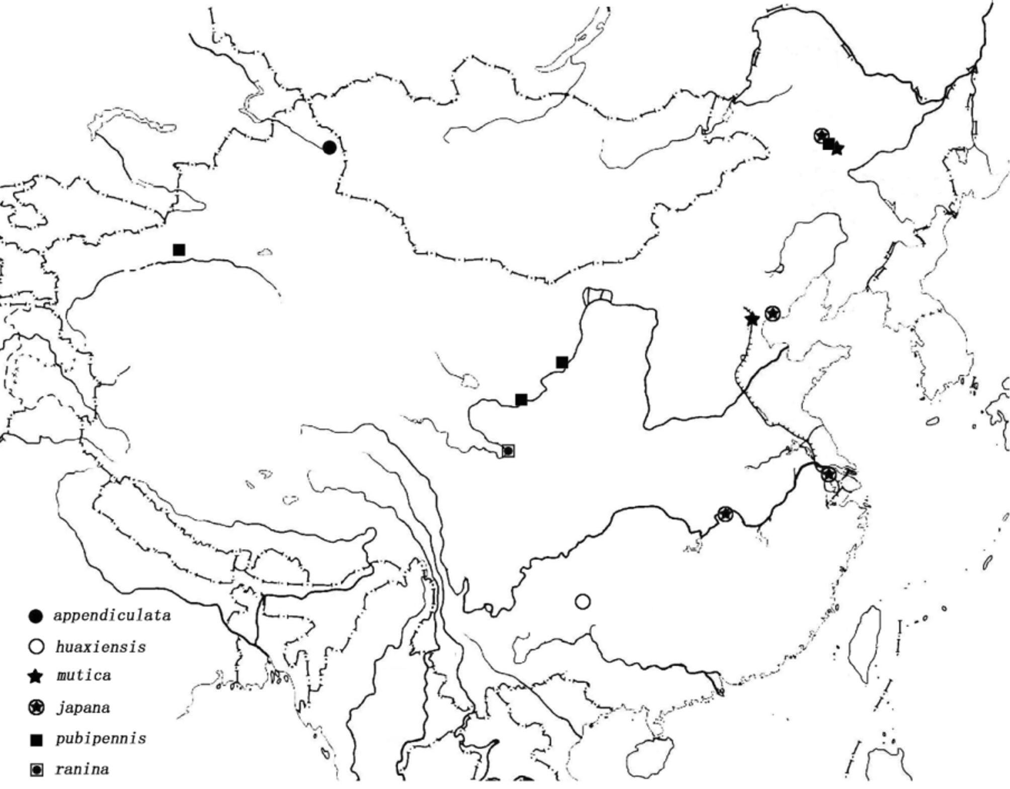
Type locality. Zoigê County, Sichuan Province, China, 33.58143° N, 102.87080° E, 3454 m.
Types. Holotype: male, “ CHINA, Sichuan Prov., Zoigê County, Keji Prairie, 33.58143°N, 102.87080°E ”/ “ 3454 m, 2007. VI.25. Hongbin Liang coll. Institute of Zoology, Chinese Acad. Sci.”/ “ Macroplea ranina sp. n., Holotype, designated by Lou & Yu, 2011” [red label]. Paratypes (total 10 specimens): 1 male and 1 female, “ CHINA, Sichuan Prov., Zoigê County, Keji Prairie, frog stomach.”/ “ 3454 m, 2005. V.6. Jindong Zhang coll. Institute of Zoology Chinese Acad. Sci.”/ “ Macroplea ranina sp. n., Paratype, designated by Lou & Yu, 2011” [yellow label]; 2 males and 2 females, “ CHINA, Sichuan Prov., Zoigê County, Keji Prairie, 33.58143°N, 102.87080°E ”/ “ 3454 m, 2007. VI.25. Hongbin Liang coll. Institute of Zoology Chinese Acad. Sci.”/ “ Macroplea ranina sp. n., Paratype, designated by Lou & Yu, 2011” [yellow label]; 2 males (Osaka Museum of Natural History), “ CHINA, Sichuan Prov., Zoigê County, Keji Prairie, 33.58143°N, 102.87080°E ”/ “ 3454 m, 2007. VI.25. T. Sota coll.”/ “ Macroplea ranina sp. n., Paratype, designated by Lou & Yu, 2011” [yellow label]; 1 male and 1 female, “ CHINA, Sichuan Prov., Zoigê County, Keji Prairie, 33.58143° N, 102.87080° E 2007.VI, Jindong Zhang coll. Institute of Zoology Chinese Acad. Sci.”/ “ Macroplea ranina sp. n., Paratype, designated by Lou & Yu, 2011” [yellow label].
Diagnosis. Pronotum and elytra glabrous ( Figs 28, 31 View FIGURES 28 – 43 ); ventral surface covered with dense long individually distinguishable setae ( Figs 32, 34 View FIGURES 28 – 43 ); antenna short, not extending beyond middle of elytron ( Figs 24–27 View FIGURES 24 – 27 ); fourth antennomere about 1.3 times as long as third ( Fig. 33 View FIGURES 28 – 43 ); mentum with very sparse setae, not bilobed ( Fig. 30 View FIGURES 28 – 43 ); pronotum wrinkled, with deep, wide, black median groove and two deep depressions near apical margin, coarsely punctate near apical and basal margins ( Figs 28, 32 View FIGURES 28 – 43 ); hypomeron with an oblong supracoxal pubescent patch ( Fig. 32 View FIGURES 28 – 43 ); elytral apex nearly truncate, without spine at apical angles, only with obtuse angulation ( Figs 24, 26 View FIGURES 24 – 27 , 35 View FIGURES 28 – 43 ); punctures along striae not arranged in paired rows, intervals with subequal width ( Figs 24, 26 View FIGURES 24 – 27 , 31 View FIGURES 28 – 43 ); apical metatarsomere as long as basal three combined ( Fig. 39 View FIGURES 28 – 43 ).
Description.
Size. Male: BL = 5.4–6.9 mm. BW = 2.2–2.7 mm; female: BL = 6.9–8.5 mm. BW = 2.6–3.3 mm.
Color. Head black; antenna dark brown or black ( Figs 24, 26 View FIGURES 24 – 27 , 33 View FIGURES 28 – 43 ); mouth parts dark brown; pronotum yellow with black median groove and black punctures ( Fig. 28 View FIGURES 28 – 43 ); scutellum black; elytra yellow with punctures along striae black and elytral suture brown ( Figs 24, 26 View FIGURES 24 – 27 ); underside black ( Figs 25, 27 View FIGURES 24 – 27 ); legs yellow, apices of femora, tibiae and tarsomeres brown ( Figs 24–27 View FIGURES 24 – 27 ).
Head. Eyes prominent, supraocular furrow distinct ( Fig. 29 View FIGURES 28 – 43 ); vertex with distinct tubercles along medial groove, with dense long yellowish setae extending over tubercles ( Figs 29, 32 View FIGURES 28 – 43 ); transverse carina behind antennal base as high as vertex, covered with long yellow setae ( Figs 29, 32 View FIGURES 28 – 43 ); frontoclypeus covered with long yellow setae ( Fig. 29 View FIGURES 28 – 43 ); anteclypeus exposed or enclosed; labrum broad, with a row of punctures at base and a few punctures at lateral sides; mentum with very sparse setae, apical margin nearly straight ( Fig. 30 View FIGURES 28 – 43 ); apical labial palpomere flattened, short, broad; labial and maxillary palpi with setae restricted to mesal surface; antenna short, not extending beyond middle of elytron ( Figs 24–27 View FIGURES 24 – 27 ); scape (antennomere 1) with mesal surface flat, smooth, glabrous, rest of scape and antennomeres 2–11 with dense short individually distinguishable setae ( Figs 29, 33 View FIGURES 28 – 43 ); length ratio of scape and antennomeres 2–4 equals 43:22:25:33.
Thorax. Pronotum ( Figs 28, 32 View FIGURES 28 – 43 ) quadrate, PL/PW = 0.92 (0.87–0.95); pronotal angles projecting, each bearing a seta (two or three setae at a single angle in a few specimens); apex slightly wider than base; apical margin convex; basal margin flat; anterolateral tubercles protruded distinctly; lateral margin narrowed in middle; posterolateral tubercle small, extending to disc, forming a conic elevation; disc shiny, glabrous, with two wrinkled depressions deep near anterolateral tubercle, and shallow posteriorly; disc with scattered coarse punctures near apical and basal margins; median groove deep, wide, slightly wrinkled at bottom. Hypomeron ( Fig. 32 View FIGURES 28 – 43 ) with wrinkles and an oblong supracoxal pubescent patch. Prosternum ( Fig. 32 View FIGURES 28 – 43 ) covered with dense long yellow setae, prosternum slightly convex in central area before procoxae. Scutellum ( Fig. 28 View FIGURES 28 – 43 ) triangular, with sharp apex.
Elytra. EL/EW = 1.78 (1.64–1.93), widest near middle, narrowed backwards; apex slightly emarginate with obtuse angulation at outer angle ( Figs 24, 26 View FIGURES 24 – 27 , 35 View FIGURES 28 – 43 ); punctures along striae relatively coarse, not arranged in paired rows; intervals nearly uniformly wide, convex, with irregular shallow transverse wrinkles, surface shiny ( Figs 24, 26 View FIGURES 24 – 27 , 31 View FIGURES 28 – 43 ), elytral suture convex, smooth ( Fig. 31 View FIGURES 28 – 43 ); sutural apex with obtuse angulation ( Fig. 35 View FIGURES 28 – 43 ); epipleuron as wide as or narrower than outermost interval, convex ( Fig. 35 View FIGURES 28 – 43 ); groove at shoulder shallow, basal depression shallow, medial depression inconspicuous ( Figs 24, 26 View FIGURES 24 – 27 ).
Abdomen. Sterna with dense long yellow setae, individual seta distinguishable, surface with large punctures, interstices microreticulate ( Figs 34, 40 View FIGURES 28 – 43 ).
Legs. Profemur with a short linearly arranged brush of setae basally on posterior surface ( Fig. 36 View FIGURES 28 – 43 ); pro- and meso-tibial spurs short, inconspicuous, metatibia slightly curved in both sex ( Figs 37, 38 View FIGURES 28 – 43 ); metatarsus ( Fig. 39 View FIGURES 28 – 43 ) with markedly reduced pubescence, apical tarsomere elongate, as long as basal three combined, first tarsomere longer than second, second longer than third, third tarsomere cylindrical, slightly bilobed in dorsal view, tarsal claws simple, elongate, with a small tooth at base.
Male genitalia. Median lobe of aedeagus acute at apex ( Figs 44, 45 View FIGURES 44 – 49 ); cap of tegmen robust at middle, with several long setae at apex ( Fig. 46 View FIGURES 44 – 49 ); endophallus with apex of MEG turned dorsal, two ELDs fused, enclosing MEG, PDS also fused, cricoid in lateral view, without pELD ( Figs 47–49 View FIGURES 44 – 49 ).
Sexual dimorphism. Male smaller than female ( Figs 24–27 View FIGURES 24 – 27 ); apical abdominal sternum with a shallow medial depression of variable depth in male ( Fig. 40 View FIGURES 28 – 43 ), while such a depression is absent in female ( Fig. 42 View FIGURES 28 – 43 ); hind margin of pygidium slightly rounded in male ( Fig. 41 View FIGURES 28 – 43 ) but the same is truncate in female ( Fig. 43 View FIGURES 28 – 43 ).
Distribution. China (Sichuan) ( Fig. 85 View FIGURE 85 ).
Host plant. Hippuris vulgaris L. ( Hippuridaceae ) (Larval host plant).
Biology. The type specimens of this species collected by the corresponding author (Liang) and Teiji Sota were from two fresh ponds ( Figs 88, 89 View FIGURES 87 – 90 ) at Keji Prairie, Sichuan Province in 2007. A number of live cocoons were found adhering to the roots of Hippuris vulgaris L. ( Hippuridaceae ) in one pond, and live adults and empty cocoons were found in another pond where Ranunculus sp. ( Ranunculaceae ) was dominant.
Etymology. The species is named after Ranidae , a family of frogs, from the stomach of which this new species was first collected.
Remarks. The new species closely resembles Neohaemonia voronovae Medvedev, 1977 (type locality: Ogii Nuur, Arkhangay, Mongolia) in having shorter antenna not extending beyond the middle of elytron ( Figs 24–27 View FIGURES 24 – 27 ); pronotum with setae at anterior and posterior angles ( Fig 28 View FIGURES 28 – 43 ); pronotum with transverse elevation at anterior quarter ( Figs 28, 32 View FIGURES 28 – 43 ); elytral striae with single rows of punctures ( Figs 24, 26 View FIGURES 24 – 27 , 31 View FIGURES 28 – 43 ); and metatarsomeres having similar length ratio ( Fig. 39 View FIGURES 28 – 43 ).
We have not examined the type of N. voronovae Medvedev. However, Dr. Medvedev kindly sent us habitus photographs of the type. According to the original description and comments and photographs from Dr. Medvedev, the new species differs from N. voronovae Medvedev in having angulation at elytral outer angle obtuse ( Fig. 35 View FIGURES 28 – 43 ) (with long and sharp spine in N. voronovae ); pronotum with median groove deep and wide ( Fig. 28 View FIGURES 28 – 43 ) (median groove on pronotum shallow and narrow in N. voronovae ); pronotum with lateral margin moderately narrowed in middle ( Fig. 28 View FIGURES 28 – 43 ) (strongly narrowed in the latter) and pronotum with coarse punctures black ( Fig. 28 View FIGURES 28 – 43 ) (punctures fulvous in N. voronovae ).
The new species differs from other Macroplea species by the following characters: antenna relatively short, not extending beyond middle of elytron ( Figs 24–27 View FIGURES 24 – 27 ); fifth metatarsomere as long as basal three combined ( Fig. 39 View FIGURES 28 – 43 ); punctures along elytral striae in single rows ( Figs 24, 26 View FIGURES 24 – 27 , 31 View FIGURES 28 – 43 ); underside with dense long individually distinguishable setae ( Figs 32, 34 View FIGURES 28 – 43 ) and hypomeron with an oblong supracoxal pubescent patch ( Fig. 32 View FIGURES 28 – 43 ). All the above characters, except the single rows of punctures along elytral striae, are attributed to species of Neohaemonia in the phylogenetic analysis by Askevold (1988). However M. ranina too shares these characters with Neohaemonia . Therefore, assignment of this new species in Macroplea has slightly expanded its generic limit. Apparently, N. voronovae Medvedev, 1977 , too could be shifted to Macroplea . However, we hesitate to place N. voronovae in Macroplea pending examination of the type.
Askevold, I. S. (1988) The genus Neohaemonia Szekessy in North America (Coleoptera: Chrysomelidae: Donaciinae): systematics, reconstructed phylogeny, and geographic history. Transactions of the American Entomological Society, 113, 360 - 430.
Medvedev, L. N. (1977) Discovery of a representative of American genus Neohaemonia Szek. (Coleoptera, Chrysomelidae) in Mongolia. Doklady Akademii Nauk CCCP, 236, 448 - 490 (in Russian, translated into English in Doklady Akademii Nauk SSSR, 236, 447 - 449).

FIGURES 24 – 27. Macroplea ranina sp. nov. 24. Paratype, male, dorsal view; 25. Paratype, male, ventral view; 26. Paratype, female, dorsal view; 27. Paratype, female, ventral view. Scale line = 5.0 mm.

FIGURES 28 – 43. Macroplea ranina sp. nov. 28 – 35, 38 – 41. Male; 36, 37, 42, 43. Female. 28. Pronotum, dorsal view; 29. Head, dorsal view; 30. Mouth parts, ventral view; 31. Left elytron, middle part; 32. Head and prothorax, left lateral view; 33. Antenna; 34. Part of abdominal sternum, showing long setae; 35. Apex of left elytron; 36. Left profemur, ventral view; 37, 38. Right metatibia, lateral view; 39. Right metatarsus, lateral view; 40, 42. Apical abdominal sternum; 41, 43. Pygidium. Scale lines: d = 0.5 mm. (Figs 28, 29, 31 – 33, 35 – 43); e = 0.5 mm. (Figs 30, 34).
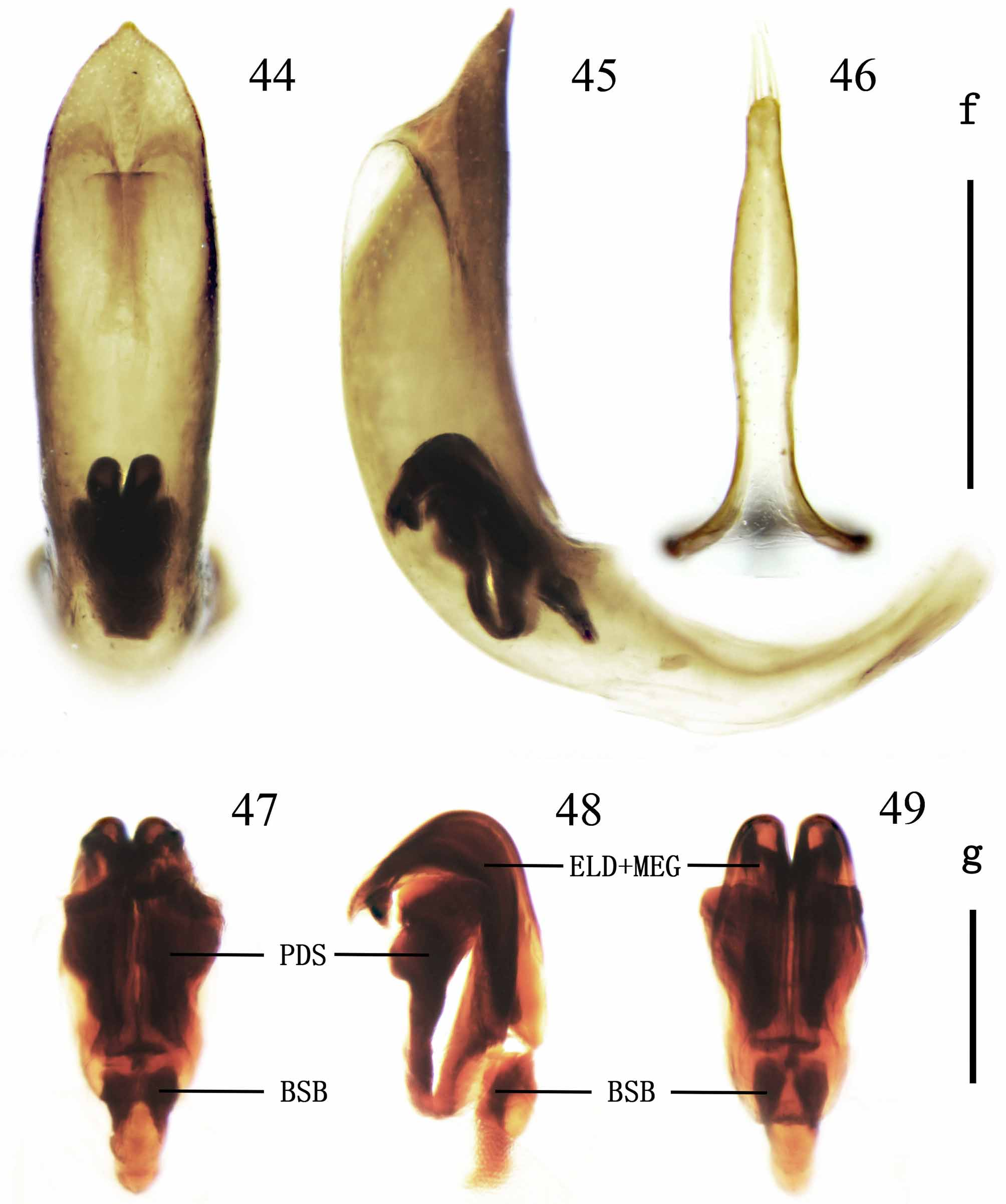
FIGURES 44 – 49. Macroplea ranina sp. nov. 44, 45. Median lobe with endophallus in it (44. Dorsal view; 45. Lateral view.); 46. Tegmen, dorsal view; 47 – 49. Endophallus (47. Dorsal view; 48. Lateral view; 49. Ventral view). Scale lines: f = 0.5 mm. (Figs 44 – 46); g = 0.2 mm. (Figs 47 – 49).
No known copyright restrictions apply. See Agosti, D., Egloff, W., 2009. Taxonomic information exchange and copyright: the Plazi approach. BMC Research Notes 2009, 2:53 for further explanation.
|
Kingdom |
|
|
Phylum |
|
|
Class |
|
|
Order |
|
|
Family |
|
|
SubFamily |
Donaciinae |
|
Genus |
1 (by plazi, 2016-04-11 02:47:03)
2 (by ImsDioSync, 2017-02-08 23:14:23)
3 (by ImsDioSync, 2017-06-16 22:20:17)
4 (by ImsDioSync, 2017-06-16 23:35:20)
5 (by ExternalLinkService, 2019-09-26 20:35:17)
6 (by ExternalLinkService, 2022-01-30 13:57:15)
7 (by ExternalLinkService, 2022-02-20 12:26:52)
8 (by plazi, 2023-10-25 19:17:20)