
Macrochaetosoma bertiscea Antić and Makarov, 2015
|
publication ID |
https://doi.org/ 10.11646/zootaxa.3948.2.1 |
|
publication LSID |
lsid:zoobank.org:pub:EC744FBA-7006-4A25-A67D-DC006AD453A8 |
|
DOI |
https://doi.org/10.5281/zenodo.6109319 |
|
persistent identifier |
https://treatment.plazi.org/id/03A2AF66-FFB9-FF9E-FF69-FE51AE68FBDD |
|
treatment provided by |
Plazi |
|
scientific name |
Macrochaetosoma bertiscea Antić and Makarov |
| status |
sp. nov. |
Macrochaetosoma bertiscea Antić and Makarov View in CoL , sp. n.
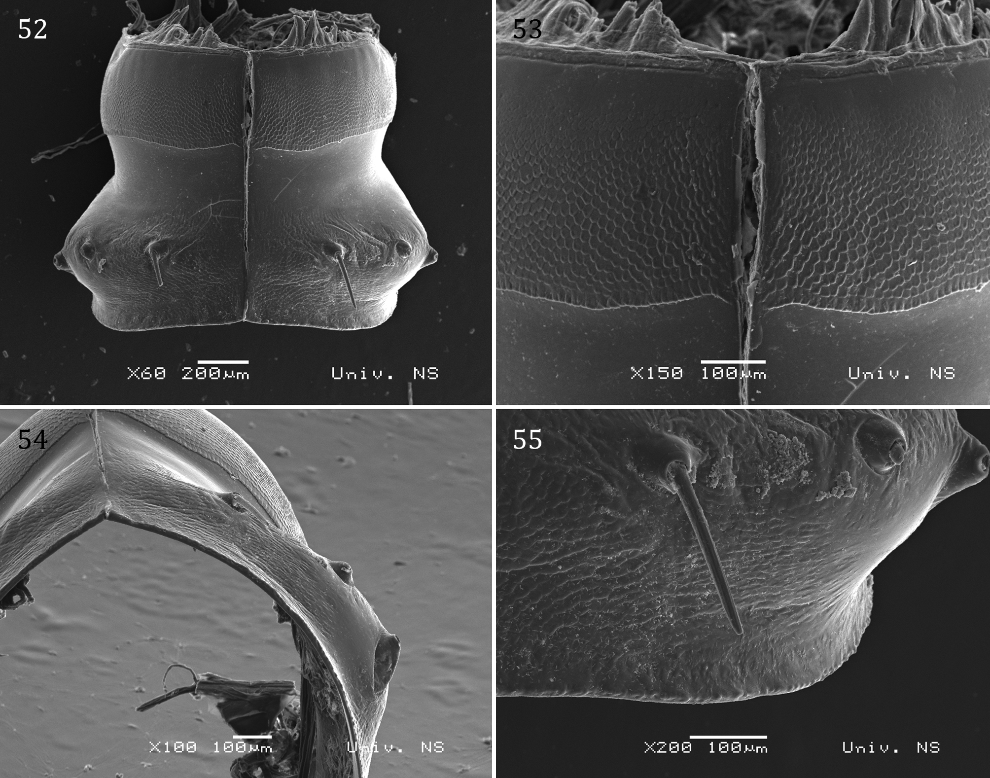
Figs 52–66 View FIGURES 52 – 55 View FIGURES 56 – 61 View FIGURES 62 – 66
Material examined. Holotype male: Gorniča Pit, ca 2000 m, Ćaf Borit, Bjelič Massif, Prokletije Mountains, Montenegro, 42°29'56.30"N, 19°53'21.70"E, 25.VII.2011, leg. I. Njunjić (IZB ANG MB 100–1). Allotype female (IZB ANG MB 100–2) and three paratype juveniles (IZB ANG MB 100–3): same data as for holotype. Further material: Juvenile from the Jama od Karimana Pit, Kučke Planine Mountains, 21.VII.2010, leg. I. Njunjić (IZB ANG MBq 100–4).
Etymology. After its type locality, Prokletije Mountains (Mons Bertiscus).
Diagnosis. The new taxon differs from the two others described in Macrochaetosoma in many characteristics of habitus and gonopodal structure such as: broad, subquadrangular, shield-like anterior coxal processes on the anterior gonopods; bifurcated medial sternal process with bilobed base on the posterior gonopods; and absence of telopodal remnants on posterior gonopods or very unusual promentum in the form of a very low and wide triangle with six setae.
Description. Body with 30 segments (including telson), in adults. Juveniles with 28 and 26 segments (including telson), respectively.
Measurements: Holotype male 27.3 mm long, vertical diameter of its largest pleurotergite 1.6 mm; allotype female 22.1 mm long, vertical diameter of its largest pleurotergite 1.7 mm.
Coloration: Pigmentless, yellowish white.
Head: Setose, frontal side in male with slight depression. Labrum (holotype ♂) with three rounded medial labral teeth, and with 2+2 labral and 8+8 supralabral bacilliform setae. Gnathochilarium (in both sexes) densely setose; promentum in the form of a low and wide triangle with six setae in one row; lingual plates wide, massive, subquadrangular. All setae very long, except setae on basal part of stipites, which are small to medium-sized. Antennae long and thin, 4.1 mm in holotype male. Length of antennomeres: I (0.16), II (0.37), III (0.91), IV (0.62), V (1.27), VI (0.46), VII (0.28), and VIII (0.03). Length/breadth ratios of antennomeres I-VII: I (1), II (1.9), III (5.3), IV (3.8), V (8.5), VI (3.1), and VII (2.5). Antennomeres II, IV, and VI slightly clavate. Antennomere V curved. Antennomeres II, IV, V, VI, and VII with one, three, one, four and one sensillum (sensilla), respectively. Blind.
Collum: Narrower than head, with six macrochaetae. Anterior side semicircular, posterior side slightly concave.
Body segments: Prozonites with hexagonal tiles on the anterior part and with smooth posterior part ( Figs 52 and 53 View FIGURES 52 – 55 ). Metazonites wrinkled ( Fig. 52 View FIGURES 52 – 55 ) with lateral keels in form of swelling ( Figs 52 and 54 View FIGURES 52 – 55 ), absent from pleurotergites XXVIII and XXIX. Better developed in male. Limbus absent. Macrochaetae on knobs. In all specimens from type material, macrochaetae were fallen off. Macrochaetal index CIX (pleurotergite 15) ~ 0.45; median index MIX (pleurotergite 15) ~ 1; paratergal index PIX (pleurotergite 15) ~ 0.35; macrochaetal angle MA (pleurotergite 15) ~160° ( Fig. 52 View FIGURES 52 – 55 ).
Telson: Epiproct with a pair of spinnerets and six setae arranged in two rows (2+2 marginal trichoid setae and 1+1 paramedial bacilliform seta). Hypoproct with two apical trichoid setae. Paraprocts with 3+3 marginal trichoid setae.
Walking legs (both sexes): Elongated and thin. Leg pairs 1 and 2 with tarsal combs, prefemora with several long and robust setae, femora and postfemora with numerous long and robust setae.
Male sexual characters: Leg pairs 3–7 greatly enlarged. Leg pair 3 with posterior excavations on all podomeres; postfemora, tibiae, and tarsi curved caudally and very flattened antero-posteriorly. Leg pair 4 with antero-posteriorly flattened podomeres with excavations; coxae with ventral protuberance, tibiae and tarsi with medio-longitudinal ridge on the oral side. All podomeres very broad and short in leg pairs 3 and 4. Leg pairs 5 and 6 are the most similar to normal walking legs, with robust femora. Coxae of leg pair 7 ventrally densely setose; tarsi broad at base and gradually tapering to last third of length, from there of the same width until claws; last third of tarsi approximately three times narrower than first third (resembling the pedipalpal chela of some pseudoscorpions with closed fingers). Leg pairs 10 and 11 with coxal glands. Leg pair 11 with posterior mesal coxal horns directed toward each other and apically touching. Sternum 10 with medial subtriangular protrusion. Coxae of leg pairs 13 and 15 with well developed, massive mesal process. This structure larger on leg pair 13 than on pair 15. Its anterior part on leg pair 13 is wider and rounded, while the posterior part is acuminating. On leg pair 15, the anterior and posterior parts are the same width, acuminate. Leg pairs 16–21 are the same size as the other walking legs.
Anterior gonopods ( Figs 56–65 View FIGURES 56 – 61 View FIGURES 62 – 66 ): Oral side with two parallel-sided, wide, almost subquadrangular, shield-like coxal processes (a) [= Cheirite sensu Verhoeff (1942); telopodites sensu Strasser (1962); innerer Coxitfortsatz sensu Strasser (1971b); or inner/paramedial main branch of the anterior coxal stem sensu Mršić (1992)]. The largest area of the oral sides of these shield-like processes is smooth, except on the top, which is wrinkled. Apically, each shield-like process carrying one outer posterior subtriangular horn (h). These shields are clearly separated from the rest of the anterior gonopods by a transverse rift. Each lateral side of the anterior gonopods with a very wide, antero-posteriorly directed lateral bristle apparatus (b1) [= äußerer Coxitfortsatz sensu Strasser (1971b); or outer/lateral branch of the anterior coxal stem sensu Mršić (1992)]. Apart from these lateral bristle apparatuses, there are two more, mesal bristle apparatuses (b2), one on each side, with numerous transversoposteriorly directed bristles. The most posterior bristles of these parts reach the cavity of the peculiar paramedial coxal vesicles (cv) [= coxite sensu Verhoeff (1942) and Strasser (1962); hinterer Basalanhang sensu Strasser (1971b); or posterior paramedial oval coxal processes sensu Mršić (1992)]. These vesicles are the most posterior parts of the anterior gonopods, giving the impression of spiral twisting when viewed distally.
Posterior gonopods ( Figs 58–61 View FIGURES 56 – 61 and 66 View FIGURES 62 – 66 ): In Mršić’s interpretation of the posterior gonopods, he noted three main structures: a medial sternite process, a paramedial coxal process, and a lateral coxal process. In our view, the posterior gonopods consist of two main structures: a medial sternal process (m) [= sternalen Mittelfortsatzes sensu Verhoeff (1942); medianen Sternitfortsatzes/mittlerer Sternitfortsatz sensu Strasser (1962, 1971b); or medial sternite process sensu Mršić (1992)]; and lateral coxal process (lcp) on both sides. The medial process is cylindrical, strongly curved posteriorly, and bifurcated distally, with a massive, bilobed basal bulge (bb). The basal and middle parts are wrinkled. In situ the bifurcated part abuts on the sternal process of leg pair 10. Both lcp consists of two parts: an inner part (cip) [= coxale Hörner sensu Verhoeff (1942); innerer Fortsatz der coxalen Seitenteile/inneren Coxitfortsatz sensu Strasser (1962, 1971b); or paramedial coxal process sensu Mršić (1992)]; and an outer part (cop) [= äußerer coxale Lappen sensu Verhoeff (1942); äußerer Fortsatz der coxalen Seitenteile sensu Strasser (1962); Seitenblatt sensu Strasser (1971b); or lateral coxal process sensu Mršić (1992)]. The inner parts of the coxal processes are the highest parts of the whole gonopodal block. These structures are curved anteriorly. The first 2/3 from anterior or posterior view are broadened, mainly smooth, while the apical 1/3 is narrower, bearing postero-mesal ribs (r) and triangular denticles (d). The outer parts of the coxal processes are wide, consisting of two smooth lobes, larger and smaller. The inner and outer parts are divided from each other by a deep gap. There are no traces of telopodal remnants; the border between the inner and outer parts is marked only with a fissure (f).
Habitat. Gorniča Pit is one of the deepest pits in Montenegro, with a depth of 516 m and length of 1629 m (http://www.speleologija.me/p/najdublje-jame-u-crnoj-gori.html). Unfortunately, no further data is available.


No known copyright restrictions apply. See Agosti, D., Egloff, W., 2009. Taxonomic information exchange and copyright: the Plazi approach. BMC Research Notes 2009, 2:53 for further explanation.